• 検索結果がありません。

Expression of Heat Shock Proteins in Response to Mild Short-term Heat Shock in Human Deciduous Dental Pulp Fibroblast-like Cells

N/A
N/A
Protected

Academic year: 2021

シェア "Expression of Heat Shock Proteins in Response to Mild Short-term Heat Shock in Human Deciduous Dental Pulp Fibroblast-like Cells"

Copied!
6
0
0

読み込み中.... (全文を見る)

全文

(1)

2021 The Hard Tissue Biology Network Association Printed in Japan, All rights reserved.

CODEN-JHTBFF, ISSN 1341-7649

Original

Expression of Heat Shock Proteins in Response to Mild Short-term Heat Shock in Human Deciduous Dental Pulp Fibroblast-like Cells

Sho Aoki

1)

, Kyoko Harada

2)

, Saki Kawai

2)

, Yoko Abe

2)

, Sachiko Nagata

2)

, Rie Imataki

2)

and Kenji Arita

2)

1)

Graduate School of Dentistry, Department of Pediatric Dentistry, Osaka Dental University, Osaka, Japan

2)

Department of Pediatric Dentistry, School of Dentistry, Osaka Dental University, Osaka, Japan (Accepted for publication, October 22, 2020)

Abstract: Appropriate heat shock results in the production of heat shock proteins (HSPs) whose expression and phosphoryl- ation contribute to repair of damaged proteins, cell proliferation, and cell recovery from shock stimuli. However, there is no information regarding the expression of HSPs in human deciduous dental pulp fibroblast-like cells (hDDPF) in response to mild short-term heat shock. The aim of this study was to investigate the cellular effects of mild short-term heat shock on hDDPF. Cells were subjected to heat shock at 43°C–49°C for 15 min, and cell proliferation was assessed by 3-(4,5-dimeth- ylthiazol-2yl)-5-(3-carboxymethoxyphenyl)-2-(4-sulfophenyl)-2H-tetrazolium assay. mRNA and protein expressions of HSP27, 70, and 90 were detected by reverse transcription PCR and western blot analysis, respectively. Phosphorylation of the AKT and ERK signaling pathways of HSP production was evaluated by western blotting. Heat shock at 43°C for 15 min increased the cell proliferation rate and the mRNA expressions of HSP27, 70, and 90 in hDDPF. Moreover, protein expres- sion of HSP70 was significantly enhanced 24 h after heat shock, and the phosphorylation of AKT was also confirmed. Be- cause HSP70 is critical in tissue repair and regeneration, mild short-term heat shock may enhance tissue repair in hDDPF.

Key words: Human deciduous dental pulp fibroblast-like cells, Heat shock proteins

Introduction

The dental pulp is a soft tissue capable of self-repair in response to various stimuli such as bacterial toxins, heat shock, and chemicals from restorative materials

1,2)

. Depending on the extent of stimuli, cells re- spond in various manners such as altering gene expression, triggering proliferation pathways, and activating cell death signaling pathways

3)

. The literature shows that prolonged and excessive heat shock exposure may damage dental pulp tissue, whereas mild heat shock is effective for cell protection, tissue repair, and tissue regeneration

4,5)

.

Heat shock results in the rapid production of heat shock proteins (HSPs) whose expression and phosphorylation contribute to the repair of damaged proteins, cell proliferation, and cell recovery from various shock stimuli

6,7)

. Based on their molecular sizes, HSPs are divided into several groups such as HSP27, HSP70, and HSP90. HSPs promote cell proliferation under shock conditions by renaturing denatured proteins, protecting shock-labile proteins, preventing protein aggregation (chap- erone function), and degrading damaged proteins (protease activity)

8,9)

.

Previous studies have demonstrated that heat shock at 42°C for 30 min not only induces HSP25 (the rodent homolog of HSP27) expression but also enhances the activity of alkaline phosphatase, an enzyme ex- pressed in the early stage of mineralization and is a marker of rat pulp cell viability

10,11)

. Furthermore, heat shock at 43°C for 45 min strongly upregulated HSP25 expression in an odontoblast-like cell line

12)

. Anoth- er study showed that heat shock at 42°C markedly induced HSP70 ex-

pression after 1 h in rat dental pulp cells and restored intercellular com- munication and alkaline phosphatase activity

13)

.

However, there is limited information regarding the expression of HSPs in human deciduous dental pulp fibroblast-like cells (hDDPF).

These cells are known to have a higher proliferation rate and larger pop- ulation than the dental pulp cells of permanent teeth

14)

. Therefore, hD- DPF may represent an ideal source of stem cells for the repair of dam- aged tooth structures, induction of bone regeneration, and treatment of neural tissue injuries or degenerative diseases

15)

. Consequently, main- taining a healthy deciduous dental pulp to the maximum possible extent is desirable to use it for regenerative medicine.

Due to their role in the repair of damaged proteins, it is possible that increasing the HSP expression would avoid dental procedures such as vital pulp amputation and pulpectomy if it is reversible pulpitis and re- sult in maintaining a healthy pulp via HSP expression

16)

. For practical clinical applications, dental pulp cells should be subjected to short-term heat shock. However, the majority of studies conducted till date report only the effects of long-term heat shock on dental pulp cells

17)

and only a few have investigated the effects of short-term heat shock.

Therefore, this study was conducted with the principal objectives of determining whether short-term heat shock can induce HSP expression in hDDPF and, if so, investigating its mechanism.

Materials and Methods Cell culture

hDDPF were obtained from the noncarious deciduous incisor of four healthy patients with orthodontic reasons at Osaka Dental University Hospital. The teeth were placed in sterile 0.01 M phosphate-buffered sa- Correspondence to: Dr. Kyoko Harada, Department of Pediatric Dentistry

School of Dentistry, Osaka Dental University, 8-1 Kuzuha Hanazono-cho,

Hirakata, Osaka 573-1121, Japan; Tel: +81-72-864-3085; Fax: +81-72-864-

3185; E-mail: kyoko-w@cc.osaka-dent.ac.jp
(2)

line (PBS). Subsequently, they were cut horizontally under sterile condi- tions, and the dental pulp tissue was gently removed, minced, and placed in 35 mm tissue culture dishes. The dental pulp tissue was cul- tured in Dulbecco’s modified Eagle’s medium (DMEM) supplemented with 10% fetal bovine serum (Wako Co., Ltd., Tokyo, Japan), 100 U/ml penicillin (Life Technologies Corp., Carlsbad, CA, USA), 100 μg/ml streptomycin (Life Technologies Corp.), and 4 mM L-glutamine (Life Technologies Corp.) at 37°C under a humidified atmosphere containing 5% CO

2

. The culture medium was replaced with fresh medium every 3 days. hDDPF were established from cells grown from the dental pulp tissue, and hDDPF at passages 2–9 were used in the experiments in this study. All experiments were approved by the Ethical Committee of Osa- ka Dental University (No 110954). Informed consent was obtained from all study participants, and the study was conducted according to the principles expressed in the Declaration of Helsinki.

Cell proliferation assay

Cell proliferation was evaluated by a CellTiter96 Aqueous One Solution Cell Proliferation Assay (Promega Corp., Madison, WI, USA) according to the manufacturer’s instructions. Briefly, hDDPF were cul- tured in 96-well plates at 1 × 10

4

cells per well for 48 h at 37°C under a humidified atmosphere containing 5% CO

2

. After 48 h, the medium was replaced with DMEM preheated to 37°C, 43°C, 44°C, 45°C, 46°C, and 49°C, and then the cells were incubated for 15 min in a water bath at the corresponding temperatures (37°C, 43°C, 44°C, 45°C, 46°C, and 49°C).

The temperature of DMEM in the well was confirmed using TP100- 100MR (ThermoPORT KK., Tokyo, Japan). After 15 min, the cells were returned to 37°C under 5% CO

2

for 24 h. Next, 3-(4,5-dimethylthi- azol-2yl)-5-(3-carboxymethoxyphenyl)-2-(4-sulfophenyl)-2H-tetrazoli- um (MTS) and phenazine methosulfate were added to the cultures. Ab- sorbance at wavelengths of 490 and 690 nm was measured using a microplate reader (Spectra Max5; Molecular Devices Inc., Downing- town, PA, USA).

Cell morphology observation

hDDPF were cultured in 6-well plates at 3 × 10

5

cells per well for 48 h at 37°C under a humidified atmosphere containing 5% CO

2

. After 48 h, heat shock was administered for 15 min using a water bath. Then, the cells were cultured at 37°C for 24 and 72 h under a humidified atmos- phere containing 5% CO

2

. Under each condition, the cell morphology was observed under a phase-contrast microscope (Olympus IX70;

Olympus Corp., Tokyo, Japan) equipped with a digital camera. Images were acquired at 300× magnification and modified by InterVideo WinD- VR (FLOVEL Filing System, FLOVEL Co., Ltd., Tokyo, Japan).

Gene expression analysis

hDDPF were cultured in 96-well plates at 1 × 10

4

cells per well for 48 h at 37°C under a humidified atmosphere containing 5% CO

2

. cDNA was synthesized from the cells using a Cells-to-CT

TM

1-Step TaqMan kit (Life Technologies Corp.) according to the manufacturer’s protocol.

Next, real-time reverse transcription PCR (RT-PCR) was performed us- ing a Step One Plus system (Life Technologies Corp.) in a total volume of 10 μl consisting of 5 μl Master Mix, 3 μl RNase-free water, 0.5 μl HSP primer (HSP27, HSP70, and HSP90), 0.5 μl glyceralde- hyde-3-phosphate dehydrogenase (GAPDH) primer, and 1 μl cDNA. In the TaqMan Gene Expression Assay (Life Technologies Corp.), the cy- cling condition was 5 min at 50°C, followed by 40 cycles of 20 s at 95°C, 3 s at 95°C, and 30 s at 60°C. To correlate the cycle threshold val- ues from the amplification plots with the copy number, a standard curve

was generated and a no-template control was included in each assay.

Protein expression analysis

hDDPF were cultured in 6-well plates at 3 × 10

5

cells per well for 48 h at 37°C under a humidified atmosphere containing 5% CO

2

. After 48 h, heat shock at 43°C was administered for 15 min using a water bath.

Then, the cells were cultured at 37°C for 24 h under a humidified atmos- phere containing 5% CO

2

. After 24 h, the culture medium was removed and cells were washed with PBS and collected using a trypsin–EDTA solution. The cell suspension was centrifuged at 1,500 rpm for 5 min at 4°C, the supernatant was removed, and the cells were washed three times with PBS. Next, 1 ml of RIPA buffer (Wako Co., Ltd., Tokyo, Ja- pan) and a Halt protease and phosphatase inhibitor cocktail (1:50, Ther- mo Scientific Inc., Carlsbad, CA, USA) were added, after which the cells were resuspended by pipetting and then incubated for 10 min on ice. Finally, the cell lysates were centrifuged at 14,500 rpm for 10 min at 4°C.

Relative expression levels of HSP27, HSP70, and HSP90 and those of the beta-actin loading control in the lysates were measured using a capillary-based, automated western blotting system (Wes, ProteinSimple Inc., Santa Clara, CA, USA). The following primary antibodies were used: rabbit anti-HSP27 (AF1580) (1:1,000, R&D Systems, Inc., Min- neapolis, MN, USA), rabbit anti-HSP70 (NBP1-77456) (1:50, Novus Biologicals LLC., Centennial, CO., USA), rabbit anti-HSP90 (NBP1- 77685) (1:50, Novus Biologicals LLC.), and mouse anti-beta-actin (#3700) (1:50, Cell Signaling Technology Inc., Tokyo, Japan). Size- based assays were performed using Wes 12 to 230-kDa rabbit and mouse master kit reagents according to the user manual (DM-001, DM- 002, ProteinSimple Inc.). Briefly, samples were diluted with a 0.1× sam- ple buffer, mixed with a master mix containing fluorescent molecular weight markers and 40 mM dithiothreitol (DTT), and heated at 95°C for 5 min. Next, the denatured samples, blocking reagent, primary antibod- ies, HRP-conjugated anti-rabbit secondary antibodies (streptavidin-HRP for VMAT2), and a chemiluminescent substrate were dispensed into the designated wells of a manufacturer-supplied microplate preloaded with separation and stacking matrices. The prepared assay plates were then loaded into the Wes system and analyzed at 37°C using the default in- strument settings. Chemiluminescent protein bands were captured by a 116 charge-coupled device (CCD) camera, and the digital images were analyzed using the Compass software (ProteinSimple Inc.). Relative band densities of all proteins were normalized to the beta-actin level for each lane.

HSP70 signaling pathway analysis

hDDPF were incubated with the PI3-K inhibitor Wortmannin (10 µM, Cayman Chemical Co., MI, USA) or the MAPK inhibitor U0126 (10 µM, Promega Corp.) for 30 min at 37°C before subjecting them to heat shock. Relative expression levels of HSP70 and those of the be- ta-actin loading control in the lysates were measured using a capil- lary-based, automated western blotting system.

hDDPF were cultured in 6-well plates at 3 × 10

5

cells per well for 48 h at 37°C under a humidified atmosphere containing 5% CO

2

. After 48 h, heat shock at 43°C was administered for 15 min using a water bath. The cells were collected after incubation at 37°C under a humidified atmos- phere containing 5% CO

2

for 15, 30, and 60 min.

The relative phospho-AKT expression levels were measured using a

capillary-based automated western blotting system. The primary anti-

bodies used were rabbit anti-AKT (#4691, 1:1,000) and rabbit anti-phos-

pho-AKT (#2965, 1:100). Size-based assays were performed using Wes

(3)

12 to 230 -kDa rabbit master kit reagents according to the user manual (DM-001, ProteinSimple Inc.). Wes was prepared as described earlier, followed by western blotting.

Statistical analysis

Results were expressed as mean ± SD. Statistical analysis was con- ducted using the t-test. Differences were considered to be significant when the P value was <0.05.

Results Heat shock affects the proliferation of hDDPF

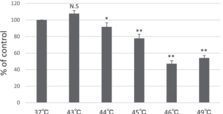
Regarding the effects of heat shock application for 15 min at 43°C, 44°C, 45°C, 46°C, and 49°C, we detected a slight increase in the prolif- eration rate of hDDPF at 43°C. In contrast, heat shock treatment for 15 min at the other temperatures, viz., 44°C, 45°C, 46°C, and 49°C, signifi- cantly reduced the proliferation rates of hDDPF compared to that in the control (Fig. 1).

Heat shock at 43°C does not affect the morphology of hDDPF hDDPF subjected to heat shock at 37°C, 43°C, and 49°C were ob- served under a phase-contrast microscope. At 24 h after heat shock at 43°C, no difference in cell morphology was found between the heat- shocked cells and the cells incubated at 37°C. However, cells subjected to 49°C heat shock exhibited cell death. At 72 h after heat shock at 43°C, the cells continued to proliferate comparable to control cells at 37°C. In contrast, there was no restoration of cell proliferation in cells heated at 49°C (Fig. 2).

Heat shock induces mRNA expression of HSPs in hDDPF

To explore the possible impact of heat shock on HSP mRNA expres- sion in hDDPF, we grew the cells at 37°C to subconfluence and then heated them at 43°C for 15 min. Samples were collected at specific time points (0, 1, 3, 6, and 24 h after heating). Results of RT-PCR analysis revealed increased HSP27 gene expressions at 3 and 6 h after heat shock. HSP70 gene expression was increased immediately after heat shock and then decreased in a time-dependent manner. Increased HSP90 gene expression was observed at 0, 1, 3, and 6 h after heat shock (Fig.

3).

Heat shock induces HSP70 protein expression in hDDPF

The protein levels of HSP27, HSP70, and HSP90 in hDDPF were determined by western blotting. hDDPF constitutively expressed HSP27, with no effects of heat shock. Low HSP70 expression was con- sistently detected, and there was almost no change immediately after heat shock. However, 24 h after heat shock, HSP70 protein expression was significantly enhanced. HSP90 expression was also observed under non-heat shock conditions with no significant change either immediately or 24 h after heat shock (Fig. 4).

PI3-K inhibitor affects HSP70 production in hDDPF

To investigate the signaling pathway, we investigated whether PI3-K and MAPK inhibitors affected HSP70 production induced by heat shock in hDDPF. Cells were incubated with the PI3-K/AKT inhibitor Wort- mannin (10 µM) or the MAPK inhibitor U0126 (10 µM) for 30 min at Figure 1. Heat shock at 43°C for 15 min slightly increases the cell prolifer-

ation rate of hDDPF. hDDPF were cultured in 96-well plates at 1×10

4

cells per well for 48 h at 37°C and then heat shocked for 15 min in a water bath set at 37°C, 43°C, 44°C, 45°C, 46°C, and 49°C. Results are expressed as mean ± SE (n = 3). *P < 0.05 and ** P < 0.01 vs. hDDPF without heat shock.

Figure 2. Heat shock of hDDPF at 43°C for 15 min does not affect cell morphology. hDDPF were cultured in 6-well plates at 3×10

5

cells per well

for 48 h at 37°C followed by heat shock for 15 min using a water bath set to 37°C, 43°C, and 49°C. Cells were then observed under a microscope

equipped with a digital camera and images were acquired at 300× magnification. Scale bar indicates 100 μm.

(4)

37°C before heat shock. Wortmannin but not U0126 inhibited the ex- pression level of HSP70 induced by heat shock (Fig. 5).

PI3-K is involved in HSP70 production in hDDPF

Heat shock at 43°C administered to hDDPF for 15 min induced AKT phosphorylation, with the maximum level at 15 min. The total AKT pro- tein level remained unaffected under the experimental conditions (Fig.

6). We next investigated whether this signaling pathway is involved in the heat-shock-induced HSP70 expression in hDDPF. It was observed that Wortmannin, a specific inhibitor of PI3-K/AKT, abolished AKT phosphorylation in heat-shocked hDDPF (Fig. 6)

Figure 3. Heat shock induces mRNA expression of HSPs in hDDPF. hDDPF were heated at 43°C for 15 min, and then HSP27, HSP70, and HSP90 gene expression levels were analysed by real-time RT-PCR. Results are expressed as mean ± SE (n = 4). *P < 0.05 and ** P < 0.01 vs.

hDDPF without heat shock.

Figure 4. Heat shock induces HSP70 protein expression in hDDPF. hDDPF were cultured in 6-well plates at 3×10

5

cells per well for 48 h at 37°C. After 48 h, cells were heat shocked at 43°C for 15 min using a water bath, fol- lowed by 24 h of culture at 37°C. The relative pixel densities of HSP27, HSP70, and HSP90 from western blot analysis were quantified and normal- ised to that of beta actin.

Figure 5. Effects of AKT and ERK inhibitor on HSP70 protein expression

in hDDPF after heat shock. hDDPF (3×10

5

) were incubated with or with-

out wortmannin (Wtmn) and U0126 for 30 min and heated at 43°C for 15

min using a water bath, followed by 24 h of culture at 37°C. The relative

pixel densities of HSP70 from western blot analysis were quantified and

normalised to that of beta actin.

(5)

Discussion

This study showed that prolonged and excessive exposure to heat shock might damage the dental pulp tissue, whereas mild heat shock is effective for cell protection, tissue repair, and tissue regeneration. Re- garding its clinical application, it is necessary to heat the dental pulp not only at the appropriate temperature but also for the shortest possible time.

Based on a previous report that heat shock for 15 min induced genet- ic changes in dental pulp cells, we also examined 15 min of heat shock, which could be applied in clinical practice

18)

. Another previous study also showed that increases in pulpal temperature of >5°C from physio- logical conditions can damage the dental pulp

19)

. Therefore, we per- formed the MTS assay to determine the temperature that affects the via- bility of hDDPF in 15 min of culture. We observed that the proliferative ability was slightly increased by heat shock at 43°C compared with con- trol (37°C). We also examined the effects 1 and 3 days after heat shock at 43°C for 15 min and observed that the cell morphology remained un- affected. Therefore, we concluded that heat shock at 43°C for 15 min is the optimum condition at which hDDPF can survive and activate their proliferative ability.

Heat shock results in a rapid production of HSPs, which participate in the repair of damaged proteins, cell proliferation, and cell recovery from various shock stimuli

6,7)

. HSP27 is present in odontoblasts and dental pulp fibroblasts, and nuclear localization and phosphorylation of HSP27 correlate with the cellular response to shock or other stimuli

20)

. HSP70 is the best-known HSP expressed in the dental pulp. This protein has been detected in the pulp during stressful conditions, including de- velopment, formation of reparative dentin, cavity preparation, and after

replantation and orthodontic tooth movement

21-23)

. HSP90 is a highly abundant cellular protein that is a key molecular chaperone associated with cell cycle regulation and apoptotic pathways

24)

. However, only a few studies have investigated HSP90 expression in deciduous dental pulp cells. Our results demonstrated that mild short-term heat shock at 43°C for 15 min affected the gene expressions of HSP27, HSP70, and HSP90 in dental pulp and dental pulp cells.

Regarding the protein expression of HSPs, we detected low levels of HSP70 expression in normal culture, and its expression did not change immediately after heat shock at 43°C for 15 min. However, 24 h after heat shock, HSP70 protein expression increased significantly. In con- trast, HSP27 protein expression was constitutive and unaltered by heat shock. Regarding HSP90, low expression was detected under normal culture conditions, and its expression remained unaffected by heat shock at 43°C for 15 min. Overall, these results suggest that only HSP70 was affected by heat shock at 43°C for 15 min after 1 h. The literature shows that the expression of HSPs other than HSP70 is also observed after prolonged heat shock

4)

. However, under the conditions of the present study, only HSP70 expression was increased, suggesting that HSP70 is the most responsive to mild short-term heat shock. As HSP70 is in- volved in cell protection, we propose that the prognosis after dental pulp treatment can be improved by applying heat shock at 43°C for 15 min in advance of treatment to increase HSP70 protein expression.

Since DNA must be translated to RNA or protein to function in vivo, genomic information is reflected in the type and amount of proteins in cells. However, the amount of mRNA does not necessarily correspond to the amount of protein, and cell-dependent modifications occur.

In this study, we only examined the changes in protein expression at 24 h after heat shock; therefore, it is necessary to investigate protein ex- pression at shorter (e.g. 6 and 12 h) or longer times (e.g. after several days) post heat shock in the further studies.

Previous studies have reported that activation of PI3-K and MAPK plays a key role in the induction of HSPs

25-28)

. We explored whether the PI3-K inhibitor Wortmannin and the MAPK inhibitor U0126 blocked HSP70 production. We observed that Wortmannin blocked HSP70 pro- duction enhanced by short-term heat shock in hDDPF. Furthermore, to investigate the signaling pathway involved, cells were preincubated with Wortmannin before heated. We observed that Wortmannin, a specific in- hibitor of AKT, abolished the activation of this kinase in hDDPF. It has been reported that the PI3-K signaling pathway plays a vital role in reg- ulating the activities of several transcriptional factors. PI3-K can phos- phorylate inositol lipids, thereby producing phosphatidylinositol 3,4,5-trisphosphate and its downstream target, and the serine/threonine kinase AKT regulates several cellular responses, including cell growth, proliferation, and migration.

In this study, we found that activation of the AKT pathway by mild short-term heat shock is involved in HSP70 expression in hDDPF. En- hanced activities of PI3-K/AKT by mild short-term heat shock induced HSP70 production, which might contribute to wound healing, cell pro- liferation, and cell recovery from various stimuli in deciduous dental pulp cells. In contrast, it has been reported that in lung fibroblast cells, HSP70 expression was detected only after chronic heat stress, and both p38 MAPK and Akt were phosphorylated

29)

. It has also been reported that long-term heat shock at 43°C for 50 min activated both MAPK and AKT in intestinal epithelial cells

30)

. Therefore, it can be suggested that differences exist in the expression of HSPs and the mechanism of HSP expression depending on the degree and type of heat shock.

Mild short-term heat shock can be used in clinical applications, where it has the potential enhance pulp repair. Therefore, it is important Figure 6. PI3K is involved in production of HSP70 in hDDPF. hDDPF

(3×10

5

) were incubated with or without wortmannin (Wtmn) for 30 min

and heated at 43°C for 15 min. Then collected 15, 30, and 60 min after in-

cubation at 37°C under a humidified atmosphere containing 5% CO

2

. The

relative pixel densities of phospho-AKT from western blot analysis were

quantified and normalised to those of AKT.

(6)

to gain better understanding of the mechanisms underlying HSP70 ex- pression induced by mild short-term heat shock for developing novel therapeutic strategies.

Acknowledgments

This work was supported by JSPS KAKENHI Grant Number JP18K17274. This study was performed at the Institute of Dental Re- search, Osaka Dental University.

Conflicts of Interest

The authors declare that they have no conflicts of interest with re- spect to the authorship or publication of this article.

References

1. Kitamura C, Nishihara T, Ueno Y, Nagayoshi M, Kasugai S and Terashita M. Thermotolerance of pulp cells and phagocytosis of ap- optotic pulp cells by surviving pulp cells following heat stress. J Cell Biochem 94: 826-834, 2005

2. Paranjpe A, Zhang H and Johnson JD. Effects of mineral trioxide aggregate on human dental pulp cells after pulp-capping. J Endod 36: 1042-1047, 2010

3. Fulda S, Gorman AM, Hori O and Samali A. Cellular stress re- sponses: cell proliferation and cell death. Int J Cell Biol 2010:

214074, 2010

4. Morotomi T, Kitamura C, Okinaga T, Nishihara T, Sakagami R and Anan H. Continuous fever-range heat stress induces thermotoler- ance in odontoblast-lineage cells. Arch Oral Biol 59: 741-748, 2014 5. Lindquist S and Craig EA. The heat-shock proteins. Annu Rev Gen-

et 22: 631-677, 1988

6. Kitamura C, Nishihara T, Ueno Y, Chen KK, Morotomi T, Yano J, Nagayoshi M and Terashita M. Effects of sequential exposure to li- popolysaccharide and heat stress on dental pulp cells. J Cell Bio- chem 99: 797-806, 2006

7. Beere HM. Death versus proliferation: functional interaction be- tween the apoptotic and stress-inducible heat shock protein path- ways. J Clin Invest 115: 2633-2639, 2005

8. Jolly C and Morimoto RI. Role of the heat shock response and mo- lecular chaperones in oncogenesis and cell death. J Natl Cancer Inst 92: 1564-1572, 2000

9. Lanneau D, de Thonel A, Maurel S, Didelot C and Garrido C. Ap- optosis versus cell differentiation. Role of heat shock proteins HSP90, HSP70 and HSP27. Prion 1: 53-56, 2007

10. Inoue T, Chen SH, Usuda J, Morohoshi Y and Shimono M. Osteo- genic activity of cells from dental pulp, periodontal ligament, bone marrow and muscle in vitro: an ultrastructural study and alka- line-phosphatase activity. Bull Tokyo Dent Coll 33: 7-12, 1993 11. Lee MW, Muramatsu T, Uekusa T, Lee JH and Shimono M. Heat

stress induces alkaline phosphatase activity and heat shock protein 25 expression in cultured pulp cells. Int Endod J 41: 158-162, 2008 12. Morotomi T, Kitamura C, Toyono T, Okinaga T, Washio A, Saito N,

Nishimura T, Terashita M and Anan H. Effects of heat stress and starvation on clonal odontoblast-like cells. J Endod 37: 955-961, 2011

13. Amano T, Muramatsu T, Amemiya K, Kubo K and Shimono M. Re- sponses of rat pulp cells to heat stress in vitro. J Dent Res 85: 432- 435, 2006

14. Harada R, Watanabe K, Shirasu S, Kato M and Daito M. DNA mi- croarray analysis of dental pulp fibroblasts exfoliated from decidu-

ous teeth. Pediatric Dent 19: 38-45, 2009

15. Gronthos S, Mankani M, Brahim J, Robey PG and Shi S. Postnatal human dental pulp stem cells (DPSCs) in vitro and in vivo. Proc Natl Acad Sci U S A 97: 13625-13630, 2000

16. Kim KK, Kim R and Kim SH. Crystal structure of a small heat- shock protein. Nature 394: 595-599, 1998

17. Okai Y, Harada K, Kawai S, Ohura K and Kato M. Expression of HSP70 in dental pulp fibroblasts from deciduous teeth. Pediatric Dent 21: 165-170, 2011

18. Soo-A K, Hong S C and Sang-G A. Pin1 induces the ADP-induced migration of human dental pulp cells through P2Y1 stabilization.

Oncotarget 7(51): 85381–85392, 2016

19. Mjör IA and Odont D. Pulp-dentin biology in restorative dentistry.

Part 2: initial reactions to preparation of teeth for restorative proce- dures. Quintessence Int 32: 537-551, 2001

20. Sens DA, McGuirt JP, Khan W, Todd JH and Howell RM. Expres- sion of heat shock protein 27 in adult human third molar dental pulp. J Oral Pathol Med 25: 382-387, 1996

21. Chen Z, Fan M, Bian Z, Zhang Q, Zhu Q and Lu P. Immunolocali- zation of heat shock protein 70 during reparative dentinogenesis.

Chin J Dent Res 3: 50-55, 2000

22. Kawagishi E, Nakakura-Ohshima K, Nomura S and Ohshima H.

Pulpal responses to cavity preparation in aged rat molars. Cell Tis- sue Res 326: 111-122, 2006

23. Shigehara S, Matsuzaka K and Inoue T. Morphological change and expression of Hsp 70, osteopontin and osteocalcin mRNAs in rat dental pulp cells with orthodontic tooth movement. Bull Tokyo Dent Coll 47: 117-124, 2006

24. McClellan AJ, Xia Y, Deutschbauer AM, Davis RW, Gerstein M and Frydman J. Diverse cellular functions of the Hsp90 molecular chap- erone uncovered using systems approaches. Cell 131: 121-135, 2007

25. Cheng H, Xia B, Su C, Chen K, Chen X, Chen P, Zou Y and Yang X.

PI3K/Akt signaling pathway and Hsp70 activate in hippocampus of rats with chronic manganese sulfate exposure. J Trace Elem Med Biol 50: 332-338, 2018

26. Rafiee P, Theriot ME, Nelson VM, Heidemann J, Kanaa Y, Horow- itz SA, Rogaczewski A, Johnson C, Ali I, Shaker R and Binion D.

Human esophageal microvascular endothelial cells respond to acidic pH stress by PI3K/AKT and p38 MAPK-regulated induction of Hsp70 and Hsp27. Am J Physiol Cell Physiol 291: C931-945, 2006 27. Wang J, Zhao Y, Ren J, Xu Y, Yang W, Lei L, Zheng Y, Qinxiu Z

and He Z. Heat shock protein 70 is induced by pepsin via MAPK signaling in human nasal epithelial cells. Eur Arch Otorhinolaryngol 276: 767-774, 2019

28. Hung JJ, Cheng TJ, Lai YK and Chang MD. Differential activation of p38 mitogen-activated protein kinase and extracellular signal-reg- ulated protein kinases confers cadmium-induced HSP70 expression in 9L rat brain tumor cells. J Biol Chem 273: 31924-31931, 1998 29. Banerjee MS, Chakraborty PK, Dey RS and Raha S. Heat stress up-

regulates chaperone heat shock protein 70 and antioxidant manga- nese superoxide dismutase through reactive oxygen species (ROS), p38MAPK, and Akt. Cell Stress Chaperones 14(6): 579-589, 2009 30. Niederlechner S, Baird C and Wischmeyer PE. P38MAP kinase, but

not phosphoinositol-3 kinase, signal downstream of glutamine-me-

diated fibronectin-integrin signaling after intestinal injury. Nutr J

12(1): 88, 2013

Figure 2. Heat shock of hDDPF at 43°C for 15 min does not affect cell morphology. hDDPF were cultured in 6-well plates at 3×10 5  cells per well  for 48 h at 37°C followed by heat shock for 15 min using a water bath set to 37°C, 43°C, and 49°C
Figure 5. Effects of AKT and ERK inhibitor on HSP70 protein expression  in hDDPF after heat shock

参照

関連したドキュメント

Although PM method has very similar smoothing results to the shock filter, their behavior has two differences: one is the PM method will not stop diffusion at corner while shock

Thus, the present study is actually quite different and can be considered as an improvement of [6] and a generalization of [3] to quasilinear (monotone operators in the gradient)

In addition to extending our existence proof there to the case of nonzero continuous drift (Theorem 1.6) and examining the effects of the order parameters 1 , 2 on e heat 1 , 2

In particular, we show that the q-heat polynomials and the q-associated functions are closely related to the discrete q-Hermite I polynomials and the discrete q-Hermite II

In section 3, we state some results on the large time behavior of subsonic solutions (for classical solutions in section 3.1 and for subsonic shock solutions in

Using the fact that there is no degeneracy on (α, 1) and using the classical result known for linear nondegenerate parabolic equations in bounded domain (see for example [16, 18]),

For strictly hyperbolic systems of conservation laws with Lipschitz contin- uous flux-functions we generalize Lax's genuine nonlinearity condition and shock ad-

“Breuil-M´ezard conjecture and modularity lifting for potentially semistable deformations after