Studies on oral colonization of periodontopathogenic bacterium Eikenella corrodens
㸦
㸦ṑ࿘ཎᛶ⣽⳦(LNHQHOODFRUURGHQVࡢཱྀ⭍ෆᐃ╔㛵ࡍࡿ◊✲㸧
㻌
㻌
FARIHA JASIN MANSUR
2017
㻌
㻌
DEDICATED TO MY BELOVED PARENTS
㻝㻌
CONTENTS
CONTENTS………. 1
LIST OF ABBREVIATIONS ………. 2
CHAPTER 1: GENERAL INTRODUCTION ………... 4
CHAPTER 2 .………. 11
2.1 ABSTRACT ………. 12
2.2 INTRODUCTION ………... 13
2.3 MATERIALS AND METHODS ……… 16
2.4 RESULTS AND DISCUSSION ……….. 21
CHAPTER 3 ………... 31
3.1 ABSTRACT ……….. 32
3.2 INTRODUCTION ……… 33
3.3 MATERIALS AND METHODS ………. 35
3.4 RESULTS AND DISCUSSION ………... 38
CHAPTER 4: GENERAL CONCLUSION ………... 44
SUMMARY ……….. 50
JAPANESE SUMMARY ………. 52
ACKNOWLEDGEMENTS ………. 54
REFERENCES ……….. 56
LIST OF PUBLICATIONS ……….. 65
㻞㻌
LIST OF ABBREVIATIONS
CE Cell envelope
GalNAc㻌 㻌 㻌 㻌 㻌 㻌 N-acetyl-D-galactosamine g Gram
g/L Gram/litre
HA㻌 㻌 㻌 㻌 㻌 㻌 㻌 㻌 Hemagglutination
∆hlyA hlyA-deficient strain H hour
IL Interleukin
IPTG Isopropyl β-D-1-thiogalactopyranoside LB㻌㻌 㻌 㻌 㻌 Luria broth
M Molar
mM Milimolar min Minute mL Mililitre
mg/mL Milligram/mililitre NaCl Sodium chloride ORF Open reading frame PBS Phosphate-buffered saline㻌 PCR Polymerase chain reaction pH㻌 㻌 㻌 㻌 㻌 㻌 㻌 㻌 㻌 㻌Potential of hydrogen
SDS–PAGE Sodium dodecyl sulfate polyacrylamide gel electrophoresis
TSB Tryptic soy broth
㻟㻌
μL Microlitre
μM Micromolar
μF Microfarad㻌
μg/mL Microgram/microlitre㻌
% Percentage
㼻C Degree Celsius Ω Ohm
㻌 㻌 㻌 㻌 㻌 㻌 㻌 㻌 㻌 㻌 㻌 㻌 㻌 㻌 㻌 㻌 㻌 㻌 㻌 㻌 㻌 㻌 㻌 㻌 㻌 㻌 㻌 㻌 㻌 㻌 㻌 㻌 㻌 㻌 㻌 㻌 㻌 㻌 㻌 㻌 㻌 㻌 㻌 㻌 㻌 㻌 㻌
㻠㻌
CHAPTER 1: GENERAL INTRODUCTION
A well-prepared army goes into battle with knowledge of the opposition, significant numbers of troops and excellent lines of communication. In some respects, bacterial pathogens are no different and in recent years it has become clear that bacterial cells are capable of exhibiting much more complex patterns of multicellular behavior than would perhaps be expected for simple unicellular micro-organisms. The ability of a single bacterial cell to communicate with its neighbors to mount a unified response that is advantageous to its survival in a hostile environment makes considerable sense. Such benefits may include improved access to complex nutrients or environmental niches, collective defense against other competitive micro-organisms or eukaryotic host defense mechanisms and optimization of population survival by differentiation into morphological forms better adapted to combating an environmental threat (Williams et al. 2000).ᴾ There are approximately ten times as many bacterial cells in the human body as human cells (Cucchiara et al. 2009). However, most of the bacteria are harmless or beneficial to the host. For example, commensal bacteria provide infection resistance, increase the overall immunological capacity of the host and act as a colonization barrier towards pathogenic microorganism (Salminen et al. 1995; Tlaskalová-Hogenová et al.
2004; Liza et al. 2010)
Though, bacterial infections affect people worldwide, and they constitute major health problems with associated morbidity and mortality. Infectious diseases are among the top ten causes of death both in low income, middle income, and high income countries (WHO Fact sheet 2008; Kim and Amar 2006). Some common infectious diseases, caused by pathogenic bacteria, are peptic ulcers and gastritis caused by Helicobacter pylori (Aspholm-Hurtig et al. 2004), periodontitis caused by
㻡㻌
Phorphyromonas gingivalis, Aggregatibacter actinomycetemcomitans and Eikenella corrodens (Asikainen et al. 2000; Hoyler et al. 2001) and dental caries associated with Streptococci and Lactobacilli (Marsh 1994).
Periodontal diseases include two general categories based on whether there is attachment or bone loss: gingivitis and periodontitis (Armitage et al. 2003). Gingivitis is induced by a bacterial plaque which forms on teeth at the gingival sulcus in the absence of effective oral hygiene (Page 1986). The initial plaque accumulation is composed mainly of viridans streptococci and actinomyces (Loesche and Syed 1978; Moore et al.
1982). Subsequently, various gram-negative bacteria and spirochetes increase in number, and gingivitis develops. The continued presence of these bacteria and the associated gingivitis may lead to periodontitis, or loss of tooth attachment (Listgarten et al. 1986).
“Peri” means around, and “odont” refers to teeth. Periodontal diseases are infections of the structures around the teeth. These include the gums, the cementum that covers the root, the periodontal ligament and the alveolar bone. In the earliest stage of periodontal disease, gingivitis, the infection affects only the gums. In more severe forms of the disease, all of the supporting tissues are involved (Colgate, on line). Periodontal disease as a risk factor for the development of various systemic conditions, such as CVD, diabetes, adverse pregnancy outcomes, and osteoporosis, is a highly researched and debated topic (Kim and Amar 2006).
In the past three decades, marked advances have occurred in our understanding of the infectious agents of periodontal disease. More than 700 different bacterial species are resident in the subgingival niche that are periodontopathogenic (Kuramitsu et al.
2007). The most frequently identified periodontal pathogens include three microaerophilic species (Actinobacillus actinomycetemcomitans, Campylobacter rectus,
㻢㻌
and Eikenella corrodens) and seven anaerobic species (Porphyromonas gingivalis, Bacteroides forsythus, Treponema denticola, Prevotella intermedia, Fusobacterium nucleatum, Eubacterium, and spirochetes). Socransky et al. (1998) divided the pathogens into two main clusters of microorganisms and deemed them the “red” and
“orange” complexes. Furthermore, they defined “green”, “yellow”, and “purple”
complexes as the bacterial colonies that formed on the tooth surface prior to the colonization of the “orange” and “red” complexes. The “red” complex consisted of three tightly related species: T. forsythensis, P. gingivalis and T. denticola. This complex is strongly related to pocket depth and bleeding on probing. Another complex (“orange” complex”) included F. nucleatum/periodonticum subspecies, P. intermedia, P.
nigrescens, Peptostreptococcus micros, C. rectus, C. gracilis, C. showae, Eubacterium nodatum, and Streptococcus constellatus, and seemed to precede colonization by species of the “red” complex. The “yellow” complex comprised six Streptococcus species: Streptococcus sp., S. sanguis, S. oralis, S. intermedius, S. gordonii, and S. mitis, while Capnocytophaga ochracea, Capnocytophaga gingivalis, Capnocytophaga sputigena, E. corrodens, and A. actinomycetemcomitans serotype a made up the “green”
complex. The fifth and final complex, the “purple” complex, consisted of Veillonella parvula, Actinomyces odontolyticus, A. actinomycetemcomitans serotype b, Selenomonas noxia, and Actinomyces naeslundii genospecies 2 (Actinomyces viscosus), but these did not constitute any cluster or ordination group.
Natural genomic transformation is present in a variety of bacterial organisms from different phyla and occupying various niches and although the mechanisms appear to be different between Gram-positive and Gram-negative organisms, the endpoint advantage is the same: to adapt to environmental stresses (Levine and Miller 1996;
㻣㻌
Hofstad and Horn 1989) and by accelerating novel virulence traits (Marks et al. 2012).
It is generally recognized that the colonization of bacteria on human mucosal tissues is an important step in the infectious process (Yamazaki et al. 1981). Bacterial pathogens have evolved a wide range of strategies to colonize and invade human body (Ribet and Cossart 2015). It has been reported that, Pneumococci lacking a gene (pspC) essential for colonization could repair its defect in the presence of a wild-type strain. In Streptococcus pneumonia, dual-strain㻌 carriage in vivo and㻌 biofilms㻌 formed㻌 in vitro㻌 can㻌 be㻌 transformed㻌 during colonization㻌 which can increase㻌 their㻌 pneumococcal㻌 fitness (Marks et al. 2012). Falush et al. provided direct evidence that, the panmictic population structure of H. pylori is caused by very frequent recombination during mixed colonization by unrelated strains resulting a highly flexible genome content and frequent shuffling of sequence polymorphisms throughout the local gene pool (Daniel㻌 et al. 2001). Previous studies of Skaar et. al. indicated that, inactivation and over expression of irg genes in the family of Neisseria gonorrhoeae have no role in pilin variation, DNA transformation or DNA repair (Eric et al. 2005). The results suggested that, colonization might have a correlation with the recombination of genomes. It has been reported that beta-hemolysin with sphingomyelinase activity from S. aureus damages keratinocytes, subsequently leading to colonization by microorganisms (Katayama et al. 2013). It has also observed that colonization of P. mirabilis isolates recovered from the urine, kidney and bladder of CBA mice with UTI showed higher hemolytic activity (Mohr et al.1999). Therefore, hemolysin might have an effect on the colonization of several kinds of pathogenic microorganisms.
Eikenella corrodens is a member of the indigenous oral microbiota; however, it can also be an opportunistic pathogen. Under dysbiotic conditions, this bacterium can
㻤㻌
be predominantly found in the subgingival plaque of patients with advanced periodontitis (Gully and Rogers 1995; Chen et al. 1996), but has also been associated with a variety of non-oral human infections, like arthritis (Chang et al. 2005), brain abscess (Karunakaran et al. 2004), liver (Yamazaki et al. 1981) and placenta (Tønjum et al. 1985). Over the past 25 years, E. corrodens has increasingly been recognized for its pathogenic potential. It was first isolated by Eiken in 1958, it was originally named Bacteroides corrodens and classified as a corroding anaerobic bacterium(Eiken 1958).
However, in1972, its current name was proposed when it was distinguished as a facultative anaerobe (Jackson and Goodman 1972; Hoyler et al. 2001). So far, its original description by Henriksen in 1948, but recently Eikenella corrodens has more gained recognition for its pathogenic potential in humans (Henriksen 1948; Brooks et al.
1974; Dorff et al. 1974). Since E. corrodens is detected in the plaque formed on teeth, it is thought to participate in the early stages of biofilm formation by specific co-aggregation with certain gram-positive and gram-negative bacteria present in human periodontal pockets. In addition, hemolysis is thought to be involved in many infections
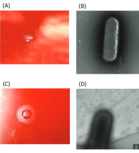
Fig. 1-1. Observation of E. corrodens strain 1073 (A and B) and strain ATCC23834 (C and D). 㻌
Colony morphologies were observed on sheep blood agar (A and C). Bacterial cells were observed by transmission electron microscope (C and D) . Strain 1073 forms corroding colonies, whereas strain ATCC23834 forms non-corroding colonies. The pilus structure was observed on the cell surface of strain ATCC 23834, whereas no pili was observed on that of strain 1073.㻌
㻥㻌
caused by pathogenic bacteria by facilitating the growth of pathogenic bacteria due to ion acquisition from erythrocytes (Otto et al. 1992). But unfortunately, there have been few reports about the hemolysis of E. corrodens.
It has been reported that GalNAc-specific lectin (large molecule and is composed of several components including 25-, 45- and 300-kDa proteins) (Yumoto et al. 1996;
Sporken et al. 1985) mediates the co-aggregation of E. corrodens with some strains of Streptococcus sanguinis and Actinomyces viscosus, which are predominant during the early stages of dental plaque formation and that it stimulates the mitogenic activity of B lymphocytes. Therefore, it can be said that the GalNAc-specific lectin contributes to the pathogenicity and virulence of E. corrodens, which can be estimated from hemagglutination (HA) activity (Azakami et al. 1996). Despite to the pathogenicity of E.
corrodens, Tonjum et al. reported the transformation of E. corrodens with chromosomal DNA from an antibiotic resistant mutant of E. corrodens (Venkatarama et al. 1993).
Based upon the results, E. corrodens, like Neisseria spp. have been characterized as natural competent for genetic transformation (Chan and McLaughlin 2000).
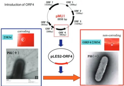
Rao and Progulske-Fox (1993) and Tønjum et.al. (1993) have been cloned 2 types IV pilin genes from E. corrodens 23834 and E. corrodens 31745. Previously, Azakami et. al. isolated a plasmid DNA (8.7 kb) from E. corrodens 1073 and designed as pMU1(Azakami et al. 2005). They found 7 open reading frames (ORFs) on pMU1 and an ORF4 which is homologous to the recombinase specific to the type IV pilin gene.
E. corrodens1073 forms large non-corroding colonies on the solid media, whereas other strains of E. corrodens form small corroding colonies. Transformation of ORF4 gene using a shuttle vector into E. corrodens strain 23834 resulted in loss of pilus structure and the morphology of the transformants changed to corroding colonies from
㻝㻜㻌
non-corroding. Furthermore, the introduction of ORF4 into strain 23834 also resulted in the elevation of growth rate, hemolytic activity, biofilm formation etc (Azakami et al.
2006a). Therefore, it is considered that ORF4-induced genomic recombination may enhance the colonization of E. corrodens to the host cells.
Fig. 1-2. Effect of the introduction of ORF4 into E. corrodens ATCC 23834 on its colony morphology and pilus structure.
Introduction of ORF4 into strain 23834 resulted in loss of pilus structure and the morphology of the transformants changed to corroding colonies from non-corroding. Furthermore, the introduction of ORF4 into strain 23834 also resulted in the elevation of growth rate, hemolytic activity, biofilm formation.
In this study, I investigated the hemolysin and genomic recombination to clarify their relationships to the colonization of E. corrodens in the oral cavity. Therefore, the objective of this study is summarized as follows:
1. To analyze the relationship between pathogenicity and hemolysis of E.
corrodens, I purified and identified the hemolysin from the periodontopathogenic bacterium E. corrodens 1073.
2. To investigate the effect of genomic recombination on the virulence factor of E.
corrodens, I transformed specific gene from E. corrodens 1073 into some other strains of E. corrodens and observed the effect on the pathogenicity of various E. corrodens strains.
㻝㻝㻌
CHAPTER 2
Purification and Characterization of Hemolysin in the
Periodontopathogenic Bacterium Eikenella corrodens Strain 1073
㻝㻞㻌 2.1 ABSTRACT
Eikenella corrodens 1073 was found to show hemolytic activity when grown on sheep blood agar. A high and dose-dependent hemolytic activity was detected in the cell envelope fraction, which was further purified by ion-exchange and gel-filtration chromatography. Consequently, a 65-kDa protein with hemolytic activity was obtained, suggesting that this protein might be a hemolysin. Its N-terminal amino acid sequence was nearly identical to that of X-prolyl aminopeptidase from E. corrodens ATCC 23834.
To confirm that X-prolyl aminopeptidase functions as a hemolytic factor, I expressed the hlyA gene, encoding X-prolyl aminopeptidase, in Escherichia coli. After induction with isopropyl β-D-1-thiogalactopyranoside, a protein of about 65 kDa was purified on a Ni column, and its hemolytic activity was confirmed. Meanwhile, a strain with a disrupted hlyA gene, which was constructed by homologous recombination, did not show any hemolytic activity. These results suggested that X-prolyl aminopeptidase might function as a hemolysin in E. corrodens.
㻌 㻌 㻌 㻌 㻌 㻌 㻌 㻌 㻌 㻌 㻌 㻌 㻌 㻌 㻌
㻝㻟㻌 2.2 INTRODUCTION
㻌
Eikenella corrodens, a gram-negative, facultative anaerobic, rod-shaped bacterium, is predominantly found in subgingival plaque samples from patients with advanced periodontitis㻌 (Tanner et al. 1979). Monoinfection of germ-free or gnotobiotic rats with E. corrodens causes periodontal disease with severe alveolar bone loss (Listgrarten et al.
1978). Previously it has been found that E. corrodens 1073 contains a cell-associated, N-acetyl-D-galactosamine (GalNAc)-specific, lectin-like substance that mediates its adherence to cell surfaces of various host tissues (Ebisu et al. 1983; Yamazaki et al.
1981; Yamazaki et al. 1988; Miki et al. 1987). In addition, it has already been reported that the GalNAc-specific lectin mediates the coaggregation of E. corrodens with some strains of Streptococcus sanguinis and Actinomyces viscosus (Ebisu et al. 1988), which are predominant during early stages of dental plaque formation, and stimulates the mitogenic activity of B lymphocytes (Nakae et al. 1994). Furthermore, E. corrodens 1073 induces KB cells (a human oral epidermoid carcinoma cell line) to express and secrete interleukin (IL)-6 and IL-8, for which no direct contact of E. corrodens with KB cells is necessarily required is also reported (Yumoto et al. 2001). Therefore, it is believed that the GalNAc-specific lectin contributes to the pathogenicity and virulence of E. corrodens, and it can be evaluated by hemagglutination (HA) activity.
Previously, a plasmid of 8.7 kb in size in strain 1073 (Azakami et al. 2005) was found and investigated its relationship to pathogenicity. As a result, it has been suggested that the type IV pilin gene locus is recombined by a recombinase, which is encoded by this plasmid (Azakami et al. 2006a). Moreover, this genomic recombination enhances the GalNAc-dependent lectin activity in parallel with the hemolytic activity of E. corrodens ATCC 23834 was also reported (Azakami et al. 2006a; Matsunaga et al.
㻝㻠㻌
2011). However, the relationship between the GalNAc-dependent lectin activity and hemolytic activity of E. corrodens has not been understood yet.
In a healthy human body, the concentration of free iron should be maintained at 10−18 M (Bullen 1981). However, bacteria require iron concentrations of 0.05–0.5 μM for their growth (Martinez et al. 1990). It has been reported that the acquisition of iron for bacterial metabolism can be facilitated by hemolysis of host erythrocytes, which can release intracellular iron (Otto et al. 1992). Moreover, it has been reported that the crevicular fluid collected from periodontitis sites had a higher iron concentration compared to that collected from gingivitis sites (Mukherjee 1985). Furthermore, most hemolysins damage host tissue, causing lysis of erythrocytes to release hemoglobin by forming pores of varying diameters in the membrane (Goebel et al. 1988).Therefore, hemolysin is regarded as a virulence factor for many gram-positive and gram-negative pathogenic bacteria and might be vital for the in vivo survival of oral bacteria (Sato et al.
2012). Hemolysin is thought to be involved in many infections caused by pathogenic bacteria since it induces bone loss owing to its cytotoxic effect and facilitates the growth of pathogenic bacteria owing to iron acquisition from erythrocytes (Chan and McLaughlin 2000). It has been reported that as extracellular toxic proteins, hemolysins are produced by many gram-negative (e.g., Escherichia coli, Serratia spp., Proteus spp., Vibrio spp., Pasteurella spp., Pseudomonas aeruginosa) and gram-positive bacteria (e.g., Streptococcus spp., Staphylococcus aureus, Listeria spp., Bacillus cereus, Clostridium tetani), all of which possess a certain pathogenic potential (Al-Shammary et al. 2012). However, there have been few reports about hemolysin production by E.
corrodens.
㻝㻡㻌
In this study, I purified and characterized a hemolytic factor from the periodontopathogenic bacterium E. corrodens 1073 to reveal the relationship between hemolysis and virulence.
㻌 㻌 㻌 㻌 㻌 㻌 㻌 㻌 㻌 㻌
㻝㻢㻌 2.3 MATERIALS AND METHODS Bacterial strains and growth conditions:
E. corrodens 1073 was provided by Dr. S. S. Socransky (Forsyth Dental Center, Boston, MA, USA), and E. corrodens ATCC 23834 was obtained from the American Type Culture Collection (Rockville, MD, USA). I constructed E. corrodens 23834 (pMU4) in a previous study (Azakami et al.2005) E. corrodens cells were grown at 37 °C either in tryptic soy broth (TSB) containing 2 mg·mL−1 KNO3 and 5 μg·mL−1 hemin or on sheep blood agar plates. Recombinant bacteria were cultured in TSB medium containing 30 μL·mL−1 kanamycin, tetracycline, or fusaric acid.
The E. coli cloning hosts, DH5α and BL21 (DE3), were grown at 37 °C with aeration in Luria–Bertani (LB) medium (10 g·L−1 tryptone, 5 g·L−1 yeast extract, and 5 g·L−1 NaCl). When necessary, LB medium was supplemented with carbenicillin (100 μg·mL−1) for plasmid selection and maintenance.
Preparation of a supernatant fraction of bacterial culture:
Cells in the late logarithmic phase of growth were harvested by centrifugation (8,000 × g, 10 min, 4 °C). The supernatant was sterile-filtered, dialyzed against 20 mM Tris–HCl buffer, and then lyophilized. After dissolving the pellet in 20 mM Tris–HCl buffer, the solution was used as a culture supernatant fraction for further investigation.
Preparation of a bacterial cell envelope fraction:
Cells in the late logarithmic phase of growth (3.6 L) were harvested by centrifugation (8,000 × g, 10 min, 4 °C), then resuspended in 20 mL of 20 mM Tris–HCl, pH 7.6, containing 0.1 mM phenylmethylsulfonyl fluoride, and sonicated for
㻝㻣㻌
30 min. After centrifugation (450 × g, 20 min, 4 °C) to remove intact cells, the supernatant was centrifuged at 10,000 × g for 40 min at 4 °C. The pellet was designated as a cell envelope (CE) fraction and used for further purification.
Purification of hemolysin:
1 g of the CE fraction was resuspended in 10 mL of 20 mM Tris–HCl, pH 7.6, containing 13.5 mM n-octyl-β-D-thioglucoside and then stirred overnight at 4 °C. After centrifugation at 40,000 × g for 60 min at 4 °C, the supernatant was dialyzed to remove the detergent and then diluted 30-fold with 20 mM Tris–HCl buffer (pH 7.6). Then, the solution was loaded onto a DEAE–Toyopearl column (Toyopearl® DEAE-650M, Tosoh, Tokyo, Japan) equilibrated with 20 mM Tris–HCl, pH 7.6, and the column was washed with the same buffer until the wash solution was free of protein. The bound proteins were eluted with a linear gradient of 0–0.15 M NaCl at a flow rate of 0.3 mL·min−1. The protein content in each fraction was determined by measuring the absorbance at 280 nm. Each peak was collected and dialyzed against distilled water.
After lyophilization and dissolution in 20 mM Tris–HCl buffer (pH 7.6), hemolytic activity of the fractions was measured. Fractions that showed hemolytic activity were loaded onto a Q-Sepharose column (Q Sepharose™ Fast Flow, GE Healthcare Life Sciences, Piscataway, NJ, USA) equilibrated with the same buffer and eluted with a linear gradient of 0–0.3 M NaCl at a flow rate of 0.3 mL·min−1. The collected fractions were tested for hemolytic activity after dialysis. Fractions that showed hemolytic activity were dissolved in 20 mM Tris–HCl buffer (pH 7.6), loaded onto a Sephadex G-75 gel filtration column (GE Healthcare Life Sciences) equilibrated with the same
㻝㻤㻌
buffer, and eluted with the same buffer at a flow rate of 0.3 mL·min−1. The fractions were tested for hemolytic activity after dialysis.
Amino acid sequencing:
After the transfer onto a polyvinylidene difluoride membrane, proteins were stained with Ponceau-S. The target protein was cut out from the membrane and applied to an automated protein sequencer (Shimadzu PPSQ-21A, Kyoto, Japan). The obtained sequence was analyzed using the BLAST program.
Hemolytic assay:
Samples (330 μL) were mixed with 560 μL of saline, 10 μL of 1 mM CaCl2, and 100 μL of rabbit blood, which was pre-washed three times with saline, and incubated at 37 °C for 24 h. Blood cells incubated with distilled water served as a positive control, and those incubated with saline served as a negative control. Following centrifugation (4,000 × g, 15 min, 4 °C), the optical density of the supernatant was measured at 540 nm in a spectrophotometer, and the hemolytic activity was expressed as a percentage of the positive control.
Hemolysis was also evaluated by the observation of colonies on sheep blood agar plate. E. corrodens strains were grown on the blood agar plates at 37 °C until colonies formed. Then, I moved the plates into refrigerator and observed clear zone of hemolysis.
Sodium dodecyl sulfate polyacrylamide gel electrophoresis:
Sodium dodecyl sulfate polyacrylamide gel electrophoresis (SDS–PAGE) was performed according to the method of Laemmli (Laemmli 1970) using a 10% or 15%
㻝㻥㻌
acrylamide separating gel. After electrophoresis, the gel was stained with Coomassie brilliant blue R-250 to visualize protein bands.
Cloning and sequencing of the gene encoding hemolysin:
Primers A (GAGTAAGATGAAGTGTGAAC) and B (AGGCTACCTGAAATT㻌 TCAGG) were designed based on the nucleotide sequence of the gene (hlyA) encoding X-prolyl aminopeptidase from E. corrodens ATCC 23834 (GenBank accession no.
NZ_EQ973321). Using these primers, polymerase chain reaction (PCR) was performed with genomic DNA of E. corrodens 1073 as a template. The amplified gene was ligated into the pGEM-T vector (Promega, Madison, WI, USA) and then introduced into cells of E. coli DH5α by electroporation. DNA was sequenced by dideoxy chain termination using a BigDye® Terminator v3.1 cycle sequencing kit (Thermo Fisher Scientific, CA, USA) and an ABI 3130 DNA sequencer (Thermo Fisher Scientific). The sequence of the hlyA gene from E. corrodens 1073 was deposited into the DDBJ/EMBL/GenBank databases under accession number LC208747.
Heterologous expression of hemolysin in E. coli:
The pET22b vector (Thermo Fisher Scientific) was used to construct a plasmid expressing the recombinant hemolysin protein with a 6 × His-tag sequence at its C-terminus. The hybrid plasmid was used to transform E. coli strain BL21 (DE3) (Thermo Fisher Scientific), which allows isopropyl β-D-1-thiogalactopyranoside (IPTG)-induced expression of T7 RNA polymerase. To confirm overexpression of the target protein, 5-mL cultures were grown in an air shaker (250 rpm) at 37 °C in LB medium containing 100 μg·mL−1 calbenicillin. At an optical density of 0.5 at 600 nm,
㻞㻜㻌
IPTG was added to a final concentration of 1 mM to induce the T7 RNA polymerase-based protein expression. Finally, expression of the target protein was confirmed by SDS–PAGE. The expressed His-tagged protein was purified using a His60 Ni gravity column (Takara Bio, Kusatsu, Japan).
Construction of a hemolysin-deficient strain:
To construct a hemolytic factor-deficient strain, a kanamycin resistance gene was inserted into the hlyA gene, and this fragment was cloned into pBR322 carrying a tetracycline resistance gene. The resultant plasmid was introduced into E. corrodens 1073 by electroporation. Tetracycline-sensitive colonies were counterselected with fusaric acid, while kanamycin- and fusaric acid-resistant colonies were selected.
Construction of the hlyA-deficient strain was confirmed using primers C (ATGAGTAATATTCACGCGCAACGTCTCGCC) and D (CATGGTAGCCATTGA㻌 GCCAAGCGGCTTCTT), which can amplify the hlyA gene, as well as primers E (CAACAAAGCCACGTTGTGTC) and F (AGCTGGCGTAATAGCGAAGA), which can amplify the kanamycin resistance gene.
Statistical analysis:
Results are shown as the mean values and standard deviation from triplicate measurements. The significance of intergroup differences was analyzed using the unpaired Student’s t-test.
㻞㻝㻌 2.4 RESULTS AND DISCUSSION
Localization of the hemolytic factor in E. corrodens 1073:
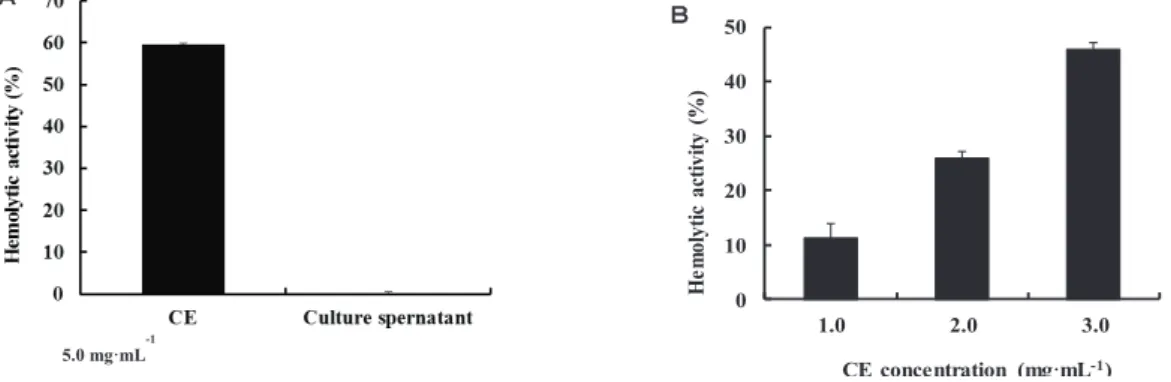
Previously, I have been observed β-hemolysis surrounding colonies of E.
corrodens 1073 on sheep blood agar media. Thus, I expected that the hemolytic factor(s) might be located on the bacterial cell surface or secreted into the extracellular space. To determine the location of the hemolytic factor(s) in E. corrodens 1073 cells, a bacterial culture was fractionated into two fractions (culture supernatant and CE fraction) as described in Materials and Methods, and a hemolytic assay was performed using each fraction. High hemolytic activity was detected in the CE fraction, whereas no activity was detected in the culture supernatant fraction (Fig. 2-1A). It was suggested that the hemolytic factor(s) might be located on the surface of bacterial cells. To further confirm the location of the hemolysin, the hemolytic assay was performed using different concentrations of the CE fraction. The hemolytic activity was increased with an increase of the CE concentration in a dose dependent manner (Fig. 2-1B). This result confirmed that the hemolytic factor is associated with the CE fraction of E. corrodens
1073. Allaker et al. (1994) have reported that hemolytic activity was observed from culture supernatants of some clinical isolates of E. corrodens. However, we could not
0 10 20 30 40 50
1.0 2.0 3.0
Hemolytic activity (%)
CE concentration (mg·mL-1)
䠝 䠞㻌
5.0 mg·mL-1㻌
Fig. 2-1. Localization of the hemolytic factor in E. corrodens 1073. 㻌
Notes: (A) Bacterial culture of E. corrodens 1073 was fractionated into two fractions, a culture supernatant fraction and cell envelope (CE) fraction. Hemolytic activity of each fraction was assayed. (B) Hemolytic activity of the CE fraction was assayed at different concentrations. The assays were repeated at least three times. Mean values and standard deviations㻌are indicated.㻌
㻞㻞㻌
observe any hemolytic activity from culture supernatant in this study. It may depend on the difference of assay conditions or the difference of used strains.
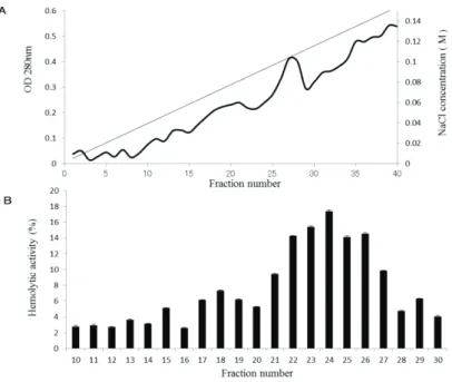
Purification of hemolysin from CE fraction:
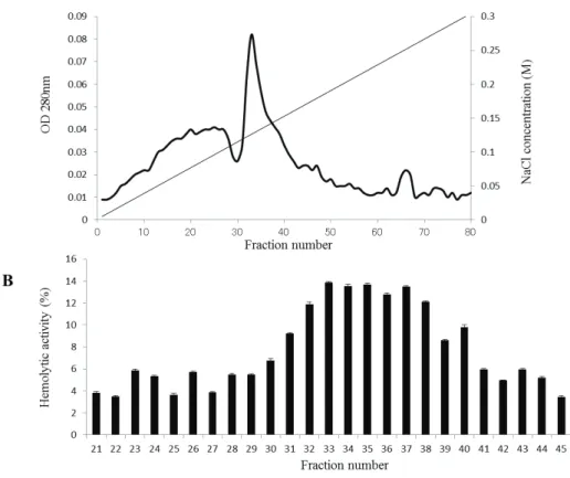
To identify the protein(s) responsible for the hemolytic activity of E. corrodens 1073, purification of the hemolysin was performed from the CE fraction. After solubilization of CE with n-octyl-β-D-thioglucoside, the solubilized proteins were passed through a DEAE–Toyopearl column. After elution with a linear gradient of 0–0.15 M NaCl, an elution pattern shown in Fig. 2-2A was observed. We measured the
hemolytic activity of each fraction. Compared to other fractions, fractions 22–26 showed higher hemolytic activity (Fig. 2-2B). Thus, it was suggested that these fractions contained the hemolytic factor.
Fig. 2-2. Purification of the hemolytic factor by ion-exchange chromatography using a DEAE–Toyopearl column. 㻌 Notes: (A) After solubilization with a detergent, the CE fraction was passed through a DEAE–Toyopearl ion-exchange column. The bound proteins were eluted with a linear gradient of 0–0.15 M NaCl at a flow rate of 0.3 mL·min−1. The protein content in each fraction was determined by measuring the absorbance at 280 nm. (B) Hemolytic activity of each eluted fraction was assayed. The assays were repeated at least three times. Mean values and standard deviations are indicated.㻌
㻞㻟㻌
For further purification, these fractions were mixed together and passed through a Q-Sepharose fast-flow column. After elution with a linear gradient of 0–0.3 M NaCl, an elution pattern shown in Fig. 2-3A was observed. I measured the hemolytic activity of each fraction. As shown in Fig. 2-3B, fractions 32–38 showed higher hemolytic activity compared with that in other samples, suggesting that these fractions contained the hemolysin.
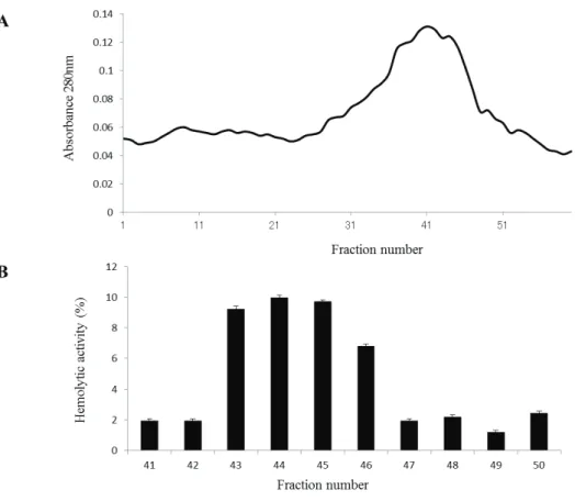
For further purification, these fractions were mixed together and separated by gel-filtration chromatography (Sephadex G-75) at a flow rate of 0.3 mL·min−1 (Fig.
2-4A). Hemolytic activity of each fraction was measured, and a high hemolytic activity was detected in fractions 43–46 (Fig. 2-4B).
Fig. 2-3. Purification of the hemolytic factor by ion-exchange chromatography on a Q-Sepharose column.㻌
Notes: (A) A fraction with a high hemolytic activity eluted after the DEAE ion-exchange chromatography was collected and passed㻌through a Q-Sepharose ion-exchange column. The bound proteins were eluted with a linear gradient of 0–0.3 M NaCl at a flow rate of 0.3 mL·min−1. The protein content in each fraction was determined by measuring the absorbance at 280 nm.
(B) Hemolytic activity of each eluted fraction was assayed. The assays were repeated at least three times. Mean values and standard deviations are indicated.㻌
㻞㻠㻌
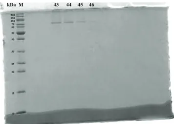
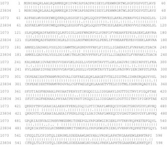
These samples were analyzed by 15% SDS–PAGE. As shown in Fig. 2-5, a single protein band was observed, with a molecular weight of about 65 kDa. Thus, it was suggested that the 65-kDa protein might be the hemolysin of E. corrodens 1073.
Identification of hemolysin protein:
Since it was suspected that the 65-kDa protein was the hemolysin, I determined its N-terminal amino acid sequence. The sequence (Ser–Asn–Ile–His–Ala–Gln–Leu–
Leu–Ala–Ala) was nearly identical to that of X-prolyl aminopeptidase from E.
corrodens ATCC 23834, except a substitution of leucine for arginine 7. Thus, it was
Fig. 2-4. Purification of the hemolytic factor by gel-filtration chromatography using a Sephadex G-75 column.
(A) A fraction with a high hemolytic activity eluted after the Q-Sepharose ion-exchange chromatography was collected and passed through a Sephadex G-75 column. The proteins were eluted with 20 mM Tris–HCl buffer (pH 7.6) at a flow rate of 0.3 mL·min−1. The protein content in each fraction was determined by measuring the absorbance at 280 nm. (B) Hemolytic activity of each eluted fraction was assayed. The assays were repeated at least three times. Mean values and standard deviations are indicated.
㻞㻡㻌
suggested that the hemolytic factor of E. corrodens 1073 might be X-prolyl aminopeptidase. As described later, I cloned and sequenced the gene encoding the X-prolyl aminopeptidase of E. corrodens 1073, which revealed that the substitution of leucine for arginine 7 was due to a misreading of the amino acid sequence.
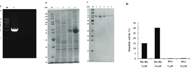
Heterologous expression of the hemolysin in E. coli:
I hypothesized that X-prolyl aminopeptidase might function as a hemolysin. To confirm this, the gene encoding X-prolyl aminopeptidase was cloned from E. corrodens 1073. Based on the nucleotide sequence of the gene encoding X-prolyl aminopeptidase from E. corrodens ATCC 23834, we designed primers A and B. Using these primers, the X-prolyl aminopeptidase gene, designated as hlyA, was amplified by PCR from the genomic DNA of E. corrodens 1073 (Fig. 2-6A). According to DNA sequencing of the hlyA gene, we found that amino acid sequence of HlyA of strain 1073 had 93.3%
identity and 99.0% similarity to that of X-prolyl aminopeptidase of strain ATCC 23834.
Fig. 2-5. SDS–PAGE analysis of the purified hemolytic factor.㻌
Notes: After gel-filtration chromatography, proteins in fractions 43 to 46 were analyzed by SDS–PAGE. M, protein molecular marker, 10–200 kDa. 㻌
㻞㻢㻌
Although there is no significant difference of amino acid sequences between 2 strains (Fig. 2-7), only strain 1073 showed hemolytic activity. It was suggested that some substitution of the coding region of X-prolyl aminopeptidase might result in the difference of hemolytic activity between 2 strains. Alternatively, it was suggested that the mutation of its promoter region might result in the difference of expression of X-prolyl aminopeptidase.
The hlyA gene was ligated into the pET22b vector. The resultant plasmid was introduced into E. coli BL21 (DE3). After induction with IPTG, overexpression of a protein about 65 kDa was observed by SDS–PAGE (Fig. 2-6B). This protein was purified by affinity chromatography using a His60 Ni gravity column. The purified 65-kDa protein was observed as a single band in a fraction eluted with imidazole (Fig.
2-6C). The purified recombinant HlyA protein showed hemolytic activity, whereas no
Fig. 2-6. Heterologous expression of the hemolysin in E. coli.㻌
Notes: (A) The hlyA gene of E. corrodens 1073 was amplified by PCR using primers specific for the hlyA gene of E. corrodens ATCC 23834 and a band of about 2.3 kb was observed. (B) The pET22b vector was used to construct a㻌plasmid expressing the recombinant HlyA protein with a C-terminal 6 × His-tag sequence. Overexpression of the HlyA protein was induced in E. coli BL21 (DE3) with IPTG. Expression of recombinant HlyA was analyzed by 10% SDS–PAGE. M, protein molecular marker; 1, crude extract of E. coli BL21 (DE3) (pET22b) without IPTG induction; 2, crude extract of E. coli BL21 (DE3) (pET22b) after induction with IPTG; 3, crude extract of E. coli BL21 (DE3) (pET22b/hlyA) without IPTG induction; 4, crude extract of E. coli BL21 (DE3) (pET22b/hlyA) after induction with IPTG. (C) After induction with IPTG, the overexpressed protein was purified by affinity chromatography using a His60 Ni gravity column. After binding of the crude extract of E. coli BL21 (DE3) (pET22b/hlyA), obtained after induction with IPTG, the target protein was eluted with 2 mL of an imidazole solution five times.
Fractions 1 to 5, respectively, eluted with imidazole, were analyzed by SDS–PAGE. (D) Hemolytic activity of purified recombinant HlyA (5 and 10 μM) was assayed. Bovine serum albumin (BSA; 5 and 10 μM) was used as a negative control. The assays were repeated at least three times. Mean values and standard deviations are indicated.㻌
㻞㻣㻌
activity was detected when using bovine serum albumin as a negative control (Fig.
2-6D). Moreover, the purified HlyA showed hemolytic activity in a dose dependent manner. Based on the data, it was suggested that the HlyA protein functions as a hemolytic factor.
Fig. 2-7. Amino acid homology of X-prolyl aminopeptidases from E. corrodens 1073 and ATCC23834.
Notes: Amino acid sequences of X-prolyl aminopeptidases from E. corrodens 1073 and ATCC23834 were aligned.
| and . show identical amino acid and similar amino acid, respectively.
∆hlyA mutation decreases hemolytic activity of E. corrodens 1073:
To further confirm HlyA involvement in hemolytic activity, a hlyA-deficient strain (∆hlyA) was constructed. After selection of kanamycin- and fusaric acid-resistant colonies, disruption of the hlyA gene was confirmed by PCR (Fig. 2-8A). As shown in Fig. 2-8B, the wild-type strain of E. corrodens 1073 showed β-hemolysis around its colony on a sheep blood agar plate. However, the ∆hlyA strain showed no hemolysis (Fig. 2-8C) on a blood agar plate. Moreover, I measured hemolytic activities of both wild-type and ∆hlyA strains (Fig. 2-8D) and showed that the hemolytic activity of the
∆hlyA strain was lower compared with that of the wild-type strain. These results
1073 1 MSNIHAQRLAALRQAMKEQKIDVWIAPSADPHISEYLPEHWRGRTWLSGFDGSVGTLAVS 60㻌 ||||||||||||||||||||||||||||||||||||||||||||||||||||||||||||㻌 23834 1 MSNIHAQRLAALRQAMKEQKIDVWIAPSADPHISEYLPEHWRGRTWLSGFDGSVGTLAVS 60㻌 㻌1073 61 ADFAELWVDSRYWEQSKRQLEGSGFILQKLGQGYPTMVESLAKRLPENAVVGIPADGLSL 120㻌 |||||||||||||||||||||||||.|||||||||||.||||.|||||.|||||||||||㻌 23834 61 ADFAELWVDSRYWEQSKRQLEGSGFVLQKLGQGYPTMIESLAERLPENSVVGIPADGLSL 120㻌 㻌1073 121 SLKQEMQADFAKKNIQLRFDIDLLNSFWHDRPGLPDNPIFVHEARFEPESASEKLARVRA 180㻌
|||.|||||||||||.||.||||||||||||||||.| ||||||||||||.|||||||||㻌 23834 121 SLKHEMQADFAKKNIHLRLDIDLLNSFWHDRPGLPENLIFVHEARFEPESVSEKLARVRA 180㻌 㻌1073 181 AMKELGADHHLVSSLDDIAWMTNLRGNDVPFNPLFISYLLIGAERATLFVNPAKLTDACR 240㻌 ||||||||.|||||||||||.|||| |||||||||||||||.|||||||||||||||||.㻌 23834 181 AMKELGADYHLVSSLDDIAWITNLRCNDVPFNPLFISYLLIDAERATLFVNPAKLTDACH 240㻌 㻌1073 241 KALNEAKIDVAEYRSVVDAVGKLSGSLLVDPDRTAVYTLGKLSAGVRIIEGINPSTLFKA 300㻌 |.|.||||||||||||||||||||||||||||||||||||||.||||.||.|||||||||㻌 23834 241 KVLSEAKIDVAEYRSVVDAVGKLSGSLLVDPDRTAVYTLGKLPAGVRVIENINPSTLFKA 300㻌 㻌1073 301 CKPEAEIEHTKNAMVRDGVALCGFFAELEQKLAAGETVTELDIDTMLIDHRSRQADYVSL 360㻌 |||||||||||||||||||||||||||||||||||||||||||||||||||||||||.||㻌 23834 301 CKPEAEIEHTKNAMVRDGVALCGFFAELEQKLAAGETVTELDIDTMLIDHRSRQADYISL 360㻌 㻌1073 361 SFDTIAGFNENAALPHYAATPEHYSTIKGQGILLIDSGAHYLDGTTDITRVIPIGQPTAE 420㻌 ||||||||||||||||||||||..|||||||||||||||||||||||||||||||.||||㻌 23834 361 SFDTIAGFNENAALPHYAATPEYHSTIKGQGILLIDSGAHYLDGTTDITRVIPIGKPTAE 420㻌 㻌1073 421 QKRDYTRVLKAHIALAEAVFPENLSGQILDTICRAPLWKEQCDYGHGTGHGVGYFLNVHQ 480㻌 |||||| |||||||||||.|||||||||||.|||||||||||||||||||||||||||||㻌 23834 421 QKRDYTLVLKAHIALAEAIFPENLSGQILDAICRAPLWKEQCDYGHGTGHGVGYFLNVHQ 480㻌 㻌1073 481 GPQRIAYNVAGIRHNVMKENMITSNEPGLYRPGKWGIRIENLVVTRRVKQPKETEFGQYL 540㻌 |||.||||..|.. | ||||||||||||||||||||.||||||| |||||||||||||||㻌 23834 481 GPQKIAYNTSGLKCNNMKENMITSNEPGLYRPGKWGFRIENLVVHRRVKQPKETEFGQYL 540㻌 㻌1073 541 CFEQLTLCPIDTQLIERSMLSDEEAAWLNGYHALVRDKLAPHTKGAAKEWLERNTRPI 598㻌 |||||||||||||||||||||||||||||.||||||.||||||.|||||||||||.||㻌 23834 541 CFEQLTLCPIDTQLIERSMLSDEEAAWLNDYHALVREKLAPHTEGAAKEWLERNTQPI 598㻌
㻞㻤㻌
suggested that X-prolyl aminopeptidase might function as a hemolytic factor in E.
corrodens 1073.
Fig. 2-8. Disruption of the hlyA gene abolished the hemolytic activity of E. corrodens 1073.
(A) Construction of a DhlyA strain of E. corrodens 1073. After selection of kanamycin- and fusaric acid-resistant colonies, disruption of the hlyA gene was confirmed by PCR. 1, PCR of genomic DNA of the wild-type strain with primers that can amplify the hlyA gene; 2, PCR of genomic DNA of the DhlyA strain with primers that can amplify the hlyA gene; 3, PCR of genomic DNA of the wild-type strain with primers that can amplify the kanamycin resistance gene; 4, PCR of genomic DNA of the DhlyA strain with primers that can amplify the kanamycin resistance gene. (B) (C)㻌Hemolysis by the wild-type strain and the DhlyA strain of E.
corrodens 1073. Colonies are shown by arrows. (D) Hemolytic activity of the wild-type and DhlyA strains of E. corrodens 1073 was assayed. The assays were repeated at least three times. Mean values and standard deviations are indicated.
So far, it has been reported that various bacteria produce hemolytic factors such as a pore-forming toxin (Los FCO et al. 2013), sphingomyelinase, and phospholipase (Flores-Diaz et al. 2016). The pore-forming toxin disrupts host cells by forming pores in the cell membrane. Sphingomyelinase and phospholipase disrupt host cells by enzymatically hydrolyzing the lipid bilayer of the cellular membrane. It has also been reported that some proteins containing an α-helix structure function as hemolysins by sticking out of the cellular membrane. Although there has been no report showing that any aminopeptidase acts as a hemolytic factor, it is suggested that the X-prolyl aminopeptidase from E. corrodens 1073 might contain an α-helix structure that can function as a hemolysin. I analyzed the amino acid sequence of X-prolyl aminopeptidase using secondary structure prediction software by Chou and Fasman (1974)and found an α-helix structure is located at its N-terminus. However, a further
㻞㻥㻌
study is necessary, since it is unknown whether X-prolyl aminopeptidase activity is required for hemolytic activity or a part of this protein is required.
Allaker et al. (1994) have reported that many strains of E. corrodens produce high levels of proline aminopeptidase and thiol-dependent hemolysin. However, they have not identified the proteins responsible for these activities and have not clarified the relationship between proline aminopeptidase and hemolysin in E. corrodens. It suggests that the hemolysin found in this study was different from other reported proteins.
Furtheremore, they suggested that reduced conditions are necessary for E. corrodens hemolytic activity. In this study, E. corrodens strains were grown in aerobic condition without shaking for aeration. In our assay, I added the calcium chloride into reaction mixture, because little activity was detected in the absence of calcium chloride in our early study. It was suggested that the reducing power of calcium ion might affect hemolytic activity of E. corrodens.
Relationship between GalNAc-dependent lectin activity and hemolytic activity:
Previously, we have reported that genomic recombination by a plasmid-mediated recombinase enhances the GalNAc-dependent lectin activity in parallel with the hemolytic activity in E. corrodens ATCC 23834 (Azakami et al 2006a; Matsunaga et al.
2011). These data suggested that the hemolytic activity is correlated with the lectin activity. To elucidate this, I measured the hemolytic activity of recombinant HlyA in the absence and presence of 3 mM GalNAc. As shown in Fig. 2-9A, the hemolytic activity was decreased by the addition of GalNAc. Since it has been reported that the lectin activity of E. corrodens 1073 was inhibited by GalNAc (Azakami et al. 2006b), this result suggested that the decrease in hemolytic activity might be due to the inhibition of
㻟㻜㻌
lectin activity. However, the lectin activity was detected in the ∆hlyA strain at the same level as in the wild-type strain (Fig. 2-9B). These results suggested that hemolysis by HlyA might require lectin activity by which bacterial cells can approach blood cells. It is suggested that X-prolyl aminopeptidase might contribute to the pathogenicitiy of E.
corrodens.
Fig. 2-9. GalNAc inhibits hemolytic activity of E. corrodens 1073.
Notes: (A) Hemolytic activity of purified recombinant HlyA (5 mM) was assayed in the absence and presence of 3 mM GalNAc.
The assays were repeated at least three times. Mean values and standard deviations are indicated. (B) Hemagglutination (HA) activity of the wild-type and DhlyA strains of E. corrodens 1073 was assayed. HA titers were expressed at the maximum dilution of the preparation that still showed HA. A representative image of three independent experiments is shown.
㻟㻝㻌
CHAPTER 3
Genomic recombination enhances pathogenic factors
in the periodontopathogenic bacterium Eikenella corrodens
㻟㻞㻌 3.1 ABSTRACT
Previously it has been reported that plasmid-mediated genomic recombination at the pilin gene locus increased hemagglutination activity, growth rate, biofilm formation, hemolytic activity, and adherence to epithelial cells in Eikenella corrodens 23834. To determine whether these enhancements were common in this bacterium, I introduced the recombinase gene ORF4 into seven clinically isolated strains. Genomic recombination at the type IV pilin gene locus was observed in strains 1080, L9B6, L8Ao3, and RV2 (group A), but not in strains 261-2, 612-L, and 257-4 (group B). Similarly, group A strains displayed changed colony morphology following loss of type IV pili, which was not observed in group B. Group A strains showed also enhanced hemagglutination activity, growth rate, hemolytic, activity and biofilm formation. These results suggest that ORF4-induced genomic recombination at the pilin gene locus is a general phenomenon in E. corrodens, which likely stimulates pathogenicity and virulence.
㻟㻟㻌 3.2 INTRODUCTION
E. corrodens, a facultative gram-negative㻌anaerobic rod, is found predominantly in subgingival plaque samples of patients with advanced periodontitis (Tanner et al.
1979). The monoinfection of germ-free or gnotobiotic rats by E. corrodens causes periodontal disease with severe alveolar bone loss (Listgarten et al. 1978). Given that E.
corrodens is detected in dental plaque (Noiri et al. 2001), it is thought that the bacterium may participate in the early stages of biofilm formation by specific coaggregation with certain gram-positive and gram-negative bacteria present in human periodontal pockets.
Previously it has been found that E. corrodens 1073 secreted cell-associated GalNAc-specific lectin-like substance that enabled adherence to various host cell surfaces (Ebisu and Okada1983; Yamazaki et al. 1981; Yamazaki et al. 1988; Miki et al.
1987). Additionally, it has found that the GalNAc-specific lectin mediated coaggregation of E. corrodens with some strains of Streptococcus sanguinis and Actinomyces viscosus (Ebisu et al. 1988), which are predominant during the early stages of dental plaque formation, while also stimulating the mitogenic activity of B lymphocytes (Nakae et al. 1994). Therefore, GalNAc-specific lectin is thought to contribute to the pathogenicity and virulence of E. corrodens, and this property can be estimated by hemagglutination (HA) activity.
On solid medium, E. corrodens 1073 forms large, non-corroding colonies, whereas other strains form small, corroding colonies due to twitching motility.
Previously, Azakami et al. (2005) identified a DNA plasmid of 8.7 kb in strain 1073 and designated it as pMU1. Upon investigating its relevance for E. corrodens pathogenicity, I identified seven ORFs on pMU1, one of which (ORF4) was
㻟㻠㻌
homologous to the recombinase specific for the type IV pilin gene. Transformants with pMU4, in which the ORF4 gene was subcloned into a shuttle vector, lost their pilus structure and formed non-corroding colonies on solid medium. Moreover, I confirmed that the introduction of the ORF4 gene into strain 23834 resulted in genomic recombination at the type IV pilin gene locus. Furthermore, it has been observed that this recombination event markedly enhanced GalNAc-specific lectin activity (Azakami et al. 2006a), as well a growth rate, biofilm formation, hemolytic activity, and adherence to epithelial cells (Azakami et al. 2006a; Matsunaga et al. 2011; Azakami et al. 2006b). In this study, I investigated the universal effect of genomic recombination in E. corrodens strains other than strain 23834.
㻟㻡㻌 3.3 MATERIALS AND METHODS Bacterial strains, plasmids, and media:
E. corrodens 1073 and 1080 were provided by S. S. Socransky (Forsyth Dental Center, Boston, MA, USA) and E. corrodens 23834 was obtained from the American Type Culture Collection (ATCC, Rockville, MD, USA). E. corrodens 612-L, 257-4, and 261-2 were isolated clinically from human supragingival plaque (Ebisu and Okada 1983). E. corrodens L8Ao3, L9B6, and RV2 were kindly provided by Dr. Giuseppe Valenza (University of Würzburg, Würzburg, Germany). Escherichia coli XL-1 Blue was used for cloning and sequencing. The E. coli/E. corrodens shuttle vector pLES2 (Stein et al. 1983) was obtained from the ATCC. E. corrodens cells were grown in tryptic soy broth (TSB) containing 2 mg/mL KNO3 and 5 μg/mL hemin, or on sheep blood agar plates at 37°C. Bacteria harboring plasmids were cultured on a medium supplemented with 50 μg/mL carbenicillin.
Transformation of E. corrodens strains:
E. corrodens was electrotransformed using a Gene Pulser electroporator (Bio-Rad, Hercules, CA, USA) with 5 to 10 μg plasmid DNA. Briefly, a 100-mL bacterial culture was grown for 12 h, washed three times in solution A (272 mM sucrose, 1 mM MgCl2
pH 7.4), and resuspended in 100 μL solution A. A 39-μL aliquot of the bacterial suspension was mixed with 1 μL DNA and electroporated at 2.1 kV, 25 μF, 200 Ω. For transformations involvingthe broad-host-range shuttle vector pLS88 and its derivatives, cells were recovered by being spun for 12 h at 37°C. After recovery, recombinantcells were cultured on sheep blood agar plates containing 50 μg/mL carbenicillin at 37°C.
㻟㻢㻌
Detection of genomic recombination at the type IV pilin gene locus:
I designed primers A (GGGAAGAAAAGGGAAGTGCT) and B (TCTTCA GGTACCGTCAGCAAAA) based on the 16S rDNA sequence of E. corrodens (GenBank accession no. AF320620), and primers C (TTTTATCCGCAATGGGTATC) and D (TACAAATCTTTGCCCTTCAC) based on the type IV pilin gene sequence of E.
corrodens 23834 (GenBank accession no. Z12609). Primers A and B allow the detection of all E. corrodens strains; whereas primers C and D are specific for those strains, in which genomic recombination occurred at the type IV pilin locus. To determine the occurrence of genomic recombination, I used these primers in combination with real-time PCR.
Real-time PCR was performed using the SYBR Green PCR Master Mix (Applied Biosystems, Waltham, MA, USA) as follows: 23 μ㻸 master mix was added to 96-well PCR plates containing genomic DNA and primers. The plates were sealed with a clear plastic sheet and placed in an ABI 7300 Sequence Detector (Applied Biosystems).
During the course of 40 cycles (94°C, 15 s and 60°C, 1 min), data were collected through optical cables connected to each well. Ct values were calculated by the ABI 7300 software and genomic recombination was estimated from the following formula:
(Ct value by primers C and D)/(Ct value by primers A and B) = Detection rate of genomic recombination.
Hemagglutination activity assay:
The HA activity assay was performed as previously described (Ebisu and Okada 1983). Erythrocytes that were obtained by the centrifugation of preserved rabbit blood were washed three times with saline before being suspended in
㻟㻣㻌
phosphate-buffered saline (PBS, pH 7.2) at a concentration of 2%. The HA assay was performed in microtiter plates (Vdispo; Nalge Nunc International, Roskilde, Denmark).
Test preparations (50 μL) were serially diluted two fold in PBS and mixed for 2 min with equal volumes of the 2% erythrocyte suspension. HA activity was examined after 1 h, and HA titers were expressed as the maximum dilution of the test preparation that exhibited HA activity.
Adherence assay for quantitation of biofilm production.
E. corrodens strains formed a macroscopically visible biofilm that was firmly attached to the wells of 96-well tissue culture plates (non-treated polystyrene, flat-bottom with lid; BD Bioscience, San Jose, CA, USA), and biofilm production was determined as described previously (Djordjevic et al. 2002). The assay measured the primary attachment and accumulation of multilayered cell clusters, and subsequent biofilm production on the polystyrene surface. Briefly, after growth in TSB for 36 h at 37°C, plates were gently washed four times with PBS, and adherent bacterial cells were fixed with methanol followed by staining with crystal violet. The optical density of the stained adherent bacterial biofilms was measured at 595 nm (OD595) with a spectrophotometer.
Statistical analysis:
Results are presented as mean values and standard deviations from triplicate measurements. The significance of intergroup differences was analyzed using Student’s t-test (unpaired t-test).
㻟㻤㻌 3.4 RESULTS AND DISCUSSION
Effect of ORF4 on colony morphology and hemolysis of E. corrodens:
It has been reported that genomic recombination increased E. corrodens 23834 pathogenicity (Azakami et al. 2005; Azakami et al. 2006a; Matsunaga et al. 2011;
Azakami et al. 2006b). To examine the effect of genomic recombination in other strains, I introduced the recombinase-encoding ORF4 gene into seven clinical isolates, 261-2, 1080, 257-4, 612-L, L9B6, L8Ao3, and RV2. I checked the colony morphology of each transformant on solid agar plates. Whereas in the absence of ORF4, the strains formed corroding colonies (Fig. 3-1A), colony morphology of strains 1080, L9B6, L8Ao3, and RV2 changed to non-corroding after introduction of ORF4 (Fig. 3-1B). This change was not observed for strains 261-2, 612-L, and 257-4 (Fig. 3-1B). Accordingly, strains were classified into two groups: A (strains 1080, L9B6, L8Ao3, and RV2), and B (strains 261-2, 612-L, and 257-4).
㻌㻌 㻌 㻌 㻌 㻌
Fig. 3-1. Colony morphology and hemolysis of E. corrodens strains.
E. corrodens strains 1080, RV2, L8Ao3, L9B6 261-2, 612-L, and 257-4 were cultured on sheep blood agar at 37°C. A) Strains transformed with empty vector pLES2. B) Strains transformed with ORF4 on pMU4. Colony morphology and hemolysis were observed on agar plates.
A)
B)
㻟㻥㻌
Next, I assessed hemolysis of E. corrodens strains on sheep blood agar media.
Hemolysis can be observed as a transparent zone surrounding the colony. Although no hemolysis was observed in the absence of ORF4 (Fig. 3-1A), strains 1080, L9B6, L8Ao3, and RV2 presented hemolytic activity after introduction of ORF4 (Fig. 3-1B).
No hemolytic activity was observed in strains 261-2, 612-L, and 257-4 after introduction of ORF4 (Fig. 3-1B). These results were consistent with those relating to colony morphology, as hemolysis was detected in all strains whose colony morphology was altered by introduction of ORF4.
In a healthy human body, the concentration of free iron should be maintained at 10-18 M (Bullen JJ 1981). However, bacteria require an iron concentration of 0.05–0.5 μM for growth (Martinez et al. 1990). To this end, bacteria can satisfy their metabolic
needs and acquire iron by applying hemolytic factors that lyse host erythrocytes and cause the release of intracellular iron (Sato et al. 2012). Therefore, hemolysis is thought to be important for the pathogenicity of many invading bacteria and increasing hemolytic activity through introduction of ORF4 might be vital for in vivo survival of oral bacteria.
Effect of ORF4 on genomic recombination at the type IV pilin gene locus:
I showed previously that introduction of ORF4 into strain 23834 resulted in genomic recombination at the type IV pilin locus. Here, I investigated whether the same occurred in other E. corrodens strains. In strains 261-2, 612-L, and 257-4, I could not detect any increased genomic recombination compared to the non-transformed strains (Fig. 3-2). Instead, introduction of ORF4 resulted in enhanced genomic recombination in strains 1080, L9B6, L8Ao3, and RV2 (Fig. 3-2). These results were consistent with
㻠㻜㻌
those pertaining to colony morphology and hemolysis. Accordingly, group A strains showed both colony morphology changes and genomic recombination at the type IV pilin locus, whereas group B strains displayed none of the above.
Fig. 3-2. Detection of genomic recombination at the type IV pilin gene locus by real-time PCR using specific primers. Ct values were calculated by specialized software and genomic recombination was evaluated based on the following formula: Ct value by primers specific for genomic recombination/Ct value by primers specific for E. corrodens = Detection rate of genomic recombination. Assays were performed at least three times. Mean values and standard deviations are reported.
Given that the pili on the cell surface are involved in the cells’ twitching motility as well as colony morphology (McMichael 1992; Villar et al. 2001), it is likely that the change from corroding to non-corroding colonies reflected a loss of pili following genomic recombination at the type IV pilin locus.
Effect of ORF4 on hemagglutination activity:
It is believed that the GalNAc-specific lectin contributes to the pathogenicity and virulence of E. corrodens. To assess the effect of introducing ORF4 on pathogenicity, I measured HA activity in seven E. corrodens strains. As shown in Fig. 3-3, HA activity was high in group A strains following introduction of ORF4, but did not change in group B strains even after introduction of ORF4.
㻠㻝㻌
I reported previously that genomic recombination by plasmid-mediated recombinase stimulated simultaneous GalNAc-dependent lectin activity and hemolytic activity in E. corrodens 23834(Azakami et al. 2006a; Matsunaga et al. 2011). Recently, I demonstrated that hemolytic activity decreased upon addition of GalNAc as described in chapter 1. Here, I show that the introduction of ORF4 enhanced both hemolytic activity and GalNAc-dependent lectin activity (Figs. 3-1 and 3-3). These findings suggest that hemolytic activity correlates with lectin activity. Moreover, I recently isolated the hemolytic factor from E. corrodens 1073 and demonstrated that in its absence lectin activity was the same in this as in the wild-type strain as described in chapter 1. It has been suggested that hemolysin and lectin are not the same protein, because absence of hemolysin does not have any effect on lectin. It is thought that once lectin mediates adhesion to the blood cell, the latter becomes susceptible to attack by hemolysins present on the bacterial surface. Therefore, enhancement of hemolytic activity following introduction of ORF4 may depend on increased lectin activity.
Fig. 3-3. Hemagglutination of rabbit erythrocytes by cell cultures of E. corrodens.
Hemagglutination titers were expressed as the maximum dilution of the preparation that still showed hemagglutination. Values shown represent means derived from three independent experiments. (A) Strains 1080, L8Ao3, L9B6, and RV2 transformed or not transformed with ORF4 on pMU4. (B) Strains 261-2, 612-L, and 257-4 transformed or not transformed with ORF4 on pMU4.
Effect of ORF4 on biofilm formation:
Previously, I reported that introduction of ORF4 into strain 23834 resulted in increased biofilm formation (Azakami et al. 2006a). Here, I investigated whether the
㻠㻞㻌
same occurred in other E. corrodens strains. As shown in Fig. 3-4, biofilm formation increased in group A strains following introduction of ORF4 on pMU4, but not in group B strains.
I suggested earlier that the GalNAc-specific lectin and other factors contributed additively to biofilm formation in some strains of E. corrodens (Azakami et al. 2006a).
In this study, I demonstrate that the introduction of ORF4 enhanced both GalNAc-specific lection activity and biofilm formation. Accordingly, the GalNAc-specific lectin might be involved in biofilm formation by E. corrodens.
Fig. 3-4. Biofilm formation by E. corrodens strains.
Strains 1073, 23834, 1080, RV2, L9B6, L8Ao3, 261-2, 612-L, and 257-4 transformed or not transformed with ORF4 on pMU4 were grown aerobically in TSB medium containing hemin and KNO3 at 37°C using polystyrene microtiter plates. Biofilm formation was quantified by measuring optical density at 595 nm (OD595). Assays were performed in at least 8 wells; mean values and standard deviations are shown.
Effect of ORF4 on the growth rate of E. corrodens:
Given that in our previous study, the introduction of ORF4 stimulated growth rate (Azakami et al. 2006a), I investigated whether the same occurred in other E. corrodens strains. As shown in Fig. 3-5, growth rate in group A strains increased after introduction of ORF4 on pMU4, whereas that in group B strains did not change. Biofilm formation is
㻠㻟㻌
thought to be a means by which pathogenic bacteria fix to a solid surface and enhance their pathogenicity (Kolenbrander et al. 2002). In this light, increased growth and biofilm formation following transformation with pMU4 might represent one of the strategies by which E. corrodens can survive in the oral cavity and, at the same time, promote pathogenicity and virulence.
Fig. 3-5. Growth rates of E. corrodens strains.
A) Strains L9B6, 1080, L8Ao3, and RV2 transformed or not transformed with ORF4 on pMU4. B) Strains 612-L, 261-2, and 257-4 transformed or not transformed with ORF4 on pMU4. Strains were grown aerobically in TSB medium containing hemin and KNO3
at 37°C. Bacterial growth was monitored spectrophotometrically by measuring optical density at 600 nm (OD600). Assays were performed at least three times. Mean values and standard deviations are reported.
㻠㻠㻌
CHAPTER 4: GENERAL CONCLUSION
Periodontal diseases are disorders of supporting structures of the teeth (example: gingivae, periodontal, ligament and supporting alveolar bone). The disease is due to an inflammatory process that results in destruction of supporting tissue of the teeth which is a result of mixed microbial infection (Haffajee et al. 2004; Kim and Amar 2006; Darveau 2010; Hasan and Palmer 2014). It has already been reported to affect up to 90% of the world’s population (Petersen et al. 2005; Borojevic 2012;
Mobeen et al. 2008). In addition, evidence has suggested for a potential role of periodontal infections in more serious systemic diseases including cardiovascular disease, respiratory infections, diabetics and low-birth weight complications (Li et al.
2000; Igari et al. 2014). E. corrodens which is a fastidious facultative anaerobic gram negative bacillus, present as a part of the normal human oral flora (Jackson et al. 1984).
Previous research has revealed that the monoinfection of germ free or gnotobiotic rats by E. corrodens causes severe periodontal disease, suggesting that E. corrodens is one of periodontopathogenic bacteria (Listgarten et al. 1978).
Otto et al. (1992) reported that addition of iron for bacterial metabolism may be facilitated by lysis of host erythrocytes through hemolysin, which release intracellular iron. Thus, hemolysis might be involved in the growth and survival of pathogenic bacteria. In chapter 2, I tried to purify and characterize the hemolysin produced by Eikenella corrodens 1073 to understand its involvement in pathogenicity.
Labischinski et al. (1985)㻌 reported that the envelope of Gram-negative bacteria has a well-defined structure: a classical protein lipid bilayer constituting the inner or cytoplasmic membrane and an outer membrane composed of an unusual asymmetric