The expression 2 in peri
of human epidermal growth factor receptor toneal metastasis of gastric cancer
Haruhiko Imamotol, Motohiro Imanol'2, Takao Satou3, Kiyotaka Okuno' and Hitoshi Shiozaki'
Tatsuki Itoh3,
'Department of Surgery and 'Pathology , Kinki University Faculty of Osakasayama Osaka 589-8511, Japan
2Ambulatory Treatment Center, Kinki University Hospital, Osakasayama Osaka 589-8511, Japan
Medicine,
Abstract
Background
The prognosis of gastric cancer patients with peritoneal metastasis (PM) is very poor. In recent report suggested that a monoclonal anti- body that targets human epidermal growth fac- tor receptor 2 (HER2) improve the prognosis of gastric cancer with HER2 overexpression and/
or gene amplification. However, HER2 overex- pression and/or gene amplification of gastric patients with PM is unclear.
Methods
HER2 expression of primary gastric lesion and peritoneal metastasis was investigated im- munohistologically and by fluorescence in-situ hybridization. Specimens were obtained from 15 patients with gastric cancer.
Results
There was no difference about the results of HER2 overexpression and gene amplification
between primary gastric lesion and peritoneal metastasis. Fifteen gastric carcinoma tissue
samples were classified as follows : 12 (80%) were scored as 0, 1 (6.7%) as 1, 2 (13.3%) as 2, and 0 (0%) as 3. Two specimens 2+ for HER-2 expression with immunohistological staining were analyzed by fluorescence in-situ hybridiza- tion. HER-2 gene amplification was not deter- mined in these samples
Conclusion
Primary gastric lesion and peritoneal metastasis of gastric cancer did not show HER2
amplification and/or overexpression. Further, prospective, larger-scale studies are needed to
evaluate the efficacy of monoclonal antibody that targets HER2 to GC patients with PM.
Key words : gastric cancer, peritoneal metas- tasis, human epidermal growth factor receptor 2
Background
Gastric cancer (GC) affects about one million people per year and is the second leading cause of cancer-related mortality worldwide.' One of the most frequent causes of death from GC is peritoneal metastasis (PM).2 For GC patients with PM, the median survival time was only 3.1 months in a multicenter prospective study.2
In recent large clinical Phase III trials, oral fluoropyrimidine-based regimens were generally accepted as the standard regimen against
advanced GC," however, looking at just PM, a few trials have been reported, e.g. methotrezate and 5-Fu sequential therapy has been reported to decrease malignant ascites in GC patients5 and another trial has shown the efficacy of modified FOLFOX-4 for GC patients with malignant ascites,6 although the one-year survival rates were only 16.2% and 27.2%, respectively. Despite such low survival rates, both regimens have been accepted as the standard chemotherapy. Addi- tionally, a multidisciplinary method, including intraperitoneal chemotherapy, hyperthermia and
Received September 28, 2011 ; Accepted October 17, 2011
19
aggressive surgery has been used to treat PM,7-9 however, these trials could not show a satisfac- tory clinical outcome. Therefore, new therapeu- tic strategies, treatment options and novel thera- peutic targets are desperately needed to enhance the current management of GC with PM.
Human epidermal growth factor receptor 2 (HER2) is a member of the epidermal growth factor receptor family (EGFR), which comprises HER1 (EGFR), HER2, HER3 (ErbB-3), and HER4 (ErbB-4). The binding of a ligand to aHER receptor family member leads to receptor dimerization and activation of intracellular signaling through receptor tyrosine kinases.1°
HER2 amplification and/or overexpression have also been observed in colon," bladder," pros- tate," pancreatic," and esophagea115 and gas- tric" carcinomas. Investigations of HER2 over- expression and the gene amplification rate in gastric and esophageal adenocarcinomas report- ed 20-30%,1748 however, there have been few reports about HER2 overexpression and gene amplification in GC with PM. In this study, we investigated HER2 overexpression and gene amplification in GC with PM.
Materials and Methods
Surgical Specimens
Biopsy samples and specimens of PM were obtained from 15 patients with GC between 2008 and 2010 in our department at the time of upper gastrointestinal endoscopy and staging laparos- copy. In accordance with Department of Surgery Kinki University Faculty of Medicine policy, written informed consent was obtained from the patients at the time of operation.
Immunohistochemical (IHC) study
Freshly obtained tissues were fixed with 4%
paraformaldehyde in 0.1 M PBS at 4°C over- night, dehydrated in graded alcohols and then embedded in paraffin. Then, 4 gm-thick serial sections were processed for immunohisto- chemical study, in addition to routine hematox- ylin and eosin staining.
The immunohistochemical reactions were real- ized by the streptavidin-biotin-peroxidase com- plex technique (StreptABC ; DAKO, Denmark).
The tissue sections were deparaffinized and in- cubated in citrate buffer in a pressure cooker for antigenic retrieval, and then endogenous perox- idase activity was blocked with 3% H202. The sections were then incubated with polyclonal
primary antibodies against HER2 (1 : 500, A0485 ; DAKO). Subsequently, they were in- cubated with the secondary biotinylated anti-
body from the LSAB+ peroxidase kit (K0690, DAKO), followed by incubation with Stre- ptavidin HRP (DAKO), and then counterstained with hematoxylin.
Immunohistochemical analysis
Immunohistochemical analyses of HER2 expression describe the intensity and staining pattern of tumor cells. As in the ToGA trial,"
immunohistochemistry scoring for HER2 classi- fied four categories : no staining, or weak stain- ing in fewer than 10% of the tumor cells (0) ; weak staining in part of the membrane in more than 10% of the tumor cells (1+) ; complete
staining of the membrane with weak or moderate intensity in more than 10% of the neoplastic cells (2+ ) ; and strong staining in more than 10%
(3+).
Fluorescence in-situ hybridization (FISH) The HER2 gene was amplified with dual-color FISH using a Passvision HER2 DNA probe kit (Vysis Inc ; Downers Grove, IL, USA) accord- ing to the manufacturer's instructions. Briefly, hybridization buffer, DNA probe, and purified water were centrifuged, and heated to 73°C for 5 min in a water bath. Slides were immersed in a denaturing bath (70% formamide 2 X SSC) for 5 min at 73°C, followed by dehydration in increas- ing ethanol concentrations, and then dried. The probe mixture was applied to each slide. The slides were placed in a 42°C incubator for 30 min, washed with 0.4 X SSC/0.3% NP-40 for 2 min, air-dried in darkness, counterstained with 4',6-diamidino-2-phenylindole (DAPI), and cov- ered with a cover-slip. HER-2/neu-spectrum orange probe contains a DNA sequence specific for the HER-2 human gene locus and hybridized to the region 17q11.2-q12 of human chromo- somes. CEP17 (chromosome enumeration probe 17)/spectrum green probe containing alpha-sat- ellite DNA that hybridizes to the D17Z1 locus (centromere region of chromosome 17) was used as a control. Nuclei were counterstained with DAPI.
FISH analysis
The slides were observed under a B X 60 fluo- rescence microscope equipped with a digital camera (DP50 ; Olympus, Tokyo, Japan) and the images were captured on a Windows PC with Viewfinder Lite software. A cell was considered to be amplified when a definite cluster or more
20
than 10 signals for HER-2 were found. Known positive and negative cells were used as controls for each FISH. Gene amplification was scored when a minimum of 20 cancer cell nuclei exhibit- ed a HER-2/CEP17 ratio z 2, or when a HER-2 signal cluster was observed.
GC were classified and interpreted according to the evaluation protocol recommended by the American Joint Committee on Cancer (AJCC) and International Union against Cancer (IUCC). For histological type, gastric cancers were also classified as intestinal type or diffuse type using The Laurens system.
Results
Patient characteristics
Patients had a median age of 59 years (range 47-75 years). There were 6 female and 9 male patients. Borrmann III and IV types accounted for the majority. The details of the main clinicopathological features of patients are
Table 1 Clinicopathological features of patients
presented in Table 1.
Expression of HER-2 protein in gastric cancer HER-2 protein status in 15 GC specimens from gastric cancer was determined with im- munohistochemical staining. Figure 1 demon- strates a representative case. There was no difference about the results of HER2 overexpres- sion and gene amplification between primary gastric lesion and peritoneal metastasis. Fifteen CG tissue samples were classified as follows ; 12 (80%) were scored as 0, 1 (6.7%) as 1, 2 (13.3%) as 2, and 0 (0%) as 3 (Fig. 2).
HER-2 gene amplification in gastric cancer Two specimens 2+ for HER-2 expression with IHC staining were analyzed by FISH (Fig. 2).
HER-2 gene amplification was not determined in these samples (Fig. 2). Finally, the HER2 over- expression and gene amplification rate was 0%
(0/15) in all PM specimens from GC.
Clinicopathological factors No. of cases
Gender Male
Female Average age (range ; Borrmann type
I II III IV Laurens system
intestinal type
diffuse type
years)
9 6 59 (47-75)
0 1 5 9
4 11
IHC score: 0 12 cases
IHC score: 1+
1 cases PM specimen
of GC: 15 cases
FISH: negative 2 cases IHC score: 2+
2 cases
FISH: positive 0 cases IHC score: 3+
0 cases
Fig. 2 Analysis of HER2 overexpression and gene amplifi- cation
HER2 overexpression and gene amplification cascade in peritoneal metastasis from gastric cancer.
PM : peritoneal metastasis, GC : gastric cancer, IHC : immunohistochemistory, FISH : Fluorescence in-situ hybridization, HER2: human epidermal growth factor receptor
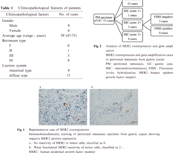
Fig. 1
a
• t: ,•::
.-• .:;„ -,;;;:* , • ; ,
• ‘1, s.. „
$.5*. '0—.1' • •
,
't • • Ili. a-. - , • „
* ' ..";`' • i• •••• 14 . 1'1/4; '
p". !;,•,S 's.
. ;-
•
• "tel. ; , , •
Representative case of HER2 overexpression
Immunohistochemistry staining of peritoneal metastasis specimen from gastric negative HER-2 protein expression.
a : No reactivity of HER2 in tumor cells, classified as 0.
b : Weak basolateral HER2 reactivity of tumor cells, classified as 2+.
HER2: human epidermal growth factor receptor
cancer showing
21
Discussion
In our data, all biopsy samples from primary lesion and PM specimens did not show HER2 overexpression and/or gene amplification. Boz- zetti et al. compared the HER2 status in primary and paired metastatic sites of GC, and concluded that high concordance was observed between HER2 results obtained by both IHC and FISH on primary tumors and metastatic lesions,2°
however, they did not mention PM. Addition- ally, common GC classified as intestinal type is more likely to be HER2-positive (16-34%) than diffuse (2-7%).18,21 PM easily developed from GC classified as diffuse type ; therefore, the PM spec- imen was suspected to not show HER2 overex- pression and/or gene amplification. However, there are no data about HER2 overexpression and/or gene amplification of primary lesion and PM. This is the first report about these findings.
The reason for not showing overexpression of HER2 in diffuse-type GC is complex and needs further investigation. The association of HER2 with a specific type suggests that intestinal- and diffuse-type gastric cancers develop along differ- ent molecular pathways and supports earlier studies showing distinct patterns of genetic alter- ations in gastric cancers with differing histopath- ologic features.22
Some similarities can be drawn with breast cancer : low expression of E-cadherin was recog- nized in lobular invasive breast carcinomas23'24 and diffuse-type gastric cancers,25 which correlat- ed with the low frequency of HER2 amplifica- tion and/or overexpression.23,24,26 These results are consistent with our results.
In this study, there were no patients with HER2 overexpression 2+ and amplification positive. In the ToGA study, probably because of 75% patients belonged to the intestinal type, so 27% patients might show HER2 overexpression 2+ and amplification positive. In contrast, 73%
of patients in our study belonged to the diffuse type. However, a small number of patients showed HER2 overexpression 1+ or 0 and amplification positive.27 To determined exact HER2 expression, HER2 expression could be tested by FISH.
ToGA trial suggested that a monoclonal anti- body that targets human epidermal growth factor receptor 2 (HER2) improve the prognosis of advanced and/or recurrent gastric cancer patients with HER2 overexpression and/or gene
amplification." However, HER2 overexpression and/or gene amplification of gastric patients with PM is unclear. Although our study size was small, PC patients with PM not showed HER2 overexpression and/or gene amplification. Fur- ther, prospective, larger-scale studies are needed to evaluate the efficacy of monoclonal antibody that targets HER2 to GC patients with PM.
In conclusion, peritoneal metastasis with gas- tric cancer did not show HER2 amplification
and/or overexpression. This is the first report about these findings. Further, prospective, lar- ger-scale studies are needed to evaluate the effi- cacy of monoclonal antibody that targets HER2 to GC patients with PM.
Acknowledgments
The authors express their appreciation to Dr. Har- umasa Ohyanagi, vice board director of the University of KinDAI Himeji for his expert comments on the manu- script. We also wish to thank Mr. Tadao Uesugi and Miss Fusako Kamada for technical assistance.
References
1. Parkin DM, Bray F, Ferlay J, Pisani P (2001) Estimating the world cancer burden : Globocan 2000.
Int J Cancer 94: 153-156
2. Sadeghi B, Arvieux C, Glehen 0, Beaujard AC, Rivoire M, Baulieux J, Fontaumard E, Brachet A, Caillot JL, Faure JL, Porcheron J, Peix JL, Francois Y, Vignal J, Gilly FN (2000) Peritoneal carcinomatosis from non-gynecologic malignancies : results of the EVOCAPE 1 multicentric prospective study. Cancer 88 : 358-363
3. Cunningham D, Starling N, Rao S, Iveson T, Nicol- son M, Coxon F, Middleton G, Daniel F, Oates J, Norman AR (2008) Capecitabine and oxaliplatin for advanced esophagogastric cancer. N Engl J Med 358 : 36-46
4. Koizumi W, Narahara H, Hara T, Takagane A, Akiya T, Takagi M, Miyashita K, Nishizaki T, Kobayashi 0, Takiyama W, Toh Y, Nagaie T, Takagi S, Yamamura Y, Yanaoka K, Orita H, Takeuchi M (2008) S-1 plus cisplatin versus S-1 alone for first-line treatment of advanced gastric cancer (SPIRITS trial) : a phase III trial. Lancet Oncol 9 : 215-221
5. Yamao T, Shimada Y, Shirao K, Ohtsu A, Ikeda N, Hyodo I, Saito H, Iwase H, Tsuji Y, Tamura T, Yamamoto S, Yoshida S (2004) Phase II study of sequential methotrexate and 5-fluorouracil chemother- apy against peritoneally disseminated gastric cancer with malignant ascites : a report from the Gastrointesti- nal Oncology Study Group of the Japan Clinical Oncology Group, JCOG 9603 Trial. Jpn J Clin Oncol 34 : 316-322
22
6. Oh SY, Kwon HC, Lee S, Lee DM, Yoo HS, Kim SH, Jang JS, Kim MC, Jeong JS, Kim HJ (2007) A Phase II study of oxaliplatin with low-dose leucovorin and bolus and continuous infusion 5-fluorouracil (modified FOLFOX-4) for gastric cancer patients with malignant ascites. Jpn J Clin Oncol 37 : 930-935 7. Imano M, Imamoto H, Itoh T, Satou T, Peng YF,
Yasuda A, Kato H, Shiraishi 0, Shinkai M, Yasuda T, Takeyama Y, Okuno K, Shiozaki H (2011) Safety of intraperitoneal administration of paclitaxel after gas- trectomy with en-bloc D2 Lymph node dissection. J Surg Oncol 105: 43-47
8. Sugarbaker PH, Yonemura Y (2000) Clinical path- way for the management of resectable gastric cancer with peritoneal seeding : best palliation with a ray of hope for cure. Oncology 58 : 96-107
9. Fujimoto S, Takahashi M, Kobayashi K, Kure M, Mutou T, Masaoka H, Ohkubo H (1993) Relation between clinical and histologic outcome of intraper- itoneal hyperthermic perfusion for patients with gastric cancer and peritoneal metastasis. Oncology 50 : 338- 343
10. Akiyama T, Sudo C, Ogawara H, Toyoshima K, Yamamoto T (1986) The product of the human c-erbB- 2 gene : a 185-kilodalton glycoprotein with tyrosine kinase activity. Science 232: 1644-1646
11. Schuell B, Gruenberger T, Scheithauer W, Zielinski C, Wrba F (2006) HER 2/neu protein expression in colorectal cancer. BMC Cancer 6 : 123
12. Eltze E, Wulfing C, Von Struensee D, Piechota H, Buerger H, Hertle L (2005) Cox-2 and Her2/neu co- expression in invasive bladder cancer. Int J Oncol 26 : 1525-1531
13. Jorda M, Morales A, Ghorab Z, Fernandez G, Nadji M, Block N (2002) Her2 expression in prostatic cancer : a comparison with mammary carcinoma. J Urol 168 : 1412-1414
14. Yamanaka Y, Friess H, Kobrin MS, Buchler M, Kunz J, Beger HG, Korc M (1993) Overexpression of HER2/neu oncogene in human pancreatic carcinoma.
Hum Pathol 24 : 1127-1134
15. Reichelt U, Duesedau P, Tsourlakis M, Quaas A, Link BC, Schurr PG, Kaifi JT, Gros SJ, Yekebas EF, Marx A, Simon R, Izbicki JR, Sauter G (2007) Fre- quent homogeneous HER-2 amplification in primary and metastatic adenocarcinoma of the esophagus. Mod Pathol 20 : 120-129
16. Jaehne J, Urmacher C, Thaler HT, Friedlander-Klar H, Cordon-Cardo C, Meyer HJ (1992) Expression of Her2/neu oncogene product p185 in correlation to clinicopathological and prognostic factors of gastric carcinoma. J Cancer Res Clin Oncol 118: 474-479 17. Yan B, Yau EX, Bte Omar SS, Ong CW, Pang B,
Yeoh KG, Salto-Tellez M (2010) A study of HER2 gene amplification and protein expression in gastric cancer. J Clin Pathol 63 : 839-842
18. Tanner M, Hollmen M, Junttila TT, Kapanen AI, Tommola S, Soini Y, Helin H, Salo J, Joensuu H,
Sihvo E, Elenius K, Isola J (2005) Amplification of HER-2 in gastric carcinoma : association with Topoisomerase IIalpha gene amplification, intestinal type, poor prognosis and sensitivity to trastuzumab.
Ann Oncol 16 : 273-278
19. Bang YJ, Van Cutsem E, Feyereislova A, Chung HC, Shen L, Sawaki A, Lordick F, Ohtsu A, Omuro Y, Satoh T, Aprile G, Kulikov E, Hill J, Lehle M, Ruschoff J, Kang YK (2010) Trastuzumab in combina- tion with chemotherapy versus chemotherapy alone for treatment of HER2-positive advanced gastric or gastro- oesophageal junction cancer (ToGA) : a phase 3, open-label, randomised controlled trial. Lancet 376 : 687-697
20. Bozzetti C, Negri FV, Lagrasta CA, Crafa P, Bas- sano C, Tamagnini I, Gardini G, Nizzoli R, Leonardi F, Gasparro D, Camisa R, Cavalli S, Silini EM, Ardizzoni A (2011) Comparison of HER2 status in primary and paired metastatic sites of gastric car- cinoma. Br J Cancer 104: 1372-1376
21. Hede K (2009) Gastric cancer : trastuzumab trial results spur search for other targets. J Natl Cancer Inst 101 : 1306-1307
22. Wu CW, Chen GD, Fann CS, Lee AF, Chi CW, Liu JM, Weier U, Chen JY (2002) Clinical implications of chromosomal abnormalities in gastric adenocar- cinomas. Genes Chromosomes Cancer 35 : 219-231 23. Perou CM, Sorlie T, Eisen MB, van de Rijn M,
Jeffrey SS, Rees CA, Pollack JR, Ross DT, Johnsen H, Akslen LA, Fluge 0, Pergamenschikov A, Williams C, Zhu SX, Lonning PE, Borresen-Dale AL, Brown PO, Botstein D (2000) Molecular portraits of human breast tumours. Nature 406: 747-752
24. Sorlie T, Perou CM, Tibshirani R, Aas T, Geisler S, Johnsen H, Hastie T, Eisen MB, van de Rijn M, Jeffrey SS, Thorsen T, Quist H, Matese JC, Brown PO, Bot- stein D, Eystein Lonning P, Borresen-Dale AL (2001) Gene expression patterns of breast carcinomas distin- guish tumor subclasses with clinical implications. Proc Natl Acad Sci U S A 98 : 10869-10874
25. Shiozaki H, Tahara H, Oka H, Miyata M, Kobayashi K, Tamura S, Iihara K, Doki Y, Hirano S, Takeichi M, et al (1991) Expression of immunoreactive E-cadherin adhesion molecules in human cancers. Am J Pathol 139: 17-23
26. Im SA, Kim JW, Kim JS, Kim MA, Jordan B, Pickl M, Han SW, Oh DY, Lee HJ, Kim TY, Kim WH, Yang HK, Bang YJ (2011) Clinicopathologic charac- teristics of patients with stage III/IV (M(0)) advanced gastric cancer, according to HER2 status assessed by immunohistochemistry and fluorescence in situ hybridi- zation. Diagn Mol Pathol 20: 94-100
27. Yang J, Luo H, Li Y, Li J, Cai Z, Su X, Dai D, Du W, Chen T, Chen M (2011) Intratumoral Heterogeneity Determines Discordant Results of Diagnostic Tests for Human Epidermal Growth Factor Receptor (HER) 2 in Gastric Cancer Specimens. Cell Biochem Biophys