INTRODUCTION
In the eyeball, a very weak electric current runs from the corneal toward the retinal side, and there is always a potential difference called the retinal rest-ing potential (RP). RP in the normal human eye was first observed by Dewar in 1877 and subsequently studied by Schott, Miles (1, 2) and Arden (3, 4), and a record of RP was termed an electrooculogram (EOG) by Carmichael and Darborn (5).
At present, EOG is used for the evaluation of ocu-lomotor abnormalities such as nystagmus, strabis-mus, and supranuclear oculomotor dysfunction (6,
7). In addition, EOG is recorded for a long time with changes in the adaptation state, and used for the examination of the function of the retinal deep area such as the retinal pigment epithelium based on changes in its amplitude (8, 9).
However, since EOG is affected by measurement conditions and environments, its reproducibility is not always high. One of the associated errors con-cerns the influence of the potential from the other eye, i.e., cross-talk.
We have performed studies to increase the ac-curacy of EOG. In conventional EOG, the potential difference between the medial and lateral canthi is measured, as shown in Fig. 1. We have reported that the eyeball can be analyzed as a battery model (10). In the battery model of the eye, the electrode potentials at the medial and lateral canthi are sepa-rately calculated, but we calculate the potential dif-ference between them to be consistent with the
con-ORIGINAL
Analyses of the characteristics of potential and cross-talk
at each electrode in electro-oculogram
Kayo Shinomiya
1), Nobuyuki Itsuki
2), Masanori Kubo
3), and Hiroshi Shiota
1) 1)Department of Ophthalmology and Visual Neuroscience, Institute of Health Biosciences, The Uni-versity of Tokushima Graduate School, Tokushima, Japan ;2)
Niihama National College of Technology, Ehime, Japan ; and 3)
Kubo Eye Clinic, Kagawa, Japan
Abstract : We placed negative electrodes on the body and positive electrodes at the me-dial and lateral canthi, measured their potentials separately, evaluated their character-istics, and analyzed cross-talk.
We recorded EOG in 6 normal subjects and found the following. The potential at the medial was lower than that at the lateral canthus in the bilateral eyes. The polarity of waves at the lateral was reverse to that at the medial canthus in the bilateral eyes. We recorded EOG in 6 patients with unilateral anophthalmia and found the following. On the anophthalmia side, the potential at the lateral was considerably lower than that at the medial canthus. The polarity of the potential was the same between the medial and lateral canthi.
The mean cross-talk to the medial canthus on the anophthalmia side was 8.7%% - 54.0%% of the potential at the medial canthus on the normal side. The mean cross-talk to the lateral can-thus on the anophthalmia side was 4.4%% -16.9%%. The influence of cross-talk of the other eye was marked at the medial but slight at the lateral canthus. In EOG recording, results with minimum errors due to cross-talk can be obtained by paying attention to the potential at the lateral canthus. J. Med. Invest. 55 : 120-126, February, 2008
Keywords : Electro-oculogram, cross-talk, contralateral effect, unilateral anophthalmia
Received for publication November 26, 2007 ; accepted Decem-ber 14, 2007.
Address correspondence and reprint requests to Kayo Shinomiya, Department of Ophthalmology and Visual Neuroscience, Insti-tute of Health Biosciences, The University of Tokushima Graduate School, Kuramoto-cho, Tokushima 770-8503, Japan and Fax : +81-88-631-4848.
ventional method. However, in EOG recording, there are influences of the potential from the other eye called cross-talk. When there is a marked differ-ence in the resting potential or ocular movements between the left and right eyes, cross-talk should be considered (11, 12). For the evaluation of cross-talk, not the potential difference between the me-dial and lateral canthi but the potentials of individ-ual electrodes should be evaluated. As shown in Fig. 2, we placed negative electrodes on the body and positive electrodes at the medial and lateral can-thi, measured their potentials separately, evaluated their characteristics, and analyzed cross-talk.
SUBJECTS AND METHODS
1. SubjectsTo evaluate the characteristics of an improved EOG recording method, examination was performed in 6 healthy subjects without organic eye disorder other than refractive errors.
Subsequently, examination was performed in 6 adults (2 males and 4 females) with anophthalmia
after unilateral enucleation due to various reasons. In all 6 subjects, the visual acuity in the normal eye was good, slit lamp biomicroscopy or funduscopy showed no abnormalities, and there was no history of operation. Table 1 shows the age, sex, side of anophthalmia, and its cause in each patient. Con-sent for cooperation was obtained from all subjects after explaining the purpose and contents of the study.
2. EOG recording method
Fig. 3 shows a scheme of the experimental sys-tem (13). We previously reported this syssys-tem, but changed the sites of electrodes in this study, plac-ing positive electrodes at the medial and lateral can-thi and negative electrodes at the earlobes so that the potential from the body was zero. EOG poten-tials were obtained using silver-silver chloride disc electrodes (small-type bioelectrode, 12 mm : Nihon Kohden, Tokyo, Japan) attached to 4 sites of the me-dial and lateral canthi using paste. The negative electrodes were attached to the earlobes. The po-tential between the medial canthus electrode and earlobe was regarded as the medial canthus poten-tial, and that between the lateral canthus electrode and earlobe as the lateral canthus potential. De-tected potentials were amplified (time constant, 3.0 sec ; high cut, 10 Hz) using an alternating cur-rent amplifier (AN601G : Nihon Kohden, Tokyo,
Fig. 2 Improved EOG
The potential of medial and lateral canthi is measured sepa-rately.
Fig. 3 Experimental system
Table 1 Characteristics of subjects with unilateral anophthalmia Case Age Sex Side of anophthalmia Cause
1 56 Female Right Trauma 2 65 Male Right Trauma 3 40 Female Left Retinoblastoma 4 66 Male Left Unknown 5 20 Female Right Glaucoma 6 73 Female Left Unknown
Fig. 1 Conventional EOG
The potential difference between the medial and lateral canthi is measured.
Japan), passed through an A/D converter (resolu-tion, 12 bits), transferred to a computer (PC9801 : NEC, Tokyo, Japan), and simultaneously recorded as analogue waves using an oscillographic recorder (Omniace RT3100, 4 channels : NEC San-ei Instru-ments, Tokyo, Japan).
In the experiment, 2 visual targets placed on a semi-cylindrical screen 50 cm in front of the eye were alternatively blinked at 1-second intervals, and potentials resulting from their tracking were meas-ured. The eye movement angle ranged from 5!to 50!and was symmetrical.
3. Cross-talk calculation method
The cross-talk value was evaluated by comparing potentials between the electrodes. The character-istics of cross-talk were analyzed in terms of the ra-tio of the potential of the medial canthus electrode on the normal side to that of the medial /lateral can-thus electrode on the anophthalmia side.
RESULTS
1. Measurement in healthy subjects
Fig. 4 shows EOG waves obtained when the
sub-jects with bilaterally normal eyes moved each eye symmetrically (20!from the front). The horizontal and vertical axis represents time and EOG poten-tial, respectively. At each angle, the potential at the medial was lower than that at the lateral canthus in the bilateral eyes. The polarity of waves at the lat-eral was reverse to that at the medial canthus in the bilateral eyes.
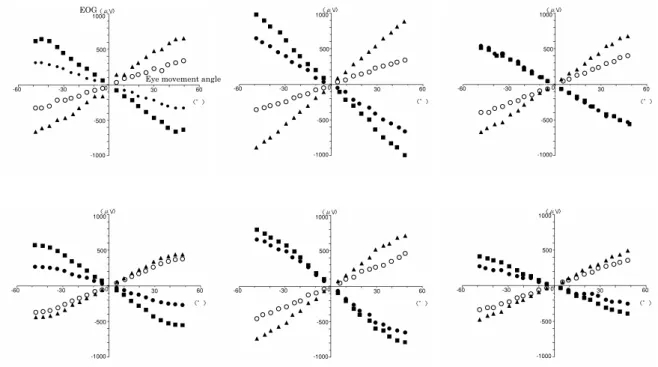
Fig. 5 shows a graph with eye movement angle on the horizontal and EOG potential on the vertical axes. In the figure, the right direction indicates plus, and the left direction does minus for ocular
move-Fig. 4 EOG waves obtained when the subjects with
bilater-ally normal eyes moved each eye symmetricbilater-ally (20!from the front).
ments. When attention is paid to the potentials of the medial and lateral canthus electrodes in the right eye, the potential detected at the medial was lower than that at the lateral canthus at each angle. In the left eye, similar findings were obtained. The polarity of the potential at the medial was reverse to that at the lateral canthus in the same eye. These findings are summarized as follows :
1. The potential detected at the medial was lower than that at the lateral canthus. 2. The polarity of the potential at the medial was
reverse to that at the lateral canthus.
2. Measurement in subjects with unilateral ano-phthalmia
Fig. 6 shows EOG waves obtained when the sub-jects with right anophthalmia moved each eye sym-metrically (50!from the front). Fig. 7 shows the results with right anophthalmia. The potential of the right lateral canthus electrode was very low and almost zero at an eye movement angle of!20!.The potential of the right medial canthus electrode was
also low but higher than that of the lateral canthus. The polarity of the potential was the same between the medial and lateral canthi. These potentials are those from the left eye (cross-talk) since the right was an anophthalmia. In the normal left eye, the polarity of the potential at the medial was reverse to that at the lateral canthus. Other cases showed the same results. The results are summarized as follows :
1. On the anophthalmia side, the potential at the lateral was considerably lower than that at the medial canthus.
2. On the anophthalmia side, the polarity of the potential was same between the medial and lateral canthi.
3. On the normal side, the polarity of the po-tential at the medial was reverse to that at the lateral canthus.
3. Analysis of the characteristics of cross-talk Table 2 shows cross-talk values during eye move-ments at 5!increments in angle. Since potentials
Fig. 6 EOG waves obtained when the subjects with right
anophthalmia moved each eye symmetrically (50!from the front). (case 2)
Fig. 7 Results with right anophthalmia(case 2)
Table 2 Cross-talk
Case
Eye movement angle Average (30∼50!) 5 10 15 20 25 30 35 40 45 50
Potential of the medial canthus electrode on the anophthalmia side/ Potential of the medial canthus electrode on the normal side 1 0.617 0.515 0.539 0.533 0.550 0.561 0.527 0.545 0.544 0.523 0.540 2 1.227 0.326 0.281 0.250 0.236 0.230 0.232 0.223 0.214 0.216 0.223 3 0.381 0.178 0.111 0.082 0.072 0.094 0.070 0.047 0.082 0.140 0.087 4 0.224 0.133 0.407 0.408 0.149 0.256 0.262 0.258 0.384 0.382 0.308 5 0.182 0.177 0.151 0.135 0.140 0.140 0.147 0.134 0.135 0.109 0.133 6 0.130 0.168 0.226 0.165 0.166 0.172 0.169 0.183 0.185 0.175 0.177
Potential of the lateral canthus electrode on the anophthalmia side/ Potential of the medial canthus electrode on the normal side 1 0.140 0.110 0.083 0.100 0.073 0.091 0.109 0.137 0.165 0.203 0.141 2 0.773 0.442 0.391 0.114 0.273 0.273 0.245 0.143 0.091 0.093 0.169 3 0.190 0.089 0.074 0.041 0.048 0.053 0.047 0.036 0.037 0.047 0.044 4 0.340 0.251 0.105 0.093 0.104 0.092 0.084 0.067 0.064 0.073 0.076 5 0.066 0.062 0.069 0.049 0.069 0.058 0.068 0.073 0.073 0.121 0.079 6 0.371 0.245 0.330 0.135 0.120 0.126 0.119 0.123 0.109 0.094 0.114
were low at an eye movement angle of!25 ",caus-ing marked errors in the ratio, the mean values from 30"to 50"are shown. The mean cross-talk to the medial canthus on the anophthalmia side was 0.087-0.540, i.e., 8.7%-54.0% of the potential at the medial canthus on the normal side. The mean cross-talk to the lateral canthus on the anophthalmia side was 0.044-0.169, i.e., 4.4%-16.9%.
DISCUSSION
Measurement in the normal subjects in this study showed a lower potential at the medial than at the lateral canthus. Since the medial canthus electrode was close to the cornea as the positive pole, we ex-pected the potential of the medial to be higher than that of the lateral canthus electrode. However, the result was the reverse of this. This phenomenon cannot be simply explained using the conventional battery model of the eye. As a cause, the influence of cross-talk can be considered. Since the polarity of the potential at the medial canthus of the normal eye was reverse to that of the anophthalmia eye, there is a possibility that the potential is reduced due to cancellation by the influence of the other eye.
Cross-talk is the influence of the potential of the other eye and is also called the contralateral effect. Miles (1, 2) observed the influence of one eye to the other based on EOG records in 9 patients with anophthalmia. Imaizumi (14, 15), Ogita (16) and Kelsey (17), et al . also reported the influence of the potential of the other eye. Cross-talk presents few problems when bilateral eyes are normal. How-ever, when there is a marked difference in the rest-ing potential or eye movements between the left and right eyes, cross-talk can not be ignored in the analysis of fine movements and should be elimi-nated in EOG recording. The methods for the meas-urement and elimination of cross-talk were first re-ported by Thijssen (18), who rere-ported a cross-talk of about 12% (-18.4 dB) in a unilateral eye movement method using convergence and about 16% (-16.0 dB) in a method for the evaluation of changes in the resting potential with the eyes placed in different adaptation situations. Kubo, et al. (11, 19) asked subjects to gaze at one point with one eye, meas-ured the potential (cross-talk) in the fixed eye dur-ing forced adduction and abduction of the other eye by applying an aspiration contact lens, and also de-vised its elimination method. They reported that the mean value by their method was -16.4 dB. Itsuki,
et al. (20) asked subjects to fix one eye on one point
and measured EOG via multiple electrodes during forced adduction and abduction of the other eye using an aspiration contact lens. As the result, they observed the influence of the electrode site in the detection of contralateral EOG, and reported that the mean cross-talk was 17.8% (-15.0 dB) when the electrode was placed 20 mm temporal to the lateral canthus.
In previous studies, EOG was recorded merely as a potential difference between electrodes without consideration of the potentials of the electrodes themselves. We previously reported that the poten-tial of each electrode can theoretically be calculated using a battery model of the eye and formula, and have compared the characteristics of potentials ob-tained using the theoretical formula and those of values obtained by measurement (10). The meas-urement and evaluation of the potential of each elec-trode allow the evaluation of changes in potential associated with eye adduction and abduction. As shown in Figs. 4 and 6, differences in the phase of waves can be clearly observed.
In this study, EOG was parallel to the eye move-ment angle until about 35",but thereafter, showed a certain degree of saturation. Assuming that the eyeball can fully rotate in the horizontal plane, the potential theoretically reaches a peak when the eye is rotated by 90",and becomes zero when it is ro-tated by 180",bringing the cornea to the site of the retina. However, eye movements!50"are consid-ered to be still linear. The saturation observed in this study may be because gazing at visual targets approaching the periphery of the visual field is dif-ficult at a visual field angle of"40".In this study, visual field angles!40"were also included in the calculation of cross-talk because the calculation of the ratio presents no problems even if eye move-ments are inadequate.
The results of this study showed the marked in-fluences of cross-talk on the potential at the medial canthus. Since the degree of influence was consid-erable (8.7% -54.0%), cross-talk in EOG analysis can-not be ignored. In contrast, the influence of cross-talk on the potential at the lateral canthus was slight. Therefore, in the analysis of cross-talk, attention only to the potential at the medial canthus is nec-essary for its calculation. When attention is paid to the potential at the lateral canthus, results with mini-mum errors due to cross-talk can be obtained.
The cross-talk value varies among subjects, due to the sites of attachment of electrodes (4) and
indi-vidual differences (21). Further studies in which the sites of electrodes are clearly mentioned are nec-essary.
CONCLUSION
We recorded EOG by an improved method in normal subjects and patients with unilateral ano-phthalmia. This method allowed the evaluation of EOG characteristics according to electrodes and was useful for EOG analysis. The influence of cross-talk of the other eye was marked at the medial but slight at the lateral canthus. In EOG recording, re-sults with minimum errors due to cross-talk can be obtained by paying attention to the potential at the lateral canthus.
ACKNOWLEDGMENTS
This study was supported in part by a Grant-in-Aid for Scientific Research (C) (No. 18500357) from the Japanese Ministry of Education, Culture, Sports, Science and Technology.
REFERENCES
1. Miles WR : The steady polarity potential of the human eye. Proc Nat Acad Sci 25 : 25-36, 1939 2. Miles WR : The steady potential of the human eye in subjects with unilateral enucleation. Proc Nat Acad Sci 25 : 349-358, 1939
3. Arden GB, Barrada A, Kelsey JH : New clinical test of retinal function based upon the standing potential oh the eye. Brit J Ophthal 46 : 449-467, 1962
4. Arden GB, Barrada A : Analysis oh the electro-oculograms of a series of normal subjects. Brit J Ophthal 46 : 468-482, 1962
5. Marg E : Development of electro-oculography, standing potential of the eye in registration of eye movement. Arch Ophthalmol 45 : 169-188, 1951
6. Ingster-Moati I, Bui Quoc E, Pless M, Djomby R, Orssaud C, Guichard JP, Woimant F : Ocu-lar motility and Wilson’s disease : a study on 34 patients. J Neurol Neurosurg Psychiatry 78 : 1199-1201, 2007
7. Melek NB, Blanco S, Garcia H : Electro-ocu-lography of smooth pursuit and optokinetic
nystagmus eye movements in type I Duane’s retraction syndrome. Binocul Vis Strabismus Q 21 : 37-44, 2006
8. Mansour AM, Uwaydat SH, Chan CC : Long-term follow-up in Bietti crystalline dystrophy. Eur J Ophthalmol 17 : 680-682, 2007
9. Renner AB, Tillack H, Kraus H, Krämer F, Mohr N, Weber BH, Foerster MH, Kellner U : Late onset is common in best macular dystro-phy associated with VMD2 gene mutations. Ophthalmology 112 : 586-592, 2005
10. Itsuki N, Kubo M, Shiraishi M, Nishikawa Y, Mimura Y : A battery model of the eyeball to calculate standing potential of the eye. J Jpn Ophthalmol Soc 99 (in Japanese) : 1012-1016, 1995
11. Kubo M, Hirai K, Mitsui Y : The measurement and elimination of Contralateral Effect(Cross-Talk) in EOG. Nihon Ganka Kiyo 30 (in Japa-nese) : 701-704, 1979
12. Shimooku J, Mimura O, Haruta R, Taoka N : How to read EOG. Neuro-ophthalmol Jpn 2 (in Japanese) : 129-133, 1985
13. Itsuki N, Kubo M, Shiraishi M, Nishikawa Y : Characteristics of the EOG potential changes generated by saccadic eye movements of large ampoitude. J Jpn Ophthalmol Soc 97 (in Japa-nese) : 514-518, 1993
14. Imaizumi K, Takahashi F, Toyama T, Horie E, Tazawa Y, Ogawa K, Shoji U : EOG in case of highly disturbed virsion. Rinshoganka 18 (in Japanese) : 333-344, 1964
15. Imaizumi K : The clinical application of electro-oculography(EOG). Proc 3rd ISCERG Symp. New York, 1966, pp.311-326
16. Ogita Y, Sotani T, Mimura O, Taniguchi I : Comparison of the amplitude of EOG depend-ing on the eye position. Ganka Rinsho Iho 73 (in Japanese) : 943-946, 1979
17. Kelsey JH : The combined use of the EOG and ERG as a routine clinical procedure. Proc 6th ISCERG Symp. Leipzig, 311-326, 1966
18. Thijssen JM, Pinckers A : Contralateral effects in the electro-oculogram. Acta Ophthalmol 52 : 441-454, 1974
19. Kubo M, Itsuki N : Development of an auto-matic measuring and recording system for the EOG employing cross-talk feed-back cancella-tion. Nihon Ganka Kiyo 32 (in Japanese) : 1496-1500, 1981
20. Itsuki N, Kubo M, Nishikawa Y, Mimura Y, Nishihara M, Minamoto Y : Reducing cross-talk
in electrooculograms made with several elec-trodes. J Jpn Ophthalmol Soc 98 (in Japanese) : 251-257, 1994
21. Toyama T : Changes in the electro-oculogram
by light and dark adaptation in case of pigmen-tary degeneration of the retina. J Jpn Ophthal-mol Soc 66 (in Japanese) : 1517-1531, 1962