p53 gene mutation in N-butyl-N-(4-hydroxybutyl)nitrosamine-induced
urinary bladder tumors and N-methyl-N-nitrosourea-induced
colon tumors of rats
Hongzhong Min
a, Eiji Kudo
a, Akiko Hino
a, Katsuhiko Yoshimoto
b,
Hiroyuki Iwahana
b, Mitsuo Itakura
b, Keisuke Izumi
a ,*
aSecond Department of Pathology, School of Medicine, The University of Tokushima, 3-18-15 Kuramoto-cho, Tokushima 770, Japan b
Otsuka Department of Clinical and Molecular Nutrition, School of Medicine, The University of Tokushima, 3-18-15 Kuramoto-cho, Tokushima 770, Japan
Received 19 March 1997; accepted 25 March 1997
Abstract
We analyzed p53 mutations in 17 N-butyl-N-(4-hydroxybutyl) nitrosamine-induced bladder transitional cell carcinomas (TCCs) with or without areas of squamous cell carcinoma (SCC) of Long–Evans Cinnamon (LEC) and F344 rats, and in 7 N-methyl-N-nitrosourea-induced colon adenocarcinomas of LEC rats by polymerase chain reaction-single strand conformation polymorphism analysis and DNA sequencing. Of these bladder tumors, one TCC with moderately differentiated SCC had a T to G transversion mutation at codon 141, leading to a Val to Gly amino acid change. No p53 mutation was found in colon adenocarcinomas. Thus a p53 gene mutation seems infrequent in these rat bladder and colon carcinogenesis models even in the late stage.1997 Elsevier Science Ireland Ltd.
Keywords: p53 gene; Bladder cancer; Rat; N-butyl-N-(4-hydroxybutyl)nitrosamine; PCR-SSCP
1. Introduction
Carcinogenesis is a multistep process which involves the accumulation of gene alterations [1–3]. Inactivation of the p53 tumor suppressor gene is one of the most common genetic alterations in many human cancers including lung, breast, urinary bladder and colon cancers [4,5]. It has been reported that the frequency of p53 mutation is approximately 30–60% in human urinary bladder cancer [6–8], and 50–75% in colon cancer [4,9,10], especially in the late stage.
Hepatocellular carcinomas (HCCs) in China and southern Africa are known to have a specific G to T transversion in codon 249 of the p53 gene [11–13], and this mutation is thought to be correlated with exposure to aflatoxins. Animal studies have shown that some carcinogens, such as alkylating N-nitroso compounds, induce a specific gene alteration. For example, N-methyl-N-nitrosourea specifically induces a G to A transition at the second nucleotide of codon 12 in the Ha-ras gene in various organs of different animals [14], as a result of O6-methylguanine DNA adduct formation [15].
The LEC rat, a model of human Wilson’s disease, develops liver [16] and kidney [17] tumors sponta-neously by copper accumulation in these organs
0304-3835/97/$17.00 1997 Elsevier Science Ireland Ltd. All rights reserved
P I I S 0 3 0 4 - 3 8 3 5 ( 9 7 ) 0 0 2 0 2 - 4
* Corresponding author. Tel.: +81 886 337065; fax: +81 886 337067.
[18]. In this study, we screened the p53 gene mutation in exons 2–9 and a part of exon 10 in carcinogen-induced rat bladder and colon tumors of LEC and F344 rats by the PCR-SSCP method.
2. Materials and methods
2.1. Animal experiments
Seven-week-old male LEC (n=3) and F344 (n=7) rats were given 0.1% BBN in their drinking water every other week from week 0–11, and then 0.05% BBN every other week from week 32–43, and were killed in week 50 to obtain bladder tumors
(Fig. 1). Six-week-old male LEC rats (n=7) were injected i.p. with MNU (50 mg/kg body weight) once a week for 6 weeks and killed in week 24 to obtain colon tumors. Seventeen bladder tumors (3– 20 mm in size) and 7 colon tumors (4–8 mm in size) were examined. Parts of the tumors were frozen in liquid nitrogen and kept at −80°C until use. The remaining portions of the tumors were fixed in 10% buffered formalin for histological examination. 2.2. DNA extraction
High molecular weight DNA was prepared from tumor tissues by proteinase K digestion and phenol/ chloroform extraction [19].
Fig. 1. Experimental design to induce bladder and colon tumors.
2.3. Oligonucleotide PCR primers
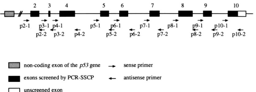
Nine pairs of primers flanking exons 2–9 and a part of exon 10 of the p53 gene were synthesized in a Model 392 DNA synthesizer (Perkin-Elmer, Nor-walk, CT). All primers except an antisense primer for exon 10 were those of intron sequences to avoid amplification of pseudogenes [20] (Fig. 2). The nu-cleotide sequences of primers are summarized in Table 1.
2.4. PCR-SSCP analysis
PCR-SSCP analysis was performed by the method of Orita et al. [21] with a minor modification. Samples of 5 ml of the PCR mixture contained 50 ng of
geno-mic DNA as a template, 5 pmol each of primers, 0.125 nmol each of dNTPs, 0.25 ml of [a-32P]dCTP (110 TBq/mmol; 370 MBq/ml) and 0.25 units of Taq DNA polymerase (Takara, Ohtsu, Japan) in the buffer supplied by Takara. Thirty cycles of the reaction at 94, 55 and 72°C each for 1 min were carried out in a thermal cycler (GeneAmpy PCR System 9600, Perkin-Elmer). The resultant PCR product was mixed with 245 ml of a formamide dye mixture (95% for-mamide, 20 mM EDTA, 0.05% xylene cya-nol and 0.05% bromophecya-nol blue) and heated at 95°C for 3 min for denaturation. Then 1 ml of the mixture was applied to 6% polyacrylamide gel containing 45 mM Tris–borate (pH 8.3) and 1 mM EDTA. All PCR products were analyzed by PCR-SSCP at glycerol concentrations of 0, 5 or 10%, respectively. Electro-phoresis was performed at 15 W for 3–5 h with cool-ing by a fan at room temperature. The gel was dried on filter paper and exposed to an X-ray film with an intensifying screen overnight at−80°C. In each case, the mobility shift on PCR-SSCP analysis was repro-ducible.
2.5. DNA sequencing
The portion of the dried polyacrylamide gel con-taining DNA with abnormal mobility found on SSCP analysis was cut out and DNA was extracted over-night from the gel with 50 ml of water at 37°C. DNA fragments re-amplified by PCR with the extracted DNAs as templates and the same primers as used in the original PCR were subcloned into TA vectors with the original TA cloningy Kit (Invitro-gen, San Diego, CA). Eight individual clones of each sample with a mobility shift were sequenced by the fluorescence-based dideoxy sequencing method with a DNA sequencer (Model 377, Perkin-Elmer).
Table 1
Oligonucleotide primers used for p53 gene analysis
Name Sequence P2-1 5′-CAGTACACAGATGTTCTCTC-3′ P2-2 5′-CAGGGGCCCTGTAAGATCCA-3′ P3-1 5′-GCCTGGGGTAAGTGAGATTC-3′ P3-2 5′-TAAGTCCCCTCTTGCCCGGC-3′ P4-1 5′-GTTCTTCTTTGGCCCATCCA-3′ P4-2 5′-AAGCAACTCTTCAGGCCCAC-3′ P5-1 5′-GATTCTTTCTCCTCTCCTAC-3′ P5-2 5′-ACAGGCAGTGCCAGTGCTCA-3′ P6-1 5′-CCCGGCCTCTGACTTATTCT-3′ P6-2 5′-CCCAACCTGGCACACAGCTT-3′ P7-1 5′-CTGTGCCTCCTCTTGTCCCG-3′ P7-2 5′-CTCCACCTTCTTTGTCCTGC-3′ P8-1 5′-AAAGTCACCCCTTGCTCTCT-3′ P8-2 5′-TAATCCAATAATAACCTTGG-3′ P9-1 5′-TGTCCTACTTCATCCTTGCT-3′ P9-2 5′-AGGCGCTGCCCCAGGTCACT-3′ P10-1 5′-CCCTCCCTTTTCTGTATTCC-3′ P10-2 5′-CAGCAGAGACCCAGCAACTA-3′ Table 2
A p53 mutation in BBN-induced bladder tumors and MNU-induced colon tumors Histology of the tumors No. of samples p53 mutation
Incidence Codon Nucleotide and amino
acid change
Bladder TCC 10 0 –
TCC with SCC 7 1 141 GTG (Val)→GGG
(Gly)
3. Results
Data are summarized in Table 2. Bladder tumors from 3 LEC and 7 F344 rats included 10 pure TCCs and 7 TCCs with areas of well to moderately differ-entiated SCC histologically. On PCR-SSCP analysis
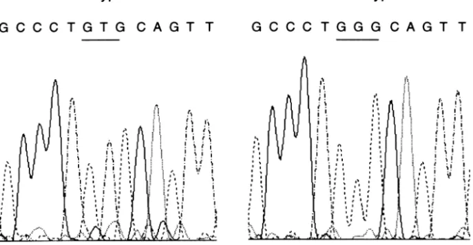
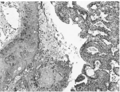
of DNA samples extracted from these bladder tumors, a band shift of exon 5 was detected in one tumor of an LEC rat (Fig. 3). Sequence analysis showed a T to G transversion mutation in codon 141, leading to a Val (GTG) to Gly (GGG) amino acid change (Fig. 4). This bladder tumor was identified as a TCC with areas of moderately differentiated SCC, and its histological grade of malignancy was the highest among the blad-der tumors examined (Fig. 5).
All colon tumors of LEC rats were adenocarcin-omas invading the submucosa and subserosa. We did not detect any p53 mutations in these colon tumors.
4. Discussion
p53 mutations generally occur within exons 5–8 in human cancers [4,5]. To test whether different hot-spots of the p53 gene exist in rodents, we screened all coding regions of the rat p53 gene except a part of exon 10. In the present study, we found a mutation in exon 5, but its frequency was lower than that reported in human bladder or colon cancer. Possible reasons for the low incidence of the p53 mutation in rats are (a) in spite of the large size of their tumors and the presence of areas of SCC the stage of tumors in rats is earlier than that in humans because usually no metastases are observed in animals with carcinogen-induced bladder
Fig. 3. Results of PCR-SSCP analysis of the p53 gene exon 5 in BBN-induced rat bladder tumors. A shifted band was observed in a TCC with moderately differentiated SCC in an LEC rat (arrow-head).
tumors and (b) there may be a species difference in the incidence of p53 mutations.
It is reported that the incidence of p53 gene muta-tions in BBN-induced bladder tumors in F344 rats is 30–67% [22,23], and that in N-[4-(5-nitro-2-furyl)-2-thiazolyl] formamide-induced tumors in F344 rats is 0–7% [24,25]. In our study, we detected a p53 muta-tion in 6% (1/17) of BBN-induced bladder tumors. One factor for this discrepancy in incidence might be a difference in the period of administration of car-cinogens, the period of administration of BBN in our study being shorter than that in the study of Masui et al. [22,23]. The GTG (Val) to GGG (Gly) mutation in codon 141 detected in the present study has been observed in BBN-induced small TCC [22] in addition to T→G mutations in other codons [5,7,22]. Although G→A and G→T mutations are common in the p53 gene, a T→G mutation is rare in human cancers [5].
In a recent study, we did not find a p53 mutation in spontaneously developed HCCs in LEC rats, but found it in diethylnitrosamine or N-ethyl-N-hy-droxyethyl-nitrosamine-induced HCCs (unpublished data). Nagao et al. [26] reported that all Apc gene mutations in rat colon tumors induced by 2-amino-1-methyl-6-phenylimidazo [4,5-b]pyridine, a hetero-cyclic amine, were specific GGGA to GGA frameshift mutations in various codons due to deletion of G, but they found no p53 mutations [27]. Animal models provide opportunities for studies on carcinogen-spe-cific or organ-specarcinogen-spe-cific mutations. As the database on
mutations in laboratory animals is limited, more stu-dies on these mutations induced by various carcino-gens are necessary.
Acknowledgements
This work was supported by Grants-in-Aid for Can-cer Research (04151053, 05151055, 06280118) from the Ministry of Education, Science and Culture, Japan.
References
[1] J.M. Bishop, The molecular genetics of cancer, Science 235 (1987) 305–311.
[2] R.A. Weinberg, Oncogenes, antioncogenes, and the molecu-lar bases of multistep carcinogenesis, Cancer Res. 49 (1989) 3713–3721.
[3] C.C. Harris, Chemical and physical carcinogenesis: advances and perspectives for the 1990s, Cancer Res. 51 (Suppl.) (1991) 5023s–5044s.
[4] M. Hollstein, D. Sidransky, B. Vogelstein, C.C. Harris, p53 mutations in human cancers, Science 253 (1991) 49–53. [5] M.S. Greenblatt, W.P. Bennett, M. Hollstein, C.C. Harris, Mutations in the p53 tumor suppressor genes: clues to cancer etiology and molecular pathogenesis, Cancer Res. 54 (1994) 4855–4878.
[6] D. Sidransky, A. Von Eschenbach, Y.C. Tsai, P. Jones, I. Summerhayes, F. Marshall, M. Paul, P. Green, S.R. Hamil-ton, P. Frost, B. Vogelstein, Identification of p53 gene muta-tions in bladder cancers and urine samples, Science 252 (1991) 706–709.
[7] K. Fujimoto, Y. Yamada, E. Okajima, T. Kakizoe, H. Sasaki, T. Sugimura, M. Terada, Frequent association of p53 gene mutation in invasive bladder cancer, Cancer Res. 53 (1992) 1393–1398.
[8] C.H. Spruck, W.M. Rideout III, A.F. Olumi, P.F. Ohneseit, A.S. Yang, Y.C. Tsai, P.W. Nichols, T. Horn, G.G. Hermann, K. Steven, R.K. Ross, M.C. Yu, P.A. Jones, Distinct pattern of p53 mutations in bladder cancer: relationship to tobacco usage, Cancer Res. 53 (1993) 1162–1166.
[9] E.R. Fearon, B. Vogelstein, A genetic model for colorectal tumorigenesis, Cell 61 (1990) 759–767.
[10] R. Kanamaru, C. Ishioka, Mutations of the p53 gene and other genes involving in human colorectal carcinogenesis, Tohoku J. Exp. Med. 168 (1992) 159–166.
[11] I.C. Hsu, R.A. Metcalf, T. Sun, J.A. Welsh, N.J. Wang, C.C. Harris, Mutational hotspot in the p53 gene in human hepato-cellular carcinomas, Nature 350 (1991) 427–428.
[12] B. Bressac, M. Kew, J. Wands, M. Ozturk, Selective G to T mutations of p53 gene in hepatocellular carcinoma from southern Africa, Nature 350 (1991) 429–431.
Fig. 5. Histology of a bladder tumor with a p53 mutation. TCC with moderately differentiated SCC (HE×99).
[13] M. Ozturk, p53 mutation in hepatocellular carcinoma after aflatoxin exposure, Lancet 388 (1991) 1356–1359. [14] S. Sukumar, V. Notario, D. Martin-Zanka, M. Barbacid,
Induction of mammary carcinomas in rats by nitroso-methy-lurea involves malignant activation of H-ras-1 locus by single point mutations, Nature 306 (1983) 658–661.
[15] A.E. Pegg, Mammalian O6-alkylguanine-DNA alkyltransfer-ase: regulation and importance in response to alkylating car-cinogenic and therapeutic agents, Cancer Res. 50 (1990) 6119–6129.
[16] M. Sasaki, M.C. Yoshida, K. Kagami, N. Takeichi, H. Kobayashi, K. Dempo, M. Mori, Spontaneous hepatitis in an inbred strain of Long–Evans rats, Rat News Lett. 14 (1985) 4–6.
[17] K. Izumi, K. Kitaura, Y. Chone, H. Tate, T. Nakagawa, Y. Suzuki, K. Matsumoto, Spontaneous renal cell tumors in Long–Evans Cinnamon rats, Jpn. J. Cancer Res. 85 (1994) 563–566.
[18] Y. Li, Y. Togashi, S. Sato, T. Emoto, J.H. Kang, N. Takeichi, H. Kobayashi, Y. Kojima, Y. Une, J. Uchino, Spontaneous hepatic copper accumulation in Long–Evans Cinnamon rats with hereditary hepatitis, J. Clin. Invest. 87 (1991) 1858– 1861.
[19] N. Blin, D.M. Stafford, Spontaneous hepatic copper accumu-lation in Long–Evans Cinnamon rats with hereditary hepatitis, Nucleic Acids Res. 3 (1976) 2303–2308. [20] J.E. Hulla, R.P. Schneider, Structure of the rat p53 tumor
suppressor gene, Nucleic Acids Res. 21 (1993) 713–717. [21] M. Orita, Y. Suzuki, Y. Sekiya, K. Hayashi, Rapid and
sen-sitive detection of point mutations and DNA polymorphisms using the polymerase chain reaction, Genomics 5 (1989) 874–879.
[22] T. Masui, I. Don, N. Takada, K. Ogawa, T. Shirai, S. Fukush-ima, p53 mutations in early neoplastic lesions of the urinary bladder in rats treated with N-butyl-N-(4-hydroxy-butyl)-nitrosamine, Carcinogenesis 15 (1994) 2379–2381. [23] T. Masui, Y. Dong, S. Yamamoto, N. Takada, H. Nakanishi,
K. Inada, S. Fukushima, M. Tatematsu, p53 mutations in transitional cell carcinomas of the urinary bladder in rats treated with N-butyl-N-(4-hydroxybutyl)nitrosamine, Cancer Lett. 105 (1996) 105–112.
[24] M. Asamoto, A.M. Mann, T.L. Macatee, S.M. Cohen, Muta-tions and expression of the p53 gene in rat bladder carcino-mas and cell lines, Mol. Carcinogenesis 9 (1994) 236–244. [25] M. Asamoto, A.M. Mann, S.M. Cohen, p53 mutation is infre-quent and might not give a growth advantage in rat bladder carcinogenesis in vivo, Carcinogenesis 15 (1994) 455–458. [26] H. Kakiuchi, M. Watanabe, T. Ushijima, M. Toyota, K. Imai, J.H. Weisburger, T. Sugimura, M. Nagao, Specific 5′ -GGGA-3′ →5′-GGA-3′mutation of the Apc gene in rat colon tumors induced by 2-amino-1-methyl-6-phenylimidazo[4,5-b]pyri-dine, Proc. Natl. Acad. Sci. USA 92 (1995) 910–914. [27] H. Makino, T. Ushijima, H. Kakiuchi, M. Onda, N. Ito, T.
Sugimura, M. Nagao, Absence of p53 mutations in rat colon tumors induced by 2-amino-6-methyldipyrido[1,2-a:3′,2′ -d]-imidazole, amino-3-methylimidazo[4,5-f]quinoline, or 2-amino-1-methyl-6-phenylimidazo[4,5-b]pyridine, Jpn. J. Cancer Res. 85 (1994) 510–514.