INTRODUCTION
18F - 2 - fluoro - 2 - deoxyglucose (FDG) positron
emission tomography/computed tomography (PET/ CT) has an expanding role in the diagnosis and stag-ing of many tumors. Durstag-ing routine oncology PET/ CT scanning we sometimes see incidental FDG up-take in blood vessel walls. Herein, we show FDG-PET/CT images and discuss cases where there was uptake of FDG in or adjacent to the vessel wall.
ARTERIOSCLEROSIS
In daily PET examination patients without a his-tory of vasculitis often exhibit a certain degree of FDG uptake in the artery walls. It is believed that
this is related to the inflammatory component of ar-teriosclerosis and that FDG is taken up by inflam-matory cells, predominantly the macrophages (1). Mild to moderate FDG accumulation in the artery walls is observed in arteriosclerosis (Fig. 1). This is quite a frequent finding and it has been reported that among 149 patients (61 males and 88 females ; age range 5-83 years) who underwent whole-body PET scans for the assessment of non-cardiovascular disorders), 145 were noted to have visible uptake in at least one segment of the large vessels (1).
ARTERIAL PLAQUE
In the case shown in Figure 2 high levels of FDG accumulation were observed in the arterial plaque and this was believed to be due to active athero-sclerotic changes in the arteries. In contrast, it has been reported that sites of calcification considered to represent inactive and stable atherosclerosis did not show FDG uptake (2). It was found that 50 out of 85 patients had at least one area of FDG uptake
REVIEW
18
F-2-fluoro-2-deoxyglucose uptake in or adjacent to
blood vessel walls
Yoichi Otomi
1, Hideki Otsuka
2, Kaori Terazawa
1, Hayato Nose
1,3, Michiko Kubo
1,
Kazuhide Yoneda
1, Kaoru Kitsukawa
1, and Masafumi Harada
1 1Department of Radiology, Institute of Health Bioscience, the University of Tokushima Graduate School, Tokushima, Japan, 2
Department of Medical Imaging, Institute of Health Bioscience, the University of Tokushima Graduate School, Tokushima, Japan,3
Department of Radiology, Oe Kyodou Hospital, Tokushima, Japan
Abstract : Incidental18F-2-fluoro-2-deoxyglucose (FDG) uptake in blood vessel walls is sometimes demonstrated during routine oncologic imaging with positron emission to-mography/computed tomography (PET/CT). FDG uptake in vessel walls can also be seen under some non-physiological conditions such as vasculitis and arteriosclerosis. Radi-ologists need to be aware of the diseases which can exhibit FDG uptake in the vessel wall for proper interpretation. J. Med. Invest. 60 : 15-20, February, 2013
Keywords : blood vessel wall,18F-2-fluoro-2-deoxyglucose, positron emission tomography/computed tomography
Received for publication December 3, 2012 ; accepted February 26, 2013.
Address correspondence and reprint requests to Yoichi Otomi, Department of Radiology, Tokushima University Hospital, 2 50 1 Kuramoto cho, Tokushima 770 8503, Japan and Fax : + 81 88 -633 - 7174.
in the thoracic aortic wall (2). Fourteen of these 50 patients showed focal FDG uptake. The FDG up-take sites were mostly distinct from the calcifica-tion sites and may possibly have been located in
areas of metabolic activity associated with athero-sclerotic changes (2). PET can reveal active arte-rial plaque.
Fig. 1. A 78 - year - old man with arteriosclerosis. (a) FDG - PET 3D MIP image, (b) coronal FDG - PET image and (c) coronal contrast- enhanced CT image. Mild to moderate FDG uptake in the artery walls is shown (arrows).
Fig. 2. A 77 - year - old man with arterial plaque. (a, b) FDG - PET 3D MIP images, (c) axial FDG - PET image, (d) sagittal CT image and (e) sagittal FDG - PET image. Focal uptake that can be seen in the thoracic descending aorta is considered to be arterial plaque (arrows). Small nodular shaped FDG uptake area visible in the left lung lobe was certified pathologically as lung cancer (arrowhead).
a
b
c
TAKAYASU DISEASE
In the case with Takayasu disease, high levels of FDG accumulation were observed in large vessels such as the thoracic and abdominal aorta, the sub-clavian artery and the common carotid artery. Con-trast CT examinations revealed wall thickening ac-companying contrast effects at locations correspond-ing to the accumulation areas as shown in Figure 3. It has been reported that FDG imaging identifies more regions of the aorta and its branches that are involved in the inflammatory process than MRI, and that FDG-PET is more reliable than MRI in the early recognition of aortitis (3, 4). If the diagnosis of Takayasu disease is late, it may become life-threat-ening or necessitate surgery. PET/CT is valuable in the recognition of the early phase of Takayasu disease.
GRAFT INFECTION
In the case in which high levels of focal FDG up-take were observed in parts of the grafted areas of the great vessels, a diagnosis of graft infection was obtained (Fig. 4). This uptake disappeared after an-tibiotic treatment and proved useful in assessing the
treatment effect (Fig. 4). However, even in cases in-volving no infection or inflammation, diffuse FDG uptake was sometimes observed along the graft due
Fig. 3. A 62 year old woman with Takayasu disease. (a) FDG -PET 3D MIP image, (b) axial FDG - -PET image and (c) axial contrast- enhanced CT image. Strong uptake can be seen in the artery walls of the aorta, the subclavian arteries, the common carotid arteries (arrows). Contrast- enhanced CT examination shows wall thickening accompanying contrast effects at locations corresponding to the uptake areas (arrowheads).
Fig. 4. A 69 - year - old man with graft infection. (a) FDG - PET 3D MIP and axial FDG - PET images before antibiotic treatment, (b) FDG - PET 3D MIP and axial FDG - PET images at 51 days after antibiotic treatment. Localized FDG uptake can be seen in part of the grafted area of the aortic arch (arrows). After antibiotic treatment, this uptake become minimal and the symptoms of fever disap-peared.
to granulation tissue in the grafted area. Although such cases are considered to indicate physiological uptake, the identification of such uptake is often difficult. To distinguish between infected and non-infected aortic grafts, the pattern of FDG uptake in the grafted areas is important. Focal or segmental FDG uptake has been shown to be associated with active infection, and diffuse uptake along the graft with nonspecific inflammation (5). Focal FDG up-take can be a sign of graft infection. In cases with a history of artery graft placement and high levels of focal uptake in grafted areas, with the symptoms and the findings of inflammation, it is necessary to consider the possibility of graft infections. FDG-PET is therefore useful for reasons such as the abil-ity to assess whether there is a focus suspected of being inflammation in any other region.
SUPERIOR VENA CAVA INVASION BY
ME-DIASTINAL LARGE B-CELL LYMPHOMA
Invasion by nearby malignant tumors can cause abnormal uptake through the vessel wall and into the lumen. We present a case of malignant lym-phoma with superior vena cava (SVC) syndrome (Fig. 5). This patient suffered face edema and ultra-sonography located a mass which had caused SVC occlusion. SVC invasion by malignant lymphoma is one factor which can cause SVC syndrome (6). Primary mediastinal large B-cell lymphoma is a
frequent cause of superior vena cava syndrome. We examined malignant tumors such as retroperitoneal leiomyosarcomas that arose primarily from the in-ferior vena cava or were secondarily involved. These lesions also showed abnormally high uptake of FDG in the vessel wall (Fig. 6).
RETROPERITONEAL FIBROSIS
Retroperitoneal fibrosis (RPF) is a rare collagen vascular disease characterized by fibro-inflammatory tissue, typically surrounding the abdominal aorta and iliac arteries. RPF is generally idiopathic but secondary RPF can be associated with certain drugs, malignant diseases, infection and surgery (7). In idi-opathic RPF, an exaggerated inflammatory response to atherosclerosis has been implicated in its patho-genesis. Therefore, some investigators use the term chronic periaortitis rather than idiopathic RPF (7). In our case, moderate FDG uptake along the abdominal aorta and iliac artery was proven to corre-spond to outgrowth of soft tissue using CT (Fig. 7). FDG-PET allows for whole-body imaging. As such, it can reveal other diseased sites such as those seen in multifocal fibrosclerosis, and it can also detect occult neoplastic or infectious processes with which RPF can be associated or secondary to. Finally, in idiopathic RPF, FDG-PET can be used to assess the full extent of vascular inflammatory involvement (8).
Fig. 5. A 74 - year - old man with SVC invasion of a large mediastinal B - cell lymphoma. (a) FDG - PET 3D MIP image, (b) axial PET/ CT fusion image and (c) axial contrast- enhanced CT image and showing high FDG uptake in the blood vessels (arrows). A tumor with soft tissue density can be seen in the superior vena cava (arrowhead).
CONCLUSION
FDG uptake in vessel walls can be seen under some non-physiological conditions. We present im-ages of various diseases which show FDG uptake
in the blood vessel walls, such as in areas associ-ated with arteriosclerosis and Takayasu disease, and in areas adjacent to blood vessel walls such as those involving invasion by nearby malignant tumors and RPF.
Fig. 6. A 63 - year - old woman with retroperitoneal leiomyosarcoma. (a) FDG - PET 3D MIP image, (b) coronal PET/CT fusion im-age and (c) coronal contrast- enhanced CT imim-age. Strong FDG uptake can be observed in the tumor progressing to the lumen of the inferior vena cava (arrows).
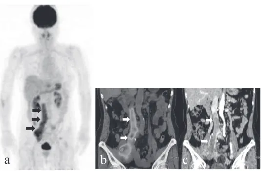
Fig. 7. A 79 - year - old man with retroperitoneal fibrosis. (a) FDG - PET 3D MIP image, (b) coronal FDG - PET image and (c) coro-nal contrast- enhanced CT image. There initially appeared to be uptake in the artery walls of the abdomicoro-nal aorta and both common iliac arteries (arrows), but comparisons with contrast- enhanced CT images revealed that there was uptake in areas demonstrating the outgrowth of soft tissue surrounding the arteries.
CONFLICT OF INTEREST
None of the authors have any conflicts of interest associated with this study.
REFERENCES
1. Bural GG, Torigian DA, Chamroonrat W, Houseni M, Chen W, Basu S, Kumar R, Alavi A : FDG-PET is an effective imaging modality to detect and quantify age-related atheroscle-rosis in large arteries. Eur J Nucl Med Mol Imaging 35 : 562-569, 2008
2. Tatsumi M, Cohade C, Nakamoto Y, Wahl RL : Fluorodeoxyglucose uptake in the aortic wall at PET/CT : possible finding for active atheroscle-rosis. Radiology 229 : 831-837, 2003
3. Meller J, Grabbe E, Becker W, Vosshenrich R : Value of F-18 FDG hybrid camera PET and MRI in early Takayasu aortitis. Eur Radiol 13 : 400-405, 2003
4. Meller J, Strutz F, Siefker U, Scheel A, Sahlmann CO, Lehmann K, Conrad M,
Vosshenrich R : Early diagnosis and follow-up of aortitis with [18F]FDG PET and MRI. Eur J Nucl Med 30 : 730-736, 2003
5. Fukuchi K, Ishida Y, Higashi M, Tsunekawa T, Ogino H, Minatoya K, Kiso K, Naito H : De-tection of aortic graft infection by fluorodeoxy-glucose positron emission tomography : com-parison with computed tomography. J Vasc Surg 42 : 919-925, 2005
6. Maryniak RK, Roszkowska-Purska K : Primary mediastinal large B-cell lymphoma is an impor-tant differential among mediastinal tumours. Pol J Pathol 53 : 139-144, 2002
7. Jansen I, Hendriksz TR, Han SH, Huiskes AW, van Bommel EF : (18)F-fluorodeoxyglucose position emission tomography (FDG-PET) for monitoring disease activity and treatment re-sponse in idiopathic retroperitoneal fibrosis. Eur J Intern Med 21 : 216-221, 2010
8. Salvarani C, Pipitone N, Versari A, Vaglio A, Serafini D, Bajocchi G, Salvo D, Buzio C, Greco P, Boiardi L : Positron emission tomography (PET) : evaluation of chronic periaortitis. Arthri-tis Rheum 53 : 298-303, 2005