R E S E A R C H A R T I C L E
Open Access
Enhancer of zeste plays an important role in
photoperiodic modulation of locomotor
rhythm in the cricket, Gryllus bimaculatus
Yoshimasa Hamada
1, Atsushi Tokuoka
1, Tetsuya Bando
2, Hideyo Ohuchi
2and Kenji Tomioka
1*Abstract
Introduction: Insects show daily behavioral rhythms controlled by an endogenous oscillator, the circadian clock. The rhythm synchronizes to daily light–dark cycles (LD) and changes waveform in association with seasonal change in photoperiod.
Results: To explore the molecular basis of the photoperiod-dependent changes in circadian locomotor rhythm, we investigated the role of a chromatin modifier, Enhancer of zeste (Gb’E(z)), in the cricket, Gryllus bimaculatus. Under a 12 h:12 h LD (LD 12:12), Gb’E(z) was constitutively expressed in the optic lobe, the site of the biological clock; active phase (α) and rest phase (ρ) were approximately 12 h in duration, and α/ρ ratio was approximately 1.0. When transferred to LD 20:4, theα/ρ ratio decreased significantly, and the Gb’E(z) expression level was significantly reduced at 6 h and 10 h after light-on, as was reflected in the reduced level of trimethylation of histone H3 lysine 27. This change was associated with change in clock gene expression profiles. The photoperiod-dependent changes inα/ρ ratio and clock gene expression profiles were prevented by knocking down Gb’E(z) by RNAi.
Conclusions: These results suggest that histone modification by Gb’E(z) is involved in photoperiodic modulation of the G. bimaculatus circadian rhythm.
Keywords: Circadian rhythm, Photoperiodic modulation, Histone modification, Clock gene, Enhancer of zeste Introduction
Most animals exhibit daily rhythms in various physio-logical functions that synchronize with daily environmen-tal cycles, such as light–dark cycles (LD), which are affected by Earth’s rotation [1]. The rhythm is generated by a circadian clock, which is an endogenous mechanism that oscillates over a period of approximately 24 h. The circadian clock’s oscillatory mechanism is based on tran-scriptional/translational molecular feedback loops [2–4]. In insects, the clock machinery has been studied most ex-tensively in the fruit fly, Drosophila melanogaster, in which the major players are Clock (Clk) and cycle (cyc) [2, 3]; their product proteins, CLK and CYC, form heterodimers and activate the transcription of period (per) and timeless (tim) during the late day to early night. The translated proteins PER and TIM accumulate in the cytoplasm
during the night, and in late night they heterodimerize and enter the nucleus to repress their own transcription by inhibiting CLK-CYC transcriptional activity. This feed-back results in a reduction of PER and TIM levels, leading to the reactivation of per and tim transcription [2, 3].
Recent studies have revealed that circadian clock cyc-ling is precisely controlled by mechanisms that include chromatin remodeling, recruitment of RNA polymer-ases, and post-transcriptional and post-translational modifications [5–8]. Chromatin remodeling plays an im-portant role in regulating the circadian clock and in its response to environmental time cues. In both mammals and insects, CLK acts as a transcriptional activator and recruits other transcription factors by binding to E-boxes at the regulatory regions of clock-controlled genes, including per and tim [9–11].
In addition to daily time-keeping, the circadian clock plays a key role in seasonal changes in physiology, including the change in active phase to rest phase ratio (α/ρ ratio), based on seasonal change in photoperiod [12–14]. The * Correspondence:[email protected]
1Graduate School of Natural Science and Technology, Okayama University, 3-1-1 Tsushima-naka, Kita-ku, Okayama 700-8530, Japan
Full list of author information is available at the end of the article
© 2016 Hamada et al. Open Access This article is distributed under the terms of the Creative Commons Attribution 4.0 International License (http://creativecommons.org/licenses/by/4.0/), which permits unrestricted use, distribution, and reproduction in any medium, provided you give appropriate credit to the original author(s) and the source, provide a link to the Creative Commons license, and indicate if changes were made. The Creative Commons Public Domain Dedication waiver (http://creativecommons.org/publicdomain/zero/1.0/) applies to the data made available in this article, unless otherwise stated.
photoperiod-dependent change in α/ρ ratio persists for many days in constant darkness and is recognized as a kind of history-dependent change in the circadian clock. History-dependent changes are also observed in the free-running period [15–17]. However, the molecular basis of photoperiod-dependent changes has remained elusive.
In the present study, we investigated the possible in-volvement of chromatin modification in photoperiod-dependent changes in circadian rhythm of the cricket, Gryllus bimaculatus. We used the cricket in this study for the following reasons: first, the cricket shows a clear photoperiodic response in locomotor activity rhythms [12, 14]; second, the circadian clock has been localized in the optic lobe [18, 19]; and third, cDNAs were previ-ously obtained for major clock genes [20]. Although number of chromatin modifiers so far known for insects including E(z), UTX, TRX, and Su(var)3-9 [21–24], we investigated the role of E(z) in photoperiodic modulation of the circadian rhythm, since its mammalian homolog EZH2 has been shown to be involved in control of the cir-cadian clock mechanism through trimethylation of histone H3 lysine 27 (H3K27) [25] and we have some experimental evidence that RNA interference (RNAi) of E(z) effectively reduces H3K27 trimethylation (H3K27me3) level in our cricket [24]. We found that knockdown of E(z) expression by RNAi prevented photoperiodic modulation. The results are discussed relative to the role of E(z) in photoperiodic modulation as well as seasonal adaptation of the circadian clock.
Materials and methods
Animals
Adult male crickets were purchased or taken from our laboratory colony, which is maintained under standard conditions of LD 12:12, at a constant temperature of 25 °C. The crickets were fed laboratory chow and water.
Measurement of mRNA levels
Quantitative real-time RT-PCR (qPCR) was used to meas-ure mRNA levels. Total RNA was extracted from optic lobes of 4–6 adult males using ISOGEN (Nippon Gene, Japan) or TRIzol Reagent (Invitrogen, California). The total RNA was treated with DNase I to remove any traces of genomic DNA. Approximately 250 ng of total RNA of each sample was reverse transcribed with random 6mers using PrimeScript RT Reagent Kit (Takara, Japan). qPCR was per-formed with an Mx3000P Real-time PCR System (Strata-gene, California) using Fast Start Universal SYBR Green Master (Roche, Japan) including SYBR Green with the fol-lowing primers for respective genes: 5′-AAGGTGC GAAAACAGGCATC-3′ and 5′-TCGTCGTTTTGGTG GATGTG-3′ for Gb’E(z) (GenBank Accession No. LC0 12934), AAGCAAGCAAGCATCCTCAT-3′ and 5′-CTGAGAAAGGAGGCCACAAG-3′ for Gb’per (GenBank
Accession No. AB375516), 5′-GATTATGAAGTCTGTG ATGATTGG-3′ and 5′-AGCATTGGAGAGAACTGAA-G AGGT-3′ for Gb’tim (GenBank Accession No. AB548625), 5′-GGCCGAAGCTCATAAAGTGG-3′ and 5′-AACCGC ACAAAGGAACCATC-3′ for Gb’cyc (GenBank Accession No. AB762416), 5′-AATGACCGTAGTCGAGAAAGT GAAG-3′ and 5′-TTGCGATGATTGAGGTTGTTG-3′ for Gb’Clk (GenBank Accession No. AB738083), and 5′-GCTCCGGATTACATCGTTGC-3′ and 5′-GC CAAATGCCGAAGTTCTTG-3′ for Gb’rpl18a (Gen-Bank Accession No. DC448653). Standard curves for the transcripts were generated by serial dilutions of amplified cDNAs and included in each qPCR run. After 40 PCR cycles, samples were subjected to melt-ing curve analysis, and a smelt-ingle expected amplicon was confirmed in each sample. The results were ana-lyzed using software associated with the instrument: quantification of mRNA levels was performed by the standard curve method, and the values were normal-ized to the values of rpl18a at each time point.
RNAi
Double-stranded RNAs (dsRNAs) were synthesized using the MEGAScript T7 Kit (Ambion, California) and
adjusted to 20 μM for DsRed2 and 2 μM for Gb’E(z) for
RNAi. For dsRNA synthesis, we used the T7 primer, 5′-TAATACGACTCACTATAGGG-3′, for Gb’E(z) and an exogenous gene, DsRed2; dsRNA lengths were 431 bp and 660 bp, respectively. In total, 700 nL of dsRNA was injected into the abdomens of the adult male crickets. As a negative control, we injected dsRNA for DsRed2.
Immunohistochemistry
Heads of adult males were fixed at zeitgeber time (ZT) 10 (ZT 0 corresponds to lights-on) with 2 % paraformalde-hyde (PFA) in phosphate-buffered saline with 0.1 % Tween 20 (PBT) for 17 h at 4 °C. The brain and optic lobe com-plex was extracted using a fine razor knife, tweezers, and micro scissors under dissecting microscope, then fat bodies and associated nerves were removed. The tissues were refixed in 4 % PFA/PBT and dehydrated in 25 %, 50 %, 75 % EtOH/PBT and 100 % EtOH. Dehydrated samples were rehydrated in 75 %, 50 %, and 25 % EtOH/PBT, washed with PBT, and blocked with 1 % normal goat serum (NGS) in PBT for 1 h. Blocked samples were incu-bated overnight at 4 °C with rabbit polyclonal anti-trimethylated H3K27 antibody (EMD Millipore, Germany; 1:500 diluted in 1 % NGS in PBT). The samples were then washed with PBT and incubated for 3 h with secondary anti-rabbit IgG antibody conjugated with Alexa Fluor 488 (Molecular Probes, Massachusetts; 1:500 diluted in 1 % NGS/PBT). Samples were washed with PBT and incubated with DAPI at 1:1000 in PBT for 15 min. The samples were mounted on a glass slide with 50 % glycerol/PBT, observed
and optical digital images were captured with a confocal laser scanning microscope (LSM-780, Zeiss, Germany).
To quantify the staining intensity of H3K27me3 and DAPI, the fluorescence intensities were quantified from digital images of six optic lobes for each treatment using the ImageJ software (freely available at http://rsb.info.nih.-gov/ij/). The H3K27me3 staining indices were shown as the ratio of staining intensity of H3K27me3 to that of DAPI.
Recording of locomotor activity
Locomotor activity of individual animals was recorded with an actograph made of a transparent plastic box (18 × 9 × 4.5 cm) with a rocking substratum, as previ-ously described [26]. A magnetic reed switch sensed rocking movements of the substratum caused by a mov-ing cricket. The number of movements was recorded every 6 min by a computerized system. Water and food were provided ad libitum. The activity chambers were placed in an incubator in which temperature was kept at 25 ± 0.5 °C and desired lighting regimens were provided by a cool white fluorescent lamp connected to an elec-tric timer. The light intensity was 600–1000 lux, which varied based on the animal’s proximity to the lamp.
The raw data were displayed as conventional double-plotted actograms to judge activity patterns, and free-running periods were calculated using theχ2periodogram [27] in ActogramJ [28]. If a peak of the periodogram was above the 5 % confidence threshold, the peak period was designated as statistically significant. The duration of the active phase, or subjective night, (α) was estimated with ActogramJ: the boundary of the active phase was defined at the time point where the moving average of activity exceeded or fell below 70 % of daily average activity; then, a linear regression line was fitted to the points for consecutive days. The rest of the time was designated as the rest phase, or subjective day, (ρ).
Statistical analysis
The one-way analysis of variance (ANOVA) followed by a post hoc Tukey-test was used to compare the differences
in means of free-running periods,α/ρ ratios, and
immu-nostaining intensity of H3K27me3 between the control and experimental groups. Significance of daily cycling of clock gene mRNA levels and difference of mRNA levels among three groups of crickets treated in different ways were tested by ANOVA followed by Tukey-test. To com-pare the means of two groups, t-test was used. In all statis-tical tests, the significance level was set at P < 0.05.
Results
Expression profile ofGb’E(z) in the optic lobe and its suppression by RNAi
We first examined whether Gb’E(z) was expressed in the optic lobe, which is known to harbor the circadian clock
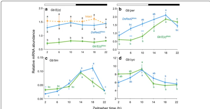
in the cricket, by using qPCR to measure Gb’E(z) mRNA levels in the tissue. Under LD 12:12 Gb’E(z) mRNA was fairly consistently expressed throughout a day in intact crickets (Fig. 1a), and no daily fluctuation was observed (ANOVA, F5, 41= 0.34, P > 0.88).
To estimate the effects of Gb’E(z)RNAi
, we measured Gb’E(z) levels in the optic lobe of Gb’E(z)RNAi
crickets
and control (DsRed2RNAi) crickets, which were treated
with dsDsRed2. DsRed2RNAi crickets showed similar
Gb’E(z) expression to that of intact crickets (Fig. 1a) and
no daily rhythm was observed (ANOVA, F5, 37= 0.52,
P> 0.76). The value at each time point did not differ from that of intact crickets (Tukey-test, P > 0.05). In Gb’E(z)RNAi crickets, Gb’E(z) expression was greatly
re-duced compared with the control and intact crickets at all time points (Tukey-test, P < 0.01). Expression reduced to approximately 53 % of that of intact and control crickets at ZT 10. These results indicate that Gb’E(z) is expressed rather constitutively in the optic lobe and could be knocked down by RNAi against Gb’E(z), and that DsRed2RNAicrickets can be used as a control.
Expression profile of clock genes in the optic lobe under LD 12:12
Expression profiles of the clock genes Gb’per, Gb’tim, and Gb’cyc were investigated in the optic lobe of
DsRed2RNAiand Gb’E(z)RNAicrickets under LD 12:12 by
qPCR. The results are shown in Fig. 1b–d. In the control crickets, the mRNA levels of Gb’per and Gb’tim showed a significant daily fluctuation, which peaked at ZT 18
under LD 12:12 (ANOVA: F5, 36= 9.01, P < 0.01 for
Gb’per, F5, 17= 67.97, P < 0.01 for Gb’tim) (Fig. 1b, c).
Gb’cyc also showed a significant daily cycling, with a peak at ZT 10 (ANOVA: F5, 41= 3.62, P < 0.01) (Fig. 1d).
Similar rhythmic expression profiles were observed for the three genes in Gb’E(z)RNAi
crickets (ANOVA: F5, 20=
15.47, P < 0.01 for Gb’per; F5, 17= 24.25, P < 0.01 for
Gb’tim; F5, 22= 4.45, P < 0.01 for Gb’cyc), but with slight
changes in pattern or phase compared with those in DsRed2RNAi crickets (Fig. 1b-d): Gb’per showed a signifi-cant reduction at ZT 6 and ZT 10 (t-test, P < 0.01), and Gb’tim showed a slight increase at ZT 2, a slight reduction at ZT 10 (t-test, P < 0.05), and a reduction at ZT 18 (t-test, P< 0.01) that resulted in a phase advance of the peak by 4 h. Moreover, Gb’cyc showed a significant increase at ZT 2 (t-test, P < 0.05).
Effects ofGb’E(z)RNAion circadian locomotor rhythms in LD 12:12 and ensuing DD
To examine the role of Gb’E(z) in regulation of circadian rhythms, we recorded the locomotor activity in Gb’E(z)RNAi
, intact and control crickets. Both intact and control crickets showed nocturnal activity rhythms that peaked just after lights-off under LD 12:12, and the rhythm persisted with a
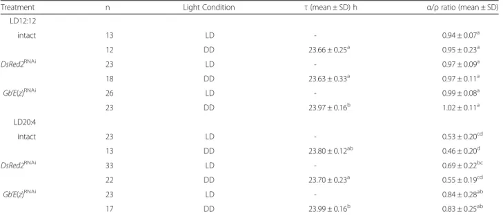
free-running period slightly shorter than 24 h in constant darkness (DD) (Fig. 2a, b, Table 1). We measured the dur-ation of active phase (α) and rest phase (ρ), and calculated α/ρ ratio to characterize the daily activity profile. The α/ρ ratios of intact and control crickets were approximately 1.0 both in LD 12:12 and DD (Table 1). These results were consistent with those of our previous study [14]. The Gb’E(z)RNAi
crickets showed nocturnal locomotor rhythms similar to those of control crickets under LD 12:12 (Fig. 2c);
their α/ρ ratios were approximately 1.0 both in LD
12:12 and DD (Table 1), and were not significantly different from those of the intact and control crickets (ANOVA followed by Tukey-test, P > 0.05). However, their average free-running period was significantly longer than that of intact and control crickets (ANOVA followed by Tukey-test, P < 0.01, Table 1), which suggests that methylation of H3K27 is involved in regulating the free-running period in DD.
Role ofGb’E(z) in photoperiodic modulation of locomotor rhythms
In the cricket, the α/ρ ratio in DD has been shown to
change depending on the preceding photoperiodic
conditions [14]. We used LD 20:4 to examine the photo-periodic modulation of the rhythm because it has been
shown to reduce the α/ρ ratio most effectively [14]. To
examine the effects of Gb’E(z)RNAi
on this photoperiodic
effect on the α/ρ ratio, we recorded locomotor activity
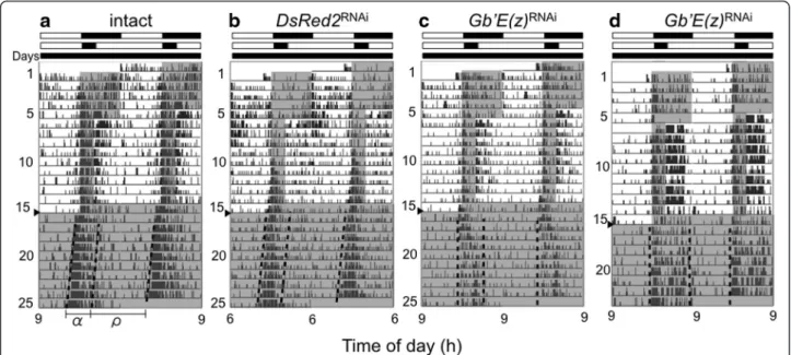
rhythm of intact, DsRed2RNAi, and Gb’E(z)RNAi crickets under LD 12:12, followed by LD 20:4 for 9 cycles, and then by DD. The representative records are shown in Fig. 3, and the locomotor rhythm parameters are sum-marized in Table 1. The intact and control crickets showed that locomotor activity was confined to a short dark phase under LD 20:4, and the shortened active phase persisted under subsequent free-running condi-tions (Fig. 3a, b).
Theα/ρ ratios of intact and DsRed2RNAicrickets under LD 20:4 and the ensuing DD were both significantly smaller than those in LD 12:12 and the ensuing DD (Table 1). There was no significant difference in the α/ρ
ratio between intact and DsRed2RNAicrickets under DD
(ANOVA followed by Tukey-test, P > 0.05). These results are consistent with those of Koga et al. [14]. Under LD
20:4 some Gb’E(z)RNAi
crickets (6/23) showed similar
changes in α/ρ ratio (Fig. 3c). However, the remaining
Fig. 1 Daily expression profiles of Gb’E(z) and clock genes in the optic lobe of the cricket Gryllus bimaculatus under LD 12:12. Blue and green symbols with solid lines indicate the mRNA levels of Gb’E(z) (a), Gb’per (b), Gb’tim (c), and Gb’cyc (d) in DsRed2RNAiand Gb
’E(z)RNAicrickets, respectively. For Gb
’E(z) data (a), intact crickets are also shown by orange symbols. In RNAi crickets, the optic lobes were collected seven days after dsRNA injection. mRNA abundance was measured by qPCR, with total RNA extracted from the optic lobes. Data collected from 3–15 independent measurements were averaged and plotted as mean ± SEM. The values shown are relative to those of Gb’rpl18a mRNA, which was used as an internal reference. For Gb’E(z), values with different lower case letters at each ZT significantly differ from each other (Tukey-test, P < 0.01). For Gb’per, Gb’tim, and Gb’cyc, *P < 0.05, **P < 0.01, t-test vs DsRed2RNAiand values
with different lower case letters of the same color differ significantly from each other (Tukey-test, P < 0.05). The Gb’E(z) mRNA showed no daily rhythms in intact, DsRed2RNAi, or Gb
’E(z)RNAicrickets, whereas the mRNA of other three genes showed a clear oscillation in both DsRed2RNAiand Gb
’E(z)RNAi
crickets (17/23) exhibited an activity pattern that con-sisted of a strong activity bout after lights-on in addition to the light-off peak under LD 20:4; the averageα/ρ ratio was 0.84 ± 0.28 (n = 23) (Table 1). Interestingly, the α/ρ ratio was maintained in the ensuing DD, and was
significantly greater than those of the intact and control crickets (ANOVA followed by Tukey-test, P < 0.01,
Table 1). The free-running period of the Gb’E(z)RNAi
crickets was again significantly longer than that of the control crickets (Tukey-test, P < 0.01). These results Fig. 2 Double-plotted actograms of locomotor activity of adult male crickets Gryllus bimaculatus under LD 12:12 and DD. a, b, and c show representative records of intact crickets (a) and those treated with dsRNA against DsRed2 (b) or Gb’E(z) (c). Arrowheads indicate the day when the crickets were transferred from LD 12:12 to DD, at a constant temperature of 25 ± 0.5 °C.α and ρ indicate active phase and rest phase,
respectively. Dotted lines indicate the onset and offset of the active phase. White and black bars above the actogram indicate the light and the dark phase, respectively. Gray area in the actogram indicates the dark phase
Table 1 Effects of RNAi against Gb’E(z) and DsRed2 on the locomotor rhythm of Gryllus bimaculatus
Treatment n Light Condition τ (mean ± SD) h α/ρ ratio (mean ± SD)
LD12:12 intact 13 LD - 0.94 ± 0.07a 12 DD 23.66 ± 0.25a 0.95 ± 0.23a DsRed2RNAi 23 LD - 0.97 ± 0.09a 18 DD 23.63 ± 0.33a 0.97 ± 0.11a Gb’E(z)RNAi 26 LD - 0.99 ± 0.08a 23 DD 23.97 ± 0.16b 1.02 ± 0.11a LD20:4 intact 23 LD - 0.53 ± 0.20cd 13 DD 23.80 ± 0.12ab 0.46 ± 0.20d DsRed2RNAi 33 LD - 0.69 ± 0.22bc 22 DD 23.70 ± 0.23a 0.55 ± 0.19cd Gb’E(z)RNAi 23 LD - 0.84 ± 0.28ab 17 DD 23.99 ± 0.16b 0.83 ± 0.25ab
ANOVA revealed significant difference in both free-running period (τ) (F5, 98= 9.01, P < 0.01) andα/ρ ratio (F11, 230= 22.0, P < 0.01). Values with different lower case letters
suggest that Gb’E(z) is involved in photoperiodic modu-lation and regumodu-lation of free-running period in the circa-dian locomotor rhythm.
Involvement ofGb’E(z) in photoperiodic modulation of clock gene expression profiles
Because Gb’E(z) is the chromatin modifier that controls gene expression through H3K27 methylation, we pre-dicted that Gb’E(z) is involved in photoperiodic
modula-tion of α/ρ ratio through alteration in clock gene
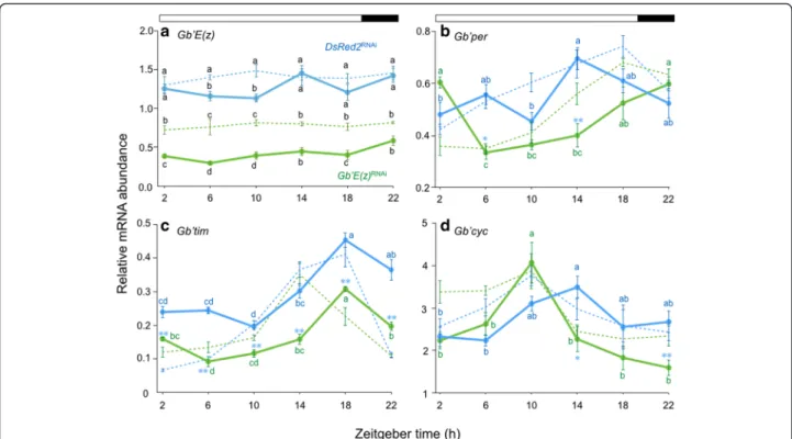
expression. Consequently, we examined whether endogen-ous Gb’E(z) mRNA levels in the optic lobe altered with a change of photoperiodic condition (Fig. 4a). In the control crickets, Gb’E(z) showed no significant daily change in its expression, but a significant reduction was found at ZT 6 and ZT 10 in LD 20:4 when compared with that in LD 12:12 (Tukey-test, P < 0.01). Gb’E(z)RNAi
highly reduced Gb’E(z) levels compared with the control at all the points in LD 20:4 (Tukey-test, P < 0.01). The reduced levels were significantly less at ZT 2–10 and ZT 18 than those with same treatment under LD 12:12 (Tukey-test, P < 0.01 for ZT 2–10 and P < 0.05 for ZT 18). These results indicate that Gb’E(z) expression level is affected by photoperiod and may be involved in photoperiodic modulation of loco-motor rhythm.
We then examined the expression profiles of the clock genes Gb’per, Gb’tim, and Gb’cyc in the optic lobe in the
control and Gb’E(z)RNAi
crickets in LD 20:4 by qPCR (Fig. 4b–d). In the control crickets, Gb’per and Gb’cyc
mRNA levels showed significant daily fluctuation
(ANOVA: F5, 26= 3.97, P < 0.01 for Gb’per; F5, 27= 3.48,
P< 0.05 for Gb’cyc). The peak phase occurred during the late day phase (ZT 14), which indicates that Gb’per and Gb’cyc oscillations advanced and delayed by approxi-mately 4 h, respectively, compared with those in the pre-ceding LD 12:12 (Fig. 4b, d). Gb’tim mRNA levels also showed significant daily cycling, with a peak at ZT 18 (ANOVA: F5, 26= 17.50, P < 0.01), retaining the phase
re-lationship to the lights-on similar to that was seen in LD 12:12 (Fig. 4c). In Gb’E(z)RNAi
crickets, mRNA levels of the three clock genes showed significant daily fluctuation (ANOVA: F5, 27= 12.09, P < 0.01 for Gb’per; F5, 18= 37.93,
P< 0.01 for Gb’tim; F5, 38= 7.58, P < 0.01 for Gb’cyc).
Gb’tim showed a phase relationship with LD 20:4 similar to that in the control crickets, but the mRNA levels were significantly reduced (t-test, P < 0.01). Interestingly, the peak phase of Gb’per and Gb’cyc was delayed and ad-vanced by approximately 8 and 4 h, respectively, relative to control crickets (Fig. 4b, d). These results suggest that expression profiles of Gb’per and Gb’cyc were altered by photoperiod and that this alteration was mediated at least in part by Gb’E(z).
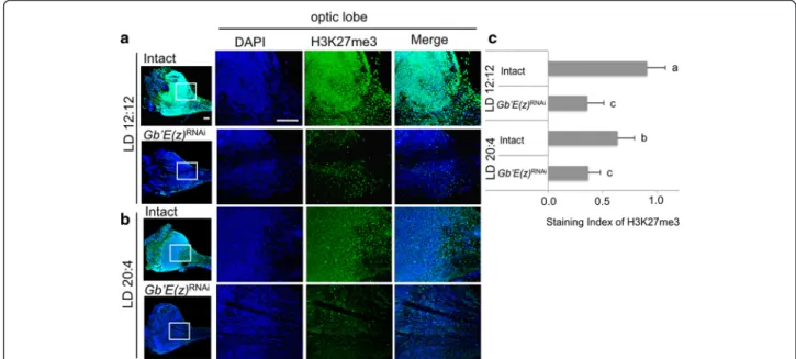
Methylation levels of histone H3K27
Since expression levels of Gb’E(z) were lower in LD 20:4 than in LD 12:12, we examined the level of histone H3 methylated at K27 at ZT 10 in the two photoperiodic con-ditions by immunohistochemistry using anti-H3K27me3 antibody. In both conditions, many nuclei were labeled by the antibody showing trimethylation of H3K27, being Fig. 3 Double-plotted actograms of locomotor activity of adult male crickets Gryllus bimaculatus under LD 12:12, LD 20:4, and DD. (a) intact cricket, (b) cricket treated with dsDsRed2, (c, d) crickets treated with dsGb’E(z). Crickets were exposed to LD 12:12 for the first 5 days, then in LD 20:4 for 9 days, and transferred to DD on the day indicated by an arrowhead, at a constant temperature of 25 ± 0.5 °C.α and ρ indicate active phase and rest phase, respectively. Dotted lines indicate onset and offset of the active phase. White and black bars above the actogram indicate the light and dark phases, respectively. Gray area in the actogram indicates the dark phase
distributed over whole optic lobe (Fig. 5a, b). The expres-sion levels in the optic lobe were significantly higher in LD 12:12 than in LD 20:4 (Fig. 5c). In the crickets treated with Gb’E(z)RNAi
, the levels of H3K27me3 in the optic lobe were significantly reduced both in LD 12:12 and LD 20:4 (Fig. 5). These results suggest that Gb’E(z) contributes to the modulation of histone H3K27me3 levels in the optic lobe. However, no significant difference was found between Gb’E(z)RNAi
crickets of the two photoperiodic conditions (Fig. 5c), although the Gb’E(z) mRNA levels significantly differed (Fig. 4a). This may be explained that the Gb’E(z) protein levels were so reduced in both cases that no signifi-cant difference was detected in H3K27me3 levels.
Discussion
Using an RNAi knockdown approach, we showed that the chromatin modifier Gb’E(z) is involved in regulating the free-running period and photoperiodic modulation of the locomotor rhythm in the cricket, G. bimaculatus.
Gb’E(z) expression in the optic lobe
Our qPCR analysis revealed that the chromatin modifier Gb’E(z) is constitutively expressed in the optic lobe, which is the location of the cricket’s circadian clock [19], which recalls the fact that most of the chromatin modifiers in-volved in circadian gene expression are present on a con-stitutive basis and recruited as needed to regulate transcription [7]. In mammals, for example, CLOCK plays a key role in the event and forms a transcriptional com-plex with BMAL1 to activate Per and Cry transcription by binding to the E-box in their upstream region [29]. Mean-while, CLOCK acts as a factor that opens up chromatin, which enables other transcriptional factors to act on target genes of CLOCK [11]. Similar molecular events may occur in transcription of per and tim in Drosophila, as CLK-CYC binding to upstream and/or intronic E-boxes of per and tim controls chromatin modifications through H3K9 acetylation and H3K4 trimethylation [10].
Interestingly, however, Gb’E(z) mRNA levels at ZT 6 and ZT 10 were higher in LD 12:12 than LD 20:4 Fig. 4 Daily expression profiles of Gb’E(z) and clock genes in the optic lobe of the cricket Gryllus bimaculatus under LD 20:4. Blue and green symbols with solid lines indicate the mRNA levels of Gb’E(z) (a), Gb’per (b), Gb’tim (c), and Gb’cyc (d) in DsRed2RNAiand Gb’E(z)RNAicrickets,
respectively. For reference, data for LD 12:12 are shown by broken lines (blue, DsRed2RNAicrickets; green, Gb’E(z)RNAicrickets). mRNA abundance
was measured by qPCR, with total RNA extracted from the optic lobes collected seven days after transfer to LD 20:4. Data collected from 3–10 independent measurements were averaged and plotted as mean ± SEM. The values shown are relative to those of Gb’rpl18a mRNA, which was used as an internal reference. In LD 20:4, Gb’E(z) mRNA showed no significant daily oscillation in DsRed2RNAicrickets (ANOVA, P > 0.05) but a
significantly lower level at ZT 6 and 10 in comparison with that in LD 12:12 (a). Gb’E(z)RNAireduced the Gb’E(z) levels which were even lower than
those of Gb’E(z)RNAicrickets in LD 12:12 at ZT 2–10 and ZT 18 (Tukey-test, P < 0.01). The values with different lower case letters at each ZT
significantly differ from each other (Tukey-test, P < 0.01, except between DsRed2RNAiin LD 20:4 and Gb’E(z)RNAiin LD 12:12 at ZT 10 and ZT 18
where P < 0.05). The mRNA levels of Gb’per (b), Gb’tim (c), and Gb’cyc (d) showed clear oscillatory profiles in both DsRed2RNAiand Gb’E(z)RNAi
crickets (ANOVA, P < 0.05 for Gb’cyc in DsRed2RNAi, and P < 0.01 for all other combinations). Values with different lower case letters differ significantly
(Fig. 4a) and the difference was reflected in the trimethy-lation levels of H3K27 (Fig. 5). To our knowledge, this is the first evidence for photoperiodic regulation of a clock-related chromatin modifier in insects. Light-induced modification of chromatin is known for H3S10 in the mammalian circadian clock in the suprachias-matic nucleus [30]; it is a result of phosphorylation and occurs within 5 min after lights-on, which is similar to c-fos induction. Gb’E(z) seems to be regulated by a differ-ent mechanism than immediate induction of phosphor-ylation, because no apparent change was observed at lights-on in Gb’E(z) mRNA levels (Figs. 1a and 4a). KIS-MET, a chromatin-remodeling enzyme, represents a can-didate regulator for this photoperiodic regulation of Gb’E(z), as it is required for normal circadian light re-sponses in Drosophila [31]. Gb’E(z) was also shown to be induced by mechanical stimulation; when a leg was in-jured, the onset of regeneration was associated with up-regulation of Gb’E(z) [24, 32]. Thus, its expression seems to be regulated by multiple pathways.
Gb’E(z) may be involved in photoperiodic modulation of the circadian rhythm
Involvement of chromatin modifiers in regulating the cir-cadian clock has been shown in animals. In Drosophila, Brahma chromatin-remodeling protein regulates CLK binding to target promoters, and hence the free-running period of the rhythm [8]. Mice that lacked Mettle3, a fac-tor that regulates RNA methylation, showed a delay in
RNA processing that leads to circadian period elongation [33]. However, our results revealed that Gb’E(z) plays a different role from those previously described regulatory models. Gb’E(z) knockdown significantly lengthened the free-running period in DD after LD 12:12 and LD 20:4 (Table 1). Another important effect of Gb’E(z)RNAi
was
elimination of the photoperiod-dependent change in α/ρ
ratio. In control crickets, the active phase was usually compressed during the dark phase in LD 20:4; conse-quently, the α/ρ ratio became much smaller (0.69 ± 0.22) than that in LD 12:12 (0.97 ± 0.09) (Table 1). Theα/ρ ratio further decreased when transferred to DD (0.55 ± 0.19) (Table 1). These results are consistent with our previous results [14]. However, compression of the active phase was only observed in a few Gb’E(z)RNAi
crickets, and the
majority of them showed no compression; the averageα/ρ
ratio was 0.84 ± 0.28 and 0.83 ± 0.25 in LD 20:4 and in the ensuing DD, respectively (Table 1). These results strongly suggest that regulation by E(z) is required in the response to a changing photoperiod through modulating the dur-ation of the active phase. The link between regulatory mechanisms and circadian waveform or free-running period should be investigated in future studies.
Gb’E(z) may contribute to response to photoperiodic changes via clock genes
This study revealed that temporal expression profiles of clock genes change in response to LD cycle. Under a long-day condition of LD 20:4, the circadian expression Fig. 5 Levels of histone H3 trimethylated at K27 (H3K27me3) in the optic lobe of the cricket Gryllus bimaculatus. (a, b) location of H3K27 in the optic lobes under LD 12:12 (a) or LD 20:4 (b). The right three columns indicate high magnification images of the optic lobe indicated by square from the low-magnification image in the left column. All images are oriented with dorsal side at the top. Scale bar, 100μm. Blue and green indicate nuclei (DAPI) and H3K27me3, respectively. (c) Staining indices of H3K27me3 in intact and Gb’E(z)RNAicrickets in LD 12:12 and LD 20:4. Average of 6 optic lobes are shown with SD. Values with different lower case letters differ significantly from each other (ANOVA followed by Tukey-test, P < 0.05). Note that level of H3K27me3 is higher in LD 12:12 than LD 20:4 and is greatly reduced in Gb’E(z)RNAicrickets
profile of the clock gene Gb’tim showed a similar pattern to that in LD 12:12, whereas Gb’per and Gb’cyc expres-sion was advanced and delayed by 4 h, respectively (Fig. 4). Gb’E(z)RNAi prevented the shift of Gb’per and
Gb’cyc rhythms in response to LD 20:4 (Fig. 4), which suggests the involvement of Gb’E(z) in photoperiod-dependent phase setting of daily clock gene expression. This function of Gb’E(z) may be mediated by trimethyla-tion of histone H3K27 because H3K27me3 levels are dependent on Gb’E(z) mRNA levels (Fig. 5) and H3K27me3 is known to regulate expression of Per1 and
Per2 in the mammalian circadian clock [25]. In the
mammalian clock, EZH2, a homologous protein of E(z), is recruited by the CLOCK/BMAL1 complex to bind to H3 in the promoter regions of Per1 and Per2 and causes di- and trimethylation of H3K27 [25]. The phase shift of Gb’cyc cycling caused by Gb’E(z)RNAi
might be attribut-able to indirect effects through the change of Gb’per rhythm, because Gb’per is involved in the transcriptional regulation of Gb’cyc [34].
The photoperiodic regulation of daily clock gene expres-sion may be caused by a change in Gb’E(z) mRNA levels (Fig. 4a). Because no daily rhythm is known in binding abil-ity of EZH2 to CLOCK: BMAL1 and H3 at the promoter regions of Per1 and Per2 [25], quantity of E(z) may affect the binding and eventually daily expression profiles of the clock genes. The change in Gb’E(z) levels may not be a sim-ple response to a given photoperiod, because theα/ρ ratio established in a photoperiod is maintained for a long period in DD [14]. The mechanism of this long-lasting change is an important issue that should be addressed in future studies.
The long-lasting response to photoperiod is reminiscent of photoperiodism regulating the seasonal physiological adaptation in insects. Many cricket species also show photoperiodic responses [35]. For example, nymphal devel-opment of Modicogryllus siamensis is photoperiodically regulated; nymphs grow faster under long-day conditions and become adults after seven moltings, whereas under short-day conditions, the nymphal period is elongated and the number of molts to adulthood increases [36]. Interest-ingly, developmental time course is determined within about 10 days after hatching [36], which indicates a long-lasting effect of photoperiod. This is consistent with the finding in this study suggesting that the long-lasting change in the clock is caused by an epigenetic mechanism, and photoperiodic response is most likely underlain by the circadian clock [37, 38]. In addition to the histone modifi-cation, we also have to consider the role of DNA methyla-tion, because a recent study showed that maternal transfer of photoperiodic information to offspring is attributable to DNA methylation in Nasonia vitripennis [39].
The means by which changes in molecular oscillation are reflected in overt activity rhythms are still being
elucidated. There are lines of evidence that molecular os-cillation of the circadian clock changes in a photoperiod-dependent manner. In Drosophila, daily per expression, i.e., phase and ratio of splicing variants, changes in re-sponse to photoperiod [40–42]. In other insects, the ex-pression profiles of clock genes also reportedly changed based on photoperiod [43, 44]. In the present study, we showed that Gb’per and Gb’cyc responded differently so that the peak phases of their daily expression rhythms be-came closer in the long photoperiod (Fig. 4). Although additional studies are necessary, this phase change might be somehow related to the shortening of the active phase under long-day conditions.
Conclusion
The present study discovered for the first time that methylation on H3K27 by Gb’E(z) is required for photo-periodic modulation of the circadian locomotor rhythm, such as length of the active phase and free-running period, which is associated with changes in expression profiles of clock component genes. These results con-tribute to molecular dissection of the mechanisms of in-sect photoperiodism and photoperiodic modulation in circadian rhythms.
Competing interests
The authors declare they have no competing interests. Authors’ contributions
YH and AT performed the experiments and YH and KT wrote the paper. KT and YH designed the experiments. TB and HO supported molecular analysis of Gb’E(z). YH, KT, TB and HO critically read the manuscript. All authors read and approved the final manuscript.
Acknowledgments
This work was supported in part by grants from JSPS [#14 J04731 to H.Y., #15 K06897 to B.T. and #15H04400 to K.T.]. H.Y. is a JSPS research fellow. Author details
1Graduate School of Natural Science and Technology, Okayama University, 3-1-1 Tsushima-naka, Kita-ku, Okayama 700-8530, Japan.2Graduate School of Medicine, Dentistry and Pharmaceutical Sciences, Okayama University, Kita-ku, Okayama 700-8558, Japan.
Received: 26 December 2015 Accepted: 14 March 2016
References
1. Dunlap JC, Loros J, DeCoursey PJ. Chronobiology: biological timekeeping. Sinauer: Sunderland; 2004.
2. Tataroglu O, Emery P. The molecular ticks of the Drosophila circadian clock. Curr Opin Insect Sci. 2015;7:51–7.
3. Hardin P. Molecular mechanisms of circadian timekeeping in Drosophila. Sleep Biol Rhythms. 2009;7:235–42.
4. Aguilar Roblero R, Díaz-Muñoz M, Fanjul-Moles ML. Mechanisms of circadian systems in animals and their clinical relevance. Heidelberg: New York, Dordrecht, London, Springer; 2015.
5. Ripperger JA, Merrow M. Perfect timing: epigenetic regulation of the circadian clock. FEBS Lett. 2011;585:1406–11.
6. Bellet MM, Sassone-Corsi P. Mammalian circadian clock and metabolism-the epigenetic link. J Cell Sci. 2010;123:3837–48.
7. Aguilar-Arnal L, Sassone-Corsi P. Chromatin landscape and circadian dynamics: spatial and temporal organization of clock transcription. Proc Natl Acad Sci U S A. 2015;112:6863–70.
8. Kwok RS, Li YH, Lei AJ, Edery I, Chiu JC. The catalytic and non-catalytic functions of the Brahma chromatin-remodeling protein collatorate to fine-tune circadian transcription in Drosophila. PLoS Genet. 2015;11:e1005307. 9. Doi M, Hirayama J, Sassone-Corsi P. Circadian regulator CLOCK is a histone
acetyltransferase. Cell. 2006;125:497–508.
10. Taylor P, Hardin P. Rhythmic E-box binding by CLK-CYC controls daily cycles in per and tim transcription and chromatin modifications. Mol Cell Biol. 2008;28:4642–52.
11. Menet JS, Pescatore S, Rosbash M. CLOCK: BMAL1 is a pioneer-like transcrption factor. Genes Dev. 2014;28:8–13.
12. Tomioka K, Chiba Y. Photoperiod during post-embryonic development affects some parameters of adult circadian rhythm in the cricket, Gryllus bimaculatus. Zool Sci. 1989;6:565–71.
13. Tomioka K, Chiba Y. Light cycle during post-embryonic development affects adult circadian parameters of the cricket (Gryllus bimaculatus) optic lobe pacemaker. J Insect Physiol. 1989;35:273–6.
14. Koga M, Ushirogawa H, Tomioka K. Photoperiodic modulation of circadian rhythms in the cricket Gryllus bimaculatus. J Insect Physiol. 2005;51:681–90. 15. Page TL. Transplantation of the cockroach circadian pacemaker. Science.
1982;216:73–5.
16. Page TL. Effects of optic tract regeneration on internal coupling in the circadian system of the cockroach. J Comp Physiol A. 1983;153:231–40. 17. Barrett RK, Page TL. Effects of light on circadian pacemaker development. I.
The freerunning period. J Comp Physiol A. 1989;165:41–9.
18. Tomioka K, Chiba Y. Effects of nymphal stage optic nerve severance or optic lobe removal on the circadian locomotor rhythm of the cricket, Gryllus bimaculatus. Zool Sci. 1984;1:385–94.
19. Tomioka K, Chiba Y. Characterization of optic lobe circadian pacemaker by in situ and in vitro recording of neuronal activity in the cricket Gryllus bimaculatus. J Comp Physiol A. 1992;171:1–7.
20. Tomioka K. Chronobiology of crickets: a review. Zool Sci. 2014;31:624–32. 21. Krauss V, Fassl A, Fiebig P, Patties I, Sass H. The evolution of the histone
methyltransferase gene Su (var) 3-9 in metazoans includes a fusion with and a re-fission from a functionally unrelated gene. BMC Evol Biol. 2006;6:18. 22. Tie F, Banerjee R, Stratton CA, Prasad-Sinha J, Stepanik V, Zlobin A, et al.
CBP-mediated acetylation of histone H3 lysine 27 antagonizes Drosophila Polycomb silencing. Development. 2009;136:3131–41.
23. Smith ER, Lee MG, Winter B, Droz NM, Eissenberg JC, Shiekhattar R, et al. Drosophila UTX is a histone H3 Lys27 demethylase that colocalizes with the elongating form of RNA polymerase II. Mol Cell Biol. 2008;28:1041–6. 24. Hamada Y, Bando T, Namamura T, Ishimaru Y, Mito T, Noji S, et al. Leg
regeneration is epigenetically regulated by histone H3K27 methylation in the cricket Gryllus bimaculatus. Development. 2015;142:2916–27. 25. Etchegaray JP, Yang X, DeBruyne JP, Peters AH, Weaver DR, Jenuwein T,
et al. The polycomb group protein EZH2 is required for mammalian circadian clock function. J Biol Chem. 2006;281:21209–15.
26. Moriyama Y, Sakamoto T, Karpova SG, Matsumoto A, Noji S, Tomioka K. RNA interference of the clock gene period disrupts circadian rhythms in the cricket Gryllus bimaculatus. J Biol Rhythms. 2008;23:308–18.
27. Sokolove PG, Bushell WN. The chi square periodogram: its utility for analysis of circadian rhythm. J Theor Biol. 1978;72:131–60.
28. Schmid B, Helfrich-Förster C, Yoshii T. A new ImageJ plug-in“ActogramJ” for chronobiological analyses. J Biol Rhythms. 2011;26:464–7.
29. Stanewsky R. Genetic analysis of the circadian system in Drosophila melanogaster and mammals. J Neurobiol. 2003;54:111–47. 30. Crosio C, Cermakian N, Allis CD, Sassone-Corsi P. Light induces
chromatin modification in cells of the mammalian circadian clock. Nat Neurosci. 2000;3:1241–7.
31. Dubruille R, Murad A, Rosbash M, Emery P. A constant light-genetic screen identifies KISMET as a regulator of circadian photoresponses. PLoS Genet. 2009;5, e1000787.
32. Bando T, Ishimaru Y, Kida T, Hamada Y, Matsuoka Y, Nakamura T, et al. Analysis of RNA-seq data reveals involvement of JAK/STAT signalling during leg regeneration in the cricket Gryllus bimaculatus. Development. 2013;140:959–64.
33. Fustin JM, Doi M, Yamaguchi Y, Hida H, Nishimura S, Yoshida M, et al. RNA-methylation-dependent RNA processing controls the speed of the circadian clock. Cell Tiss Res. 2013;155:793–806.
34. Uryu O, Karpova SG, Tomioka K. The clock gene cycle plays an important role in the circadian clock of the cricket Gryllus bimaculatus. J Insect Physiol. 2013;59:697–704.
35. Tauber TJ, Tauber CA, Masaki S. Seasonal Adaptations of Insects. New York: Oxford University Press; 1986.
36. Taniguchi N, Tomioka K. Duration of development and number of nymphal instars are differentially regulated by photoperiod in the cricket Modicogryllus siamensis (Orthoptera: Gryllidae). Eur J Entomol. 2003;100:275–81.
37. Saunders DS. Insect photoperiodism: seeing the light. Physiol Entomol. 2012;37:207–18.
38. Goto SG. Roles of circadian clock genes in insect photoperiodism. Entomol Sci. 2013;16:1–16.
39. Pegoraro M, Bafna A, Davies NJ, Shuker DM, Tauber E. DNA methylation changes induced by long and short photoperiods in Nasonia. Genome Res. 2016;26:203–10.
40. Majercak J, Sidote D, Hardin PE, Edery I. How a circadian clock adapts to seasonal decreases in temperature and day length. Neuron. 1999;24:219–30. 41. Majercak J, Chen W-F, Edery I. Splicing of the period gene 3′-terminal intron is regulated by light, circadian clock factors, and phospholipase C. Mol Cell Biol. 2004;24:3359–72.
42. Collins BH, Rosato E, Kyriacou CP. Seasonal behavior in Drosophila melanogaster requires the photoreceptors, the circadian clock, and phospholipase C. Proc Natl Acad Sci U S A. 2004;101:1945–50. 43. Syrova Z, Dolezel D, Saumann I, Hodkova M. Photoperiodic regulation of
diapause in linden bugs: are period and Clock genes involved? Cell Mol Life Sci. 2003;60:2510–5.
44. Sakamoto T, Uryu O, Tomioka K. The clock gene period plays an essential role in photoperiodic control of nymphal development in the cricket Modicogryllus siamensis. J Biol Rhythms. 2009;24:379–90.
• We accept pre-submission inquiries
• Our selector tool helps you to find the most relevant journal
• We provide round the clock customer support
• Convenient online submission
• Thorough peer review
• Inclusion in PubMed and all major indexing services
• Maximum visibility for your research Submit your manuscript at
www.biomedcentral.com/submit