Local Vascular Response Induced by Arm Position Change: The Related Myogenic Mechanism and Veno-arteriolar Reflex in Humans
8
0
0
全文
(2) Local Vascular Response Induced by Arm Position Change : The Related Myogenic Mechanism and Veno-arteriolar Reflex in Humans Junichi MAEDA', Takashi TAKEMIYA~, Masahiro SHIMODA~, Junichi SUZUKI~and Hideaki KOMIYA~ I. Faculty of Education, Miyagi University of Education, Aramaki Aza Aoba, Aoba-ku, Sendai 980, Japan 2 Institute of Health and Sport Sciences, University of Tsukuba, 1-1-1 Tennodai, Tsukuba, Zbaraki 305, Japan "~esearchand Education Center for Winter Sports, Hokkaido University of Education, Sapporo 002, Japan 4 Faculty of Education, Utsunomiya University, Utsunomiya 321, Japan. Abstract MAEDA, J. TAKEMIYA, T. SHIMODA, M. SUZUKI, J and KOMIYA, H. Local Vascular Response lnduced by Arm Position Change : The Related Myogenic Mechanism and Veno-arteriolar Reflex in Humans. Adv. Exerc. sports Physiol., Vo1.3, ~ 0 . 2pp.67-73, 1997. The local vascular reactlon to the change In transmural pressure of vascular bed5 plays a slgnlficant role In the circulatory ad~ustmentsdurlng orthostatlc stress. The placement of a limb below the level of the heart markedly Increases the vascular resistance of the deoendent llmb The contrlbutlons of the myogenlc mechanisms and the veno-arter~olarreflex to the Increase In the tonus of the arteriolar smooth muscles in dependent limbs are not clear, especially under normal innervation conditions. We evaluated the myogenic responses and veno-arteriolar reflex in the dependent fingers of 7 healthy male subjects using the differential digital photoplethysmogram ( A DPG) during vertical arm position change and moderate venous occlusion. The DC component of the digital photoplethysmogram, which represents volume changes in the finger, increased by 277.0+50.0 mV (n=7) during venous occlusion at 40 mmHg and by 279.0+56.2 mV (n =7) in the 40 cm arm lowering below the heart level, with no significant difference between the increases. The mean digital arterial blood pressure increased by 5.14+0.59 mmHg (n=7) during venous occlusion, and this increase was significantly (P<0.001) smaller than the 30.2k1.8 mmHg (n=7) decrease observed in lowering of the arm by 40 cm. The decrease in compliance index from the control value during venous occlusion was significantly smaller (P<0.01) than that observed in lowering of the arm by 40 cm (4.32+ 1.17 vs 7.20+ 1.83 mVIVlseclmmHg, n=7). These results suggest that the myogenic mechanism is more important than the veno-arteriolar reflex in the increase in the arteriolar smooth muscle tonus of dependent fingers. Key words: myogenic response, veno-arteriolar reflex, differential digital photoplethysmogram. Introduction The local vascular reaction to the change in transmural pressure of vascular beds plays a significant role in the circulatory adjustments during Address a correspondence: Junichi MAEDA Faculty of Education, Miyagi University of Education, Aramaki Aza Aoba, Aoba-ku, Sendai 980, Japan. Phone: 022-214-3455, Fax: 022-214-3455 e-mail: j-mae@,ipc.miyakyo-u.ac.jp. orthostatic stress. The placement of a limb below the level of the heart markedly increases the vascular resistance of t h e dependent limb. Veno-arteriolar reflex associated with venous distension has been considered to be the primary mechanism of this increase in the vascular resistance, with the myogenic responses induced by an increase in the arteriolar blood pressure (transmural pressure) playing a minor role (2,7, 22). However, increases in the vascular resistance associated with lowering of the limb were observed in subsequent studies using the skin of a free flap in which the sympathetic nerves were completely isolated, and there have been reports suggesting the involvement of myogenic responses (24). In these controversial studies, both the venous volume and the arterial blood pressure were increased simultaneously upon lowering of the limb below the heart level, and the responses of vascular smooth muscle were evaluated indirectly by measuring the blood flow by the 1 4 4 ~ eclearance method. The veno-arteriolar reflex is a local axonal reflex that causes contraction of arterial smooth muscle associated with an increase in the venous volume (7), and myogenic response is a mechanism of inducing changes in the arteriolar smooth muscle tonus associated with changes in the vascular transmural pressure ( 0 , l l 1 5 ) Therefore, concerning the mechanism of the increase in vascular resistance observed in lowering of the limb to below the heart level, the parameters of responses of vascular smooth muscle must primarily be evaluated by separating the effects of changes in the venous volume from those of changes in the transmural pressure. There.
(3) 68. J. MAEDA et al.. have been few studies evaluating the contributions of the myogenic mechanisms and the veno-arteriolar reflex to the increase in the tonus of the arteriolar smooth muscles in the dependent arms by such a method, especially under normal innervation conditions. Recently, Takemiya et a1.(21) developed a method of inducing a gravitational potential energy change (GPEC), in which changes in the finger volume are measured non-invasively and continuously using a modified differential digital photoplethysmograph ( A DPG). This GPEC- A DPG method provides information as to the local vascular response of arteriolar smooth muscle of the finger associated with the change in the transmural pressure producing by raising and lowering of the finger in relation to the heart level (13,14,20,21). The purpose of this study was to identify the separate effects of the veno-arteriolar reflex and of the myogenic response of arteriolar smooth muscle on the increase in vascular resistance observed in dependent fingers. The arm position change method, which simultaneously causes changes in the venous volume and arterial transmural pressure, and the venous occlusion method, which primarily causes an increase in the venous volume, were used to discriminate between the effects of venous volume and transmural pressure. Methods. Subjects The subjects were 7 healthy adult males (age range, 18-22 years) with no history of cardiovascular disorders, or of numbness or cold sensation at the finger tips. The subjects were informed as to the procedures and purpose of the study in detail, and gave their consent prior to participation in the study. Differential digital photoplethysmography and DC component of digital photoplethysmography Photoplethysmograms were recorded from the index finger using a modified conventional photoplethysmograph (MLV-2201, NIHONKOHDEN Co.) . A light-transmitting type pick-up for photoplethysmography was attached to the palmar side of the finger with two-sided adhesive tape to minimize its fluctuation during movements of the arm. Differential digital photoplethysmograms were recorded by reintroducing photoplethysmograms into an AC amplifier with a time constant of 0.03 second. The standard light absorption (DC component), which represents the digital tissue volume, was recorded simultaneously with the conventional photoplethysmograms and differential photoplethysmograms,. using a conventional photoplethysmograph modified for direct recording of the DC component from the tissue volume detection circuit.. Measurement of the digital arterial blood pressure The digital arterial blood pressure was measured non-invasively and continually using a Finapres (OHMEDA Co.) at the middle finger of the same hand as examined in photoplethysmography. Calculation of the arteriolar compliance index The volume of the finger, represented by the amplitude of the digital photoplethysmogram and differential digital photoplethysmogram, changes with the arteriolar smooth muscle tonus of the finger and the arterial pulse pressure. In this study, to correct for the effect of the arteriolar pulse pressure on the volume change, we calculated the quotient of changes in the volume of the finger and the pulse pressure (amplitude of the differential digital photoplethysmogram / pulse pressure) as an index of the finger tip arteriolar compliance. Arm position changes The arm position was changed by using the Takemiya procedure (20,21). The hands and forearms were placed comfortably in the sitting position on a flat horizontal board (armrest) of 40 cm width and 60 cm length. This armrest was supported between two adjustable height props and could be pivoted upon its proximal edge by a slinglpulley system. First, the forearm was fixed in the horizontal position on the armrest that allowed flexion and extension of the elbow, and the level of the fingers was set to the same level as the heart. The level of the heart (right atrium) was taken to be 7 cm below the cranial end of the manubrio-sternam in the sitting subject (12). The elbow was flexed or extended passively from this baseline position to raise or lower the fingers by 20 and 40 cm. This maneuver increased or decreased the blood pressure of the finger in proportion to the distance from the heart level (Fig. 1). The finger position was changed 5 times to each position in relation to the heart level in each subject, and the mean value of the 5 measurements in each position was calculated. Brachial venous occlusion To evaluate the effect of the veno-arteriolar reflex on changes in the amplitude of differentia1 digital photoplethysmogram, venous occlusion was applied to the brachium placed horizontally at the heart level to increase the venous volume. After completion of measurements by the arm position change method,.
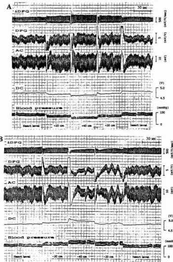
(4) 69. Local Arteriolar Responses to Arm Position Chang. a cuff for sphygmomanometry was tied around the brachium, and pressure of 40 mmHg was applied. This pressure had been confirmed in a preliminary experiment (unpublished data) not to cause significant changes in the arterial blood pressure but to significantly increase the tissue volume. This brachial venous occlusion was performed 5 times in each subject, and the mean of the 5 measurements was calculated as the value for that subject.. Statistical analyses All measured values were expressed as the mean f SE (number of samples). The data was excluded from statistical analyses when sudden changes in the photoplethysmogram or marked decreases in its amplitude due to psychological disturbance or fatigue were observed during the measurements. Differences between mean values of each arm positions were evaluated by one-way repeated measures analysis of variance. Comparisons of the changes in measured values produced by the venous occlusion and the 40 cm arm lowering were made with the paired t-test. Differences were considered significant at P<0.05. Results Arm position change Figure 1 shows representative of changes in variables upon raising and lowering of the arm. When the arm was raised, the amplitude of the ADPG, DPG, and AC component of the DPG increased, but the tissue volume (DC component of the DPG) and arterial pressure of the finger decreased, in accordance with the distance of the finger from the heart level (Fig. 1A). When the arm was lowered, ADPG, DPG and AC decreased, but the tissue volume and arterial pressure of the finger increased, again in accordance with the distance of the finger from the heart level (Fig. 1B). The systolic, diastolic and mean digital arterial blood pressures all significantly (P<0.001) decreased upon raising and increased upon lowering of the arm (Fig. 2). Because the systolic and diastolic blood pressures showed parallel changes, a slight ----C- SBP - 0 - DBP. .--. - - A - -MBP - - 4 - - Pulse oressure. Fig. 1. Responses of ADPG (differential digital photoplethysmogram), D P G (digital photoplethysmogram), AC (AC component of DPG), DC (DC component of DPG) and arteriolar blood pressure for 20 and 40 cm upward (A) and downward (B) arm position changes in relation to the heart level. When the arm was raised, the amplitude of ADPG, DPG and A C increased, but the tissue volume (DC) and the blood pressure decreased in accordance with the distance of the level of the fingers in relation to the heart level. When the arm was lowered, the ADPG, DPG and AC decreased, but the tissue volume (DC) and the blood pressure increased.. Arm position from heart level (cm) Fig. 2. Relationship between arm position in relation to the heart level and changes in blood pressure. All of the systolic, diastolic and mean arterial blood pressures decreased after arm raising and increased after arm lowering, but the pulse pressure showed no significant changes..
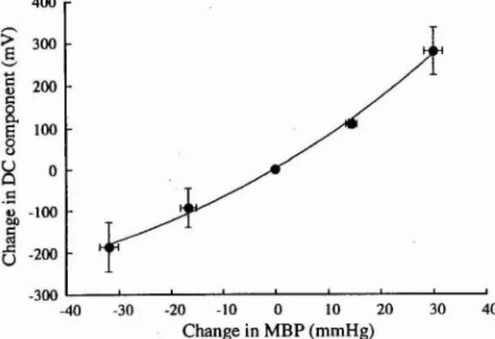
(5) 70. J. MAEDA et al.. changes were observed in the pulse pressure. The DC component of the digital photoplethysmogram, which represents the tissue volume of the finger, showed significant changes (P<0.001) proportionate to those of the digital blood pressure with arm position changes (Fig. 3). It decreased by 94.0k48.0 mV (n=7) and 187.3k58.2 mV (n=7) when the arm was raised to the +20 cm and +40 cm positions, respectively, and increased by 109.3 k10.2 mV (n=7) and 279.0k56.2 mV (n=7) when it was lowered to the -20 cm and -40 cm positions, respectively. 400. -300. r. I. -30. -20. -10. 0. 10. 20. 30. 40. Change in MBP (rnrnHg) Fig. 3. -3. 25r. E. $. +. Brachial venous occlusion Figure 5 shows representative traces of A DPG, volume of the finger (DC), and arterial blood pressure of the finger observed during venous occlusion by compression of the brachium with a cuff at 40 mmHg. The DC component increased by 277. O f 50.0 mV (n=7) as compared with the control value during venous occlusion, and this increase was not significantly different from the increase by 279.0k56.2 mV (n=7) observed with the 40 cm lowering of the arm (Fig. 6A). On the other hand,. 1 -40. The arteriolar compliance index of the finger calculated by dividing the amplitude of ADPG by the arteriolar pulse pressure of the finger showed significant changes (P<0.001) inverse to those in the arterial blood pressure of the finger with arm position changes (Fig. 4). It increased by 8.93L2.43 mVNlseclmmHg (n=7) with the 20 cm raising and by 16.5 5.80 mVN/sec/mmHg (n =7) with the 40 cm raising above the heart level. It decreased by 5. 06k1.63 mV/V/sec/mmHg (n=7) with the 20 cm lowering and by 7.20+1.83 mV/V/sec/mmHg (n=7) with the 40 cm lowering.. nnmr(rrmmrlmrrss(ImmnFnlllll(mlrmr(lr I. Relationship between changes in MBP and changes in D C component. The DC component of the digital photoplethysmogram showed changes proportionate to those of the digital arterial blood pressure induced by arm position changes.. Time (set). -. A DPG. -. 5.0. DC. 20-. 0. rT". w. Blood pressure. C .-. 6. (mmHg) -15 -40. I. I. I. I. I. I. I. I. -30. -20. -10. 0. 10. 20. 30. 40. Change in MBP (rnrnHg) Fig. 4. Relationship between changes in mean blood pressure (MBP) and changes in compliance index. The digital arteriolar compliance index calculated by dividing the amplitude of ADPG by the digital arteriolar pulse pressure was inversely related to the digital arterial blood pressure induced by arm position changes.. Cuff pressure Fig. 5. +40. mmHg. [:"". A simultaneous recording of ADPG, DC and arteriolar blood pressure responses produced by venous occlusion cuff pressure applied at the upper arm at the heart level. The D C component of DPG apparently increased, but ADPG and blood pressure showed only slight changes when the cuff pressure was increased by 40 rnmHg..
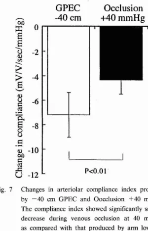
(6) Local Arteriolar Responses to Arm Position Chang. N.S.. Fig. 6. GPEC. Occlusion. -40 cm. +40 mmHg. " GPEC -40 cm. Occlusion +40 mmHg. Changes in D C component (A) and changes in MBP (B) produced by lowering the arm to -40 cm below the heart level (GPEC -40 cm) and by loading of 40 mmHg venous occlusion pressure at the upper arm at the heart level (Occlusion +40 mrnHg). The changes in the DC component showed no significant difference (N.S.) in GPEC -40 cm and in Occlusion 1 4 0 mmHg, but the changes in MBP showed significant difference in GPEC -40 cm and in Occlusion f 4 0 mmHg.. the mean arterial blood pressure increased by only 5.14*0.59 mmHg (n=7) as compared with the control value, and the increase was significantly smaller (P<0.001) than the 30.2k1.8 mmHg increase (n=7) observed with the 40 cm lowering of the arm below the heart level (Fig. 6B). The compliance index, which represents the vascular tonus of the arteriolar smooth muscle of the finger, decreased by 4.32 1.17 mVlV/sec/mmHg (n=7) during venous occlusion by compression at 40 mmHg. However, this decrease was significantly smaller (P <0.01) than the decrease observed upon the 40 cm lowering of the arm (7.20f 1.83 mVNlseclmmHg), which induced a similar increase in the tissue volume (Fig. 7).. GPEC. Occlusion. -40 cm. +40 mmHg. *. Discussion The major finding of this study is that the decrease in the compliance index of the finger vasculature produced by the 40 cm lowering of the arm in relation to the heart level is greater than the decrease observed during venous occlusion produced by compression at 40 mmHg with no significant difference in the volume of the finger. In the follow-. Fig. 7. Changes in arteriolar compliance index produced by -40 cm GPEC and Oocclusion +40 mmHg. The compliance index showed significantly smaller decrease during venous occlusion at 40 mmHg, as compared with that produced by arm lowering by 40 cm..
(7) 72. J. MAEDA et al.. ing discussion, the responses of the differential digital photoplethysmogram to arm position changes and to venous occlusion are compared and contrasted. When the level of the fingers is moved upward or downward in relation to the heart level, the intravascular hydrostatic pressure of the fingers changes in proportion to the vertical distance of the upward or downward movement (4). In raising of the level of the fingers, the arterial and venous pressures of the fingers decrease, and the blood in the venous system returns to the heart with a decrease in the volume of the fingers. In lowering of the level, on the other hand, both the arterial and venous pressures increase, and the tissue volume of the fingers increases due to retention of blood in the venous system (1, 5, 17). Takemiya (20,21) devised the arm position change method for non-invasive and continuous analysis of local vascular responses to changes in the arteriolar pressure of the fingers in humans using ADPG. Studies using this arm position change method have demonstrated opposite changes in the amplitude of ADPG and transmural pressure (13, 14,20,21), and the results of the present study are consistent with these observations. The mechanism producing changes in the amplitude of A DPG associated with arm position changes can be explained on the basis of the myogenic response of the arteriolar smooth muscle (3, 10, 11, 15). In raising of the arm, the arterial pressure (transmural pressure) of the finger decreases, and the tonus of the arteriolar smooth muscle of the finger is reduced by the myogenic response with consequent dilation of the arterioles. At this time, no significant changes are observed in the arterial pulse pressure, but the change in the volume per unit change in the pressure (volume changelpulse pressure), i.e., compliance (6), increases, and this is recorded as an increase in the amplitude of A DPG. In lowering of the arm, the vascular transmural pressure of the finger increases, the tonus of vascular smooth muscle of the finger increases due to the myogenic mechanism, and the vessels are constricted. At this time, as at the time of raising, no significant changes are observed in the arterial pulse pressure, and the compliance decreases. This decrease in the compliance is recorded as a decrease in the amplitude of ADPG. However, it is possible that the veno-arteriolar reflex might contribute to the constricting of the arterioles of the finger when the arm is lowered to below the heart level. Because the venous volume as well as the transmural pressure change with arm position changes, the tonus of the arteriolar smooth. muscle may be modified by the axonal veno-arteriolar reflex mechanism (5, 7, 22,23). The axonal veno-arteriolar reflex is a mechanism that increases the tonus of arteriolar smooth muscle through the local axonal reflex caused by venous distension (5, 7). To what extent the veno-arteriolar reflex and myogenic response contribute to the arteriolar constriction during arm lowering is not yet well understood (2, 7, 8, 9, 24). In the present study, to clarify this point, we induced increased venous volume by venous occlusion without inducing a marked increase in the arterial blood pressure, and we compared the results with those of arm position changes. The mean digital arterial blood pressure increased by only 5.14*0.59 mmHg during venous occlusion, which was a significantly smaller increase than the 30.211.8 mmHg increase observed with the 40 cm lowering of the arm position. The DC component of DPG, which indicates changes in the volume of the finger, increased by 277.0150.0 mV during the venous occlusion at 40 mmHg, and this increase was not significantly different from the 279.0156.2 mV increase observed in the 40 cm lowering of the arm. Therefore, venous occlusion at 40 mmHg increased the venous volume of the finger to a similar degree, but increased the arteriolar blood pressure of the finger significantly less, as compared with the 40 cm lowering of the arm. During brachial venous occlusion, the arteriolar compliance index of the finger showed a decrease of only 4.3211.17 mV/Vlsec/mmHg, which was significantly smaller than the 7.20 1.83 mVIViseclmmHg decrease observed in the 40 cm lowering of the arm position. The similar increase in the volume but with significantly smaller decrease in the arteriolar compliance index during venous occlusion at 40 mmHg as compared with that during lowering of the arm by 40 cm suggests that the changes in the compliance associated with position changes are related to changes in the arteriolar blood pressure rather than those in the venous volume. Therefore the changes in the amplitude of ADPG with arm position changes are considered to reflect changes in the arteriolar smooth muscle tonus due to the myogenic response of vascular smooth muscle associated with changes in the arteriolar blood pressure (transmural pressure) of the finger more so than they reflect the veno-arteriolar reflex mechanism associated with changes in the venous volume of the finger. The myogenic response was enhanced during sympathetic activation (16), while the veno-arteriolar reflex was attenuated (18). The response of the A. *.
(8) Local Arteriolar Responses to Arm Position Chang. DPG amplitude to arm position change was enhanced during sympathetic activation by cold water stimulation (19). These findings support our notion that the myogenic response is more important than the veno-arteriolar reflex in the increase in the arteriolar smooth muscle tonus of intact dependent fingers. In conclusion, our results indicate that changes in the amplitude of the differential digital photoplethysmogram during arm position changes reflect changes in the tonus of arteriolar smooth muscle due to the myogenic mechanism associated with changes in the arteriolar blood pressure (transmural pressure) of the finger more so than they reflect the veno-arteriolar reflex associated with changes in the venous volume of the finger. References 1) Almond NE, Jones D P and Cooke E D (1988) Noninvasive measurement of the human peripheral circulation: Relationship between laser Doppler flowmeter and photoplethysmograph signals from the finger. Angiology 39: 819-829. 2) Ahmad A , Hassdn K and Tooke J E (1988) Mechanism of the postural vasoconstrictor response in the human foot. Clin Sci 75: 379-387. 3) Bayliss WM (1902) On the local reactions of the arterial wall to changes of internal pressure. J Physiol 28: 220-231. 4) Ganong WF (1995) Dynamics of blood & lymph flow. In: Review of medical physiology 17th editions. Appleton & Lange, East Norwalk, Connecticut, pp. 525-541. 5) Gaskell P and Burton AC (1953) Local postural vasomotor reflexes arising from the limb veins. Circ Res 1: 27-39. 6) Goerke J and Mines A H (1988) Cardiovascular Physiology. Raven Press, Ncw York. pp. 80-84. 7) Henr~kscn 0 (1977) Local sympathetic reflex mechanism in regulation of blood flow in human subcutaneous adipose tissue. Acta Physiol Scand 101 (Suppl 450): 1-48. 8) Jepsen H and Gaehtgens P (1993) Postural vascular response in human skin: passive and active reactions to alteration of transmural pressure. Am J Physiol 265: H949-H958. 9) Jepsen H and Gaehtgens P (1995) Postural vascular response vs. sympathetic vasoconstriction in human skin during orthostasis. Am J Physiol 269: H53-H61.. 73. 10) Johnson PC (1980) The myogenlc response In Hdndbook of Physiology, Sect 2, The Card~ovascularSystem, Vol 11, Vascular Smooth Muscle, eds Bohr DF, Somolyo AP dnd Sparks H V , Society, Bethesda, Maryland, pp 409-442 American Phys~olog~cal 11) Johnson PC (1981) Myogenic mechanisms of blood flow rcgulation In Vasodilatation, eds Vanhoutte PM and Leuse I , Raven Press, New York pp 255-262 12) Levick JR and Michel CC (1978) The effects of position and 5k1n temperdture on the capillary pressures in the fingers dnd toes J Physiol 274 97-109 13) Maeda J , Ando S , Takemiya T and Nagashimd C (1989) D~fferential digital plethysmographic dnalysis of Bayliss effect In Microcirculation annual 1988, eds Tsuchiya M , Asano M and Mishima Y, N~hon-Igakukan,Tokyo, Japan pp 39-40 14) Maeda J and Takemiya T (1990) Laser Doppler and ultrasonic Doppler flowmetric analysi\ of microvascular responses in the finger to change In arm position Jpn J Phys Fitness Sports Med 39 106-113 (In Japanese with Enghsh abstract) 15) Mellander S (1988) Myogenic mechanisms in local vdscular control Acta Physiol Scand 133 (Suppl 571) 25-42 16) Ping P and Johnson P C (1992) Role of myogenic response In enhanc~ngautoregulation of flow during sympathetic nerve st~mulation Am J Phys~ol263 H1177-H1184 17) Richardson D (1988) Effects of gravity on reg~onal and capillary blood flows In the human toe Microvasc Res 35 334-340 18) Skagen K (1983) Sympathetic reflex control of blood flow In human subctaneous tissue during orthostat~cmaneuvres Danish Med Bull 30 229-241 19) Suzuk~J , Maeda J and Takemlya T (1994) Analysis of microvascular responses in the finger to change in arm position during cold water stimulation Jpn J Physiol 44 181-191 20) Takemiya T, Maeda J , Ando S and Miyazaki J (1989) Differentlal digital plethysmographic analysis of microvascular response produced by the gravitat~onalpotential energy change Jpn J Phys Fitness Sports Med 38 64-70 ( In Japanese with English abstract) 21) Takem~ya T , Maeda J, Suzuki J, N~shihira Y and Shimoda M (1996) Differential digital photoplethysmographic observations of finger vascular exponential response to the arm positlon changes in humans Adv Exerc Sports Phys~ol2 83-90 e pressure on blood flow 22) Yamada S (1954) Effects of p o s ~ t ~ vtissue of the finger J Appl Physiol 6 495-500 23) Yamada S and Burton AC (1954) Effect of reduced tissue pressure on blood flow of the fingers, the veni-vasomotor reflex J Appl Physiol 6 501-505 24) Zoltie N, Young C, Earls I and Tan E (1989) The veno-arteriolar reflex in free skin flaps Clin Physiol 9 183-188 (Received 25 May 1997, accepted 7 July 1997).
(9)
図
関連したドキュメント
We present sufficient conditions for the existence of solutions to Neu- mann and periodic boundary-value problems for some class of quasilinear ordinary differential equations.. We
In Section 13, we discuss flagged Schur polynomials, vexillary and dominant permutations, and give a simple formula for the polynomials D w , for 312-avoiding permutations.. In
Analogs of this theorem were proved by Roitberg for nonregular elliptic boundary- value problems and for general elliptic systems of differential equations, the mod- ified scale of
The commutative case is treated in chapter I, where we recall the notions of a privileged exponent of a polynomial or a power series with respect to a convenient ordering,
Then it follows immediately from a suitable version of “Hensel’s Lemma” [cf., e.g., the argument of [4], Lemma 2.1] that S may be obtained, as the notation suggests, as the m A
Definition An embeddable tiled surface is a tiled surface which is actually achieved as the graph of singular leaves of some embedded orientable surface with closed braid
Correspondingly, the limiting sequence of metric spaces has a surpris- ingly simple description as a collection of random real trees (given below) in which certain pairs of
[Mag3] , Painlev´ e-type differential equations for the recurrence coefficients of semi- classical orthogonal polynomials, J. Zaslavsky , Asymptotic expansions of ratios of
