PROTACs and Other Chemical Protein Degradation
Technologies for the Treatment of
Neurodegenerative Disorders
著者
Shusuke Tomoshige, Minoru Ishikawa
journal or
publication title
Angewandte Chemie International Edition
volume
59
page range
2-11
year
2020-05-14
URL
http://hdl.handle.net/10097/00131806
Recent Progress in PROTACs and Other Chemical Protein Degradation Technologies for the Treatment of Neurodegenerative Disorders
Shusuke Tomoshige[a] and Minoru Ishikawa*[a]
[a] Dr. S. Tomoshige, Prof. Dr. M. Ishikawa Graduate School of Life Sciences
Tohoku University
2-1-1 Katahira, Aoba-ku, Sendai 980-8577, Japan E-mail: [email protected]
[author profile]
Shusuke Tomoshige completed his Ph.D. at The University of Tokyo in 2016 under the supervision of Professor Yuichi Hashimoto. After his postdoctoral training for two years at the University of Notre Dame, under the guidance of Professor Shahriar Mobashery, he joined Tokyo University of Science, working with Professor Kouji Kuramochi. Since September 2019, he has been an Assistant Professor at Graduate School of Life Sciences, Tohoku University. His research focuses on the degradation of biopolymers, including proteins and bacterial cell wall.
Minoru Ishikawa received his M.Eng. (1996) from Tokyo Institute of Technology, then became a Researcher at the Medicinal Chemistry Research Labs in Meiji Seika Kaisha, Ltd., Japan (1996−2008). During that time, he received his Ph.D. degree from The University of Tokyo (2006). He subsequently joined The University of Tokyo as an Assistant Professor (2008), and was subsequently promoted to Lecturer (2012) and Associate Professor (2013). He then moved to Tohoku University as a Full Professor (2019). His research interests are medicinal chemistry and chemical biology.
Abstract: Neurodegenerative disorders (NDs) are a group of diseases that cause neural 1
cell damage, leading to motility and/or cognitive dysfunctions. One of the causative 2
agents is misfolded protein aggregates, which are considered as undruggable in terms of 3
conventional tools, such as inhibitors and agonists/antagonists. Indeed, there is currently 4
no FDA-approved drug for the causal treatment of NDs. However, emerging 5
technologies for chemical protein degradation are opening up the possibility of selective 6
elimination of target proteins through physiological protein degradation machineries, 7
which do not depend on the functions of the target proteins. Here, we review recent 8
efforts towards the treatment of NDs using chemical protein degradation technologies, 9
and we briefly discuss the challenges and prospects. 10
1. Introduction 1
1.1. Neurodegenerative Disorders and Treatment Approaches 2
Neurodegenerative disorders (NDs) are a series of diseases characterized by 3
progressive impairments in motility and/or cognitive function, leading in some cases to 4
death.[1] Alzheimer’s disease (AD) is the most common cause of dementia: 10-30% of
5
people >65 years of age are estimated to live with AD.[2] Onset of the major NDs, such
6
as AD, Parkinson’s disease (PD), and polyglutamine diseases (polyQDs), is associated 7
with the accumulation of aggregation-prone misfolded proteins (amyloid β (Aβ), tau, α-8
synuclein, and proteins with abnormally expanded polyglutamine repeats, respectively, 9
in the above diseases).[3] These misfolded proteins accumulate as insoluble fibrillar
10
aggregates via soluble oligomeric intermediates, which are currently considered as the 11
real villain in the pathogenesis[4] (Figure 1). Note that the misfolded proteins often show
12
unusual protein-protein interactions (PPIs) independently of their intrinsic functions. 13
The unusual PPIs cause dysfunctions in specified compartment including nucleus and 14
mitochondria and lead to neuronal cell death.[5] Therefore, the conventional drug
15
discovery program with modification of the intrinsic functions of pathogenic proteins is 16
not suitable for the treatment of NDs. 17
1
Figure 1. Schematic illustration of the process of misfolded protein aggregation. 2
Many attempts have been made to develop NDs treatments, generally by employing 3
chemical or biological techniques to eliminate the toxic oligomeric species from 4
neuronal cells. Medicinal chemistry studies have yielded various small molecules that 5
modulate aggregation pathways. Early aggregation modulators were aromatic planar 6
molecules that inhibit aggregate formation by interfering with the interaction of the 7
planar β-sheet surfaces of misfolded proteins to disrupt their stacking.[6] On the other
8
hand, in 2012, an aggregation enhancer was discovered that reduces the population of 9
oligomeric species and increases that of fibrillar aggregates.[7] But, despite this
long-10
established strategy, only one aggregation modulator is currently in clinical trial.[8]Gene
11
silencing techniques, such as RNA interference (RNAi), antisense oligonucleotides, and 12
genome editing, have also attracted attention.[9] Indeed, some in vivo applications for
13
NDs have already been reported, exploiting adeno-associated virus (AAV) or non-viral 14
delivery systems, and clinical trials for amyotrophic lateral sclerosis (ALS) and 15
Huntington’s disease (HD, one of the nine polyQDs) are ongoing.[10] Nevertheless,
16
delivery is still problematic, because non-viral delivery systems are invasive and less 17
effective, while viral delivery systems pose safety issues. The possibilities of off-target 1
effects and interference with endogenous RNAi pathways are also concerns.[11] Passive
2
immunization therapy is another approach to reduce misfolded proteins, albeit it works 3
extracellularly. Several antibodies for Aβ are currently evaluated in phase III clinical 4
studies.[12] Biogen and Eisai announced one of the Phase III study of their aducanumab,
5
an monoclonal antibody against Aβ, met its primary endpoint in 2019, and they are now 6
preparing to submit Biologics License Application to the FDA.[13] However, passive
7
immunization is costly and shows poor BBB permeability (typically ~0.1% of the 8
injected antibody cross BBB).[12] Indeed, aducanumab requires high-dose administration.
9
1.2. Emerging Prospect: Chemical Protein Degradation 10
Today, chemical inhibitors, agonist/antagonists, and ion channel openers/blockers 11
are widely used for various diseases. On the other hand, the misfolded proteins in NDs 12
are generally considered as ‘undruggable’ in that, for example, they lack ligand-binding 13
sites that could be targets for inhibitors or modulators, and the neuronal cell death is 14
induced independently of the intrinsic functions of them. Thus, novel therapeutic 15
strategies are required. One such strategy is to lower the levels of target proteins by 16
using small molecules or peptides to promote their degradation.[14–17] The chemical
17
protein degradation strategy aims to direct eukaryotic protein degradation machineries, 18
including the ubiquitin-proteasome system (UPS; for the details, see section 1.3) or 19
autophagy (for the details of autophagy, see section 3), towards a protein of interest 20
(POI) by modulating the relevant protein-protein interactions. The concept of hybrid 21
molecules with a dual mode of action provided a clue to the development of the 22
chemical protein degradation technologies;[18] one of the technologies this purpose
developed for is UPS induction using hybrid molecules ‘Proteolysis Targeting Chimeras 1 (PROTACs).[19] 2 1.3. Development of PROTACs 3
In UPS, a ubiquitin ligase (E3) repeatedly labels its protein substrate with ubiquitins, 4
which are activated by a ubiquitin-conjugating enzyme (E2), to form poly-ubiquitin 5
chain on lysine residues of the protein substrate. Subsequently, a large protease complex 6
proteasome recognizes the ubiquitin chain and hydrolyzes the substrate.[20]
7
In 2001, Crews and co-workers pioneered the development of PROTACs[21] which are
8
hetero-bifunctional molecules comprised of a ligand for an E3 linked to a ligand for the 9
POI. These hybrid molecules serve to bring the POI and the E3 into close proximity and 10
enable the POI to be ubiquitinated even though it is not an endogenous substrate of the 11
E3, thereby leading to proteasomal degradation (Figure 2). 12
13
Figure 2. The concept of PROTACs-mediated protein degradation. 14
The first-generation PROTACs are peptide-based molecules that employ β-TrCP or 15
von Hippel-Lindau (VHL) recognizing peptides as E3 ligands. However, their cell-16
permeability is problematic and these PROTACs require micro injection or 17
incorporation of cell-penetrating peptide (CPP) sequences for use in living cells.[21,22]
1
To address these problems, Crews et al. developed a small hybrid molecule consisting 2
of a ligand for androgen receptor (AR) and nutlin-3, a small-molecular murine double 3
minute 2 (MDM2, an E3) inhibitor.[23] This hybrid molecule has been described as the
4
first small-molecular PROTAC, but this may not be strictly accurate,[24] because AR is
5
actually an endogenous substrate of MDM2.[25] In addition, nutlin-3 itself enhances
6
MDM2-mediated AR degradation.[26]. Taking account of these questions, our group
7
focused on the induction of non-physiological protein degradation by small molecules, 8
and in 2010 we reported cell-permeable, small-molecular PROTACs (also known as 9
SNIPERs: Specific Non-IAP-dependent Protein Erasers) which recruit inhibitor of 10
apoptosis protein (IAP) family members possessing E3 activity.[27] We subsequently
11
applied IAP-mediated protein degradation to various proteins located in cytosol, nucleus,
12
cell membrane, and mitochondria.[28,29] In 2015, the Crews group and the Bradner group
13
independently developed VHL- and cereblon (CRBN)-based small-molecular 14
PROTACs, respectively.[30,31] These PROTACs were the first to achieve potent
15
degradation of the POI with DC50 values of nanomolar order in cells. These
16
achievements dramatically accelerated the advance of the technology,[32] like a
17
“Cambrian explosion,” and led to multiple applications, including HaloTag-fused 18
proteins,[33,34] bromodomain-containing proteins,[35–37] kinases,[38–40] and
19
phosphodiesterase,[41] as well as in vivo studies.[30,31,42] Further exploration of E3 for
20
PROTACs is attractive, because only a limited number of E3s has been utilized so 21
far.[43] Besides IAP, VHL and CRBN, five E3s have been exploited for small-molecular
22
PROTACs to date;[44–48] however, this corresponds to only a few percent of E3s.
23
Mechanistic studies have shown the unique aspects of PROTAC technology. For 24
example, studies using promiscuous warhead as a ligand for POI revealed that 1
accessibility to ternary complex formation involves in their selectivity, suggesting that 2
“PROTACization” of promiscuous drugs might be an idea to improve their 3
selectivity.[49] Since late 2019, optical control of PROTACs have attracted attention and
4
more than five papers were published so far.[50]
5
In the past few years, PROTACs technology has attracted commercial interest, with 6
the major focus being on PROTACs for cancer therapy.[51,52] Our group reported double
7
degradation of IAP and an oncogenic protein by a hybrid molecule employing an IAPs 8
pan antagonist in 2012,[53] suggesting that double protein degradation of oncogenic
9
IAPs and oncogenic proteins is a promising approach for cancer treatment. We believe 10
that this feature affords a major advantage over other PROTACs that utilize the 11
ubiquitin ligases VHL and CRBN. In 2019, a similar approach targeting oncogenic 12
proteins with PROTACs employing MDM2 inhibitors resulted in synergistic 13
activity.[44,54] The structural insights into ternary complex (POI-PROTAC-E3)[55]
14
reported in 2017 have facilitated rational molecular design of PROTACs for cancer 15
therapy. Two PROTACs from Arvinas, Inc. entered phase I clinical studies for certain 16
cancers in 2019.[56]
17
18
2. Proteolysis-Targeting Chimeras for NDs therapy 19
2.1. Peptide PROTACs Aimed at AD Therapy 20
In 2016, the group led by Chen and Li reported a tau-targeting PROTAC with 21
potential for AD treatment; this was the first attempt to apply PROTACs to the 22
treatment of NDs.[57] They designed the tau-targeting all-peptide PROTAC 1 (Figure 3);
1
this is a 32-a.a. peptide consisting of, from the N-terminus, a motif for tau recognition, a 2
linker peptide, a motif for VHL recognition, and D-Arg8 as the CPP. They successfully
3
demonstrated 1-mediated degradation of tau through UPS in cell cultures and in vivo. 4
Notably, 1 also ameliorated the neurotoxicity of Aβ. 5
Another peptide PROTAC for AD was developed by Jiang, You and colleagues.[58]
6
It is noteworthy that they harnessed CRLKeap1 by using a 9-a.a. peptide sequence for
7
Keap1 recognition, which was identified by the same group.[59] Their peptide PROTAC
8
2 (Figure 3) induced UPS-mediated degradation of tau protein in cell lines. 9
10
Figure 3. Structures of tau-targeting peptide-based PROTACs 1 and 2. 11
2.2. Small-Molecular PROTACs for NDs therapy 12
The greatest obstacle to developing small-molecular PROTACs for NDs is that no 13
selective small-molecular ligand for NDs-related proteins has yet been discovered. To 14
address this problem, we exploited small-molecular binders to misfolded protein 15
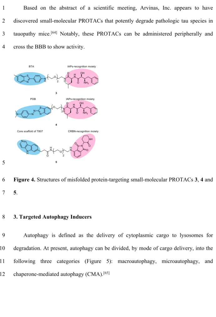
aggregates, and developed compounds 3 and 4 (Figure 4) as the first all-small-molecular 16
PROTACs targeting mutant huntingtin (mHtt, an aggregate-prone neurotoxic protein 17
involved in HD) in 2017.[60,61] In the design of 3 and 4, we used benzothiazoles BTA
18
and PDB, which are PET tracers for misfolded protein aggregates, as aggregate binders, 19
and linked them to ligands for IAP (therefore these compounds can be categorized as 1
SNIPERs). Compounds 3 and 4 successfully induced a UPS-mediated decrease of mHtt 2
in primary cells from HD patients, as well as in HeLa cells transfected with mHtt exon-3
1 bearing a long polyQ repeat. In brief, i) mechanistic analysis established that 3 did not 4
decrease HTT mRNA, ii) an artificial complex between IAP and aggregates was 5
detected by means of ELISA iii) a negative control compound without affinity for IAP 6
did not reduce the mHtt level, and iv) involvement of proteasomal degradation of mHtt 7
was confirmed by co-treatment with a proteasome inhibitor. Furthermore, 3 also 8
decreased the amount of mHtt aggregates in cells. We observed the degradation of wild-9
type Htt as well, but not that of green fluorescent protein (GFP) as a control, and we 10
concluded that wild-type Htt also forms small oligomers that can be recognized by 11
aggregate binders, leading to PROTAC-mediated degradation. Targeting protein 12
aggregates seems to be a promising strategy to develop PROTACs targeting pan-13
misfolded proteins. Indeed, we found that compounds 3 and 4 also reduce the levels of 14
mutant ataxin-3, mutant ataxin-7 and mutant atrophin-1, which are misfolded proteins 15
implicated in other polyQDs.[62] In 2019, Gray and Haggarty’s group reported,
16
independently of our group, another small-molecular PROTAC 5 targeting protein 17
aggregates (Figure 4).[63] Their compound contains the core 5H-pyrido[4,3-b]indole
18
scaffold of T807, a PET tracer for tau aggregates, as an aggregate binder, and this is 19
linked to pomalidomide for recruitment of CRLCRBN. They demonstrated that 5 triggers
20
UPS-mediated tau clearance in neurons derived from patients with frontotemporal 21
dementia (FTD), as well as promoting recovery of FTD neurons from tau-mediated 22
stress vulnerability. 23
Based on the abstract of a scientific meeting, Arvinas, Inc. appears to have 1
discovered small-molecular PROTACs that potently degrade pathologic tau species in 2
tauopathy mice.[64] Notably, these PROTACs can be administered peripherally and
3
cross the BBB to show activity. 4
5
Figure 4. Structures of misfolded protein-targeting small-molecular PROTACs 3, 4 and 6
5. 7
3. Targeted Autophagy Inducers 8
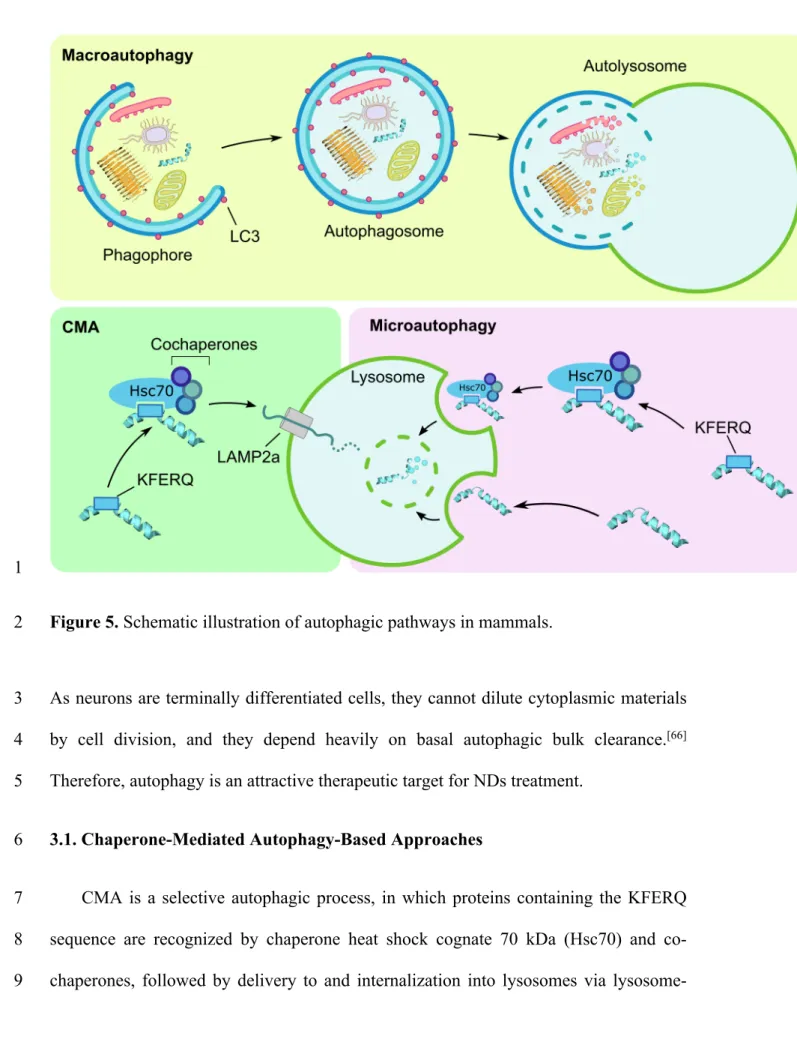
Autophagy is defined as the delivery of cytoplasmic cargo to lysosomes for 9
degradation. At present, autophagy can be divided, by mode of cargo delivery, into the 10
following three categories (Figure 5): macroautophagy, microautophagy, and 11
chaperone-mediated autophagy (CMA).[65]
1
Figure 5. Schematic illustration of autophagic pathways in mammals. 2
As neurons are terminally differentiated cells, they cannot dilute cytoplasmic materials 3
by cell division, and they depend heavily on basal autophagic bulk clearance.[66]
4
Therefore, autophagy is an attractive therapeutic target for NDs treatment. 5
3.1. Chaperone-Mediated Autophagy-Based Approaches 6
CMA is a selective autophagic process, in which proteins containing the KFERQ 7
sequence are recognized by chaperone heat shock cognate 70 kDa (Hsc70) and co-8
chaperones, followed by delivery to and internalization into lysosomes via lysosome-9
associated membrane protein type 2a (LAMP2a), leading to lysosomal degradation. 1
Besides the consensus Hsc70 binding motif KFERQ, α-synuclein has the unique Hsc70 2
binding motif VKKDQ, which is also directed to the CMA machinery (Figure 5).[67]
3
Hence, these two sequences can be utilized as CMA-targeting warheads. Note that 4
Hsc70 also involves in microautophagy but LAMP2a does not. The following two 5
reports have proven the involvement of LAMP2a. 6
In 2010, Nukina and co-workers reported targeted protein degradation by hijacking 7
the CMA machinery for selective clearance of mHtt.[68] The authors designed a DNA
8
construct coding a 46-a.a. peptide 6 (Figure 6), consisting of KFERQ and VKKDQ 9
sequences linked to two copies of polyglutamine binding peptide 1 (QBP1). This 10
reduced mHtt in mouse brain, ameliorated motor dysfunction, and improved survival 11
ratio without decreasing the body weight of wild-type mice. Additionally, 6 conjugated 12
to monomeric red fluorescent protein (mRFP) as a reporter can decrease accumulation 13
and aggregation of other polyQ proteins, such as mutant ataxin-3 and mutant AR, 14
suggesting the potential of this approach for pan-polyQDs treatment. As for synthetic 15
peptides, Wang’s group developed a cell-permeable peptide 7 as a CMA inducer 16
targeting α-synuclein.[69] Compound 7 is a 35-a.a. peptide consisting of, from the
N-17
terminus, a TAT sequence from HIV as a CPP, an α-synuclein-binding motif, and 18
CMA-targeting motifs (Figure 6). The α-synuclein-binding motif they employed is a 10-19
a.a. stretch from β-synuclein that is known to interact with α-synuclein (Kd = 1 μM,
20
fluorescence polarization).[70] The authors demonstrated that the addition of synthetic 7
21
to the culture medium of primary neurons successfully reduced the level of wild-type α-22
synuclein, as well as A53T mutant, a cause of familial PD, in a lysosome-dependent 23
manner. 24
1
Figure 6. Structures of CMA inducers 6 and 7. CTM: CMA-targeting motifs. 2
3.2. Macroautophagy-Based Approach 3
Macroautophagy is the best-characterized machinery of autophagy, in which the 4
cytoplasmic cargoes are sequestered in autophagosomes, which are double-membrane 5
vesicles formed by elongation of the phagophore, a cup-shaped membrane. The fusion 6
of an autophagosome with endosome or lysosome leads to degradation of the cargo 7
(Figure 5).[71] Microtubule-associated proteins light chain 3B (LC3B), attached to the
8
inner membrane surface of the phagophore, acts as a receptor for macroautophagy, for 9
encapsulation of the cargo.[72]
10
In 2019, Li et al. performed a small-molecule microarray (SMM)-based screening 11
to identify compounds that tether mHtt and LC3B together to encapsulate mHtt in 12
autophagosomes.[73] Excluding hits in the screening with wtHtt, the authors identified
13
small molecules 8 and 9 (Figure 7) that induce allele-selective clearance of mHtt in an 14
autophagy-dependent manner. Further, all the compounds they identified successfully 15
rescued HD-relevant phenotypes in vivo, e.g. resulting in prolongation of lifespan in 16
Drosophila HD models and amelioration of motor dysfunction in mouse HD models. It
17
is noteworthy that the identified compounds are not hetero-bifunctional molecules but 18
molecular glues, suggesting that the SMM-based screening could be used to identify 19
BBB-permeable compounds with the same activity. Further structure-activity 20
relationship studies of the compounds identified in the report should uncover the core 1
structure for binding to LC3B without perturbation of its activity. 2
3
Figure 7. Structures of molecular glues 8 and 9 that bring mHtt and LC3B together. 4
4. Hydrophobic Tagging 5
Eukaryotic cells operate a protein quality control system to remove misfolded 6
proteins and their aggregates. Key players in this process are molecular chaperones 7
called heat-shock proteins (HSPs) that refold the misfolded proteins or facilitate their 8
degradation through UPS or autophagy.[74] HSPs recognize misfolded proteins through
9
their exposed hydrophobic residues, which are buried inside proteins in their native 10
folding. This system can be utilized to degrade POIs. For example, fulvestrant is 11
composed of an endogenous ER ligand and a hydrophobic alkylsulfinyl group as a 12
HSPs recruiting moiety; this enables it to degrade ER, and it is clinically used to treat 13
ER-positive metastatic breast carcinomas. The Crews group has generalized this 14
hydrophobic tagging technique by designing small molecules, termed hydrophobic tags 15
(HyTs), consisting of a hydrophobic adamantyl group linked to a ligand for a POI to 16
induce HSP-mediated protein degradation.[75,76]
17
Li and his co-workers have reported two series of HyTs aimed at the treatment of 18
AD (in 2017) and ALS (in 2019), employing tau- and TDP-43-targeting peptides, 19
respectively, as warheads for the POI.[77,78] Compound 10, the tau-targeting HyT shown
1
in Figure 8, reduced tau in living cells in a proteasome-dependent manner. Further, the 2
intravenous administration of this HyT successfully degraded tau in brains of AD model 3
mice, suggesting that 10 is BBB-permeable. As for the TDP-43-targeting HyT, the 4
authors designed a repertoire of peptides consisting of adamantane(s), linker peptides, 5
TDP-43-targeting peptides, and CPPs. Among those peptides, 11, containing two 6
adamantyl groups as hydrophobic groups, was the most effective, reducing the TDP-43 7
levels in living cells as well as in TDP-43-overexpressing Drosophila models. But, 8
although these HyTs showed degradation activity in vivo, high doses (20-150 μM in the 9
cell) were required, and slight cytotoxicity was observed. 10
11
Figure 8. Structures of HyTs 10 and 11. 12
5. Summary and Outlook 13
Drug discovery for NDs faces at least two problems: (1) aggregation-prone proteins 14
cause diseases independently of their intrinsic functions, and (2) drugs that can cross the 15
BBB are required. Point 1 means that conventional drug discovery strategies, which rely 16
on the modulation of functions of proteins, e.g., with inhibitors or agonists/antagonists, 17
are not appropriate. Point 2 means that gene silencing of pathologic proteins is 1
problematic due to the difficulty of delivering nucleic acids into the patient’s brain, in 2
addition to safety concerns. Chemical protein degradation has already overcome the 3
point 1 because this approach eliminates pathologic proteins by utilizing common 4
structural features of misfolded proteins, i.e., β-sheet-rich structure, but not the inherent 5
function or structure of the pathogenic protein. The phase III result of aducanumab, 6
which decreases extracellular Aβ aggregates, also encourages chemical protein 7
degradation approaches towards ND therapy. Aducanumab are close to clinical use but 8
this approach has yet to solve point 2. In fact, it requires high-dose administration 9
(aducanumab required 10 mg/kg) and occurs brain swelling (edema) in a dose-10
dependent manner.[79] On the other hand, small-molecular protein degraders decrease
11
intracellular aggregation-prone proteins such as tau and polyQ proteins, and might solve 12
point 2. Although few BBB-permeable chemical degraders have been reported so far, 13
improvement of their bioavailability should be more feasible than antibodies. Their 14
hetero-bifunctional structure results in a fairly high molecular weight (MW), which is 15
unfavorable for bioavailability due to violations of ‘Lipinski’s rule of 5 (Ro5)’.[80]
16
However, the number of orally-available agents which are out of Ro5 (so-called 17
‘beyond Ro5’ drugs) has recently been increasing. Notably, an orally-active small-18
molecular PROTAC was also reported in 2019.[81] Analysis of these successes has been
19
run by several groups and is expected to offer principles to design bioavailable chemical 20
protein degraders. Moreover, to lower their MW, click-formed PROTACs (CLIPTACs) 21
technology should be suitable. CLIPTAC is a technique to form PROTACs in cells 22
through bio-orthogonal click reactions of the warheads for E3 and POI; this technique 23
should make it possible to assemble PROTAC molecules in situ in the brain from the 24
two distinct bioavailable small molecular warheads administered separately.[82]
1
Furthermore, use of HyT or discovery of druglike E3 ligands also holds a key for a good 2
bioavailability. HyT possessing less hydrogen bonding acceptors/donors and lower MW 3
might be suitable for a central nervous system (CNS) drug, but specificity by HyT 4
technology should be carefully examined. Molecular glue-type protein degraders are 5
likely ideal for CNS drugs from the perspective of Ro5 although rational design and 6
identification of the molecules are difficult. 7
A potential issue for the protein degradation strategy is that the protein degradation 8
machineries might be impaired in NDs, although this remains controversial: some 9
reports suggest that misfolded protein aggregates inhibit UPS and autophagy, though 10
opposite results have also been reported.[83,84] Therefore chemical protein degradation
11
approaches for NDs will need to be evaluated carefully. Even so, the overexpression of 12
proteins implicated in UPS or autophagy pathway was reported to reduce misfolded 13
protein aggregates,[83,85] so the impairment, if it exists, may be overcome by artificial
14
enhancement of the efficacy of the protein degradation systems. 15
Each chemical protein degradation technology described above is currently not 16
ideal; they have advantages and drawbacks as summarized in Table 1. Of these 17
chemical degraders, as of writing this minireview, molecular glues and small molecular 18
PROTACs are likely to be promising approaches in the view of potency and CNS 19
druglikeness. However, the premature judgment should not be made because these 20
technologies still have been evolving. For example, autophagy-targeting chimeras 21
(AUTACs), a novel small-molecular technology inducing autophagy, were reported in 22
2019, which is a breakthrough towards the rational design of small molecular autophagy 23
inducers.[86] Other emerging approaches to the treatment of NDs are also feasible. For
instance, in 2020, Nakatani and co-workers reported a novel polyQDs therapeutic 1
strategy based on the use of a small-molecular binder to the hairpin structure of 2
abnormally expanded CAG-repeat DNA (coding polyQ).[87] This small molecule
3
prevented further expansion of the CAG repeat and instead induced its contractions, 4
resulting in the decrease in misfolded protein aggregates in cells and in mice.Existing 5
and prospective technologies decreasing the levels of aggregation prone proteins seem 6
to offer considerable potential for the treatment of NDs in the future. 7
8
Table 1. Summary of advantages and drawbacks of chemical protein degradation 9
technologies described in this minireview. 10
Technologies Advantages Drawbacks
PROTACs Relatively potent activity (0.1-10 μM) Well-examined technology Violations of rule of 5
CMA inducers Misfolded protein aggregates are physiological substrates of autophagy Poly-peptides Weak activity
Molecular glues Small molecules with low MW Potent activity (10-75 nM) Weak degradation efficacy (up to 50%) Rare lead identification/optimization
HyTs CNS druglike property of the tag moiety Poly-peptides; weak activity (100 μM) Low specificity
11
Acknowledgements 12
The work described in this minireview article was partially supported by Grants in 13
Aid for Scientific Research from The Ministry of Education, Culture, Sports, Science 14
and Technology, Japan, and the Japan Society for the Promotion of Science (KAKENHI 15
Grant No. 17K19476 (M.I.), and 18H05502 (M.I.)), and The Uehara Memorial 1
Foundation (201920310). 2
Keywords: drug discovery • neurodegenerative disorders • protein degradation • 3
PROTACs • autophagy inducers 4
5
[1] M. G. Erkkinen, M. O. Kim, M. D. Geschwind, Cold Spring Harb. Perspect. Biol. 6
2018, 10, a033118. 7
[2] C. L. Masters, R. Bateman, K. Blennow, C. C. Rowe, R. A. Sperling, J. L. 8
Cummings, Nat. Rev. Dis. Prim. 2015, 1, 15056. 9
[3] C. Soto, S. Pritzkow, Nat. Neurosci. 2018, 21, 1332–1340. 10
[4] M. P. Lambert, A. K. Barlow, B. A. Chromy, C. Edwards, R. Freed, M. Liosatos, 11
T. E. Morgan, I. Rozovsky, B. Trommer, K. L. Viola, et al., Proc. Natl. Acad. Sci. 12
U. S. A. 1998, 95, 6448–6453.
13
[5] C. G. Chung, H. Lee, S. B. Lee, Cell. Mol. Life Sci. 2018, 75, 3159–3180. 14
[6] M. Biancalana, S. Koide, Biochim. Biophys. Acta - Proteins Proteomics 2010, 15
1804, 1405–1412.
16
[7] J. Bieschke, M. Herbst, T. Wiglenda, R. P. Friedrich, A. Boeddrich, F. Schiele, D. 17
Kleckers, J. M. Lopez Del Amo, B. A. Grüning, Q. Wang, et al., Nat. Chem. Biol. 18
2012, 8, 93–101. 19
[8] M. Medina, Int. J. Mol. Sci. 2018, 19, 1160. 20
[9] R. Ghosh, S. J. Tabrizi, Alzheimers. Res. Ther. 2017, 9, 82. 21
[10] S. J. Tabrizi, R. Ghosh, B. R. Leavitt, Neuron 2019, 101, 801–819. 22
[11] B. M. D. C. Godinho, M. Malhotra, C. M. O’Driscoll, J. F. Cryan, Drug Discov. 1
Today 2015, 20, 50–64.
2
[12] T. Wisniewski, F. Goñi, Neuron 2015, 85, 1162–1176. 3
[13] C. Morrison, Nat. Biotechnol. 2020, 38, 126–131. 4
[14] B. Boland, W. H. Yu, O. Corti, B. Mollereau, A. Henriques, E. Bezard, G. M. 5
Pastores, D. C. Rubinsztein, R. A. Nixon, M. R. Duchen, et al., Nat. Rev. Drug 6
Discov. 2018, 17, 660–688.
7
[15] P. P. Chamberlain, L. G. Hamann, Nat. Chem. Biol. 2019, 15, 937–944. 8
[16] Y. Wang, X. Jiang, F. Feng, W. Liu, H. Sun, Acta Pharm. Sin. B 2020, 10, 207– 9
238. 10
[17] D. L. Buckley, C. M. Crews, Angew. Chem. 2014, 126, 2344–2363; Angew. 11
Chem. Int. Ed. 2014, 53, 2312–2330.
12
[18] B. Meunier, Acc. Chem. Res. 2008, 41, 69–77. 13
[19] G. M. Burslem, C. M. Crews, Cell 2020, 181, 102–114. 14
[20] M. H. Glickman, A. Ciechanover, Physiol. Rev. 2002, 82, 373–428. 15
[21] K. M. Sakamoto, K. B. Kim, A. Kumagai, F. Mercurio, C. M. Crews, R. J. 16
Deshaies, Proc. Natl. Acad. Sci. 2001, 98, 8554–8559. 17
[22] J. S. Schneekloth, F. N. Fonseca, M. Koldobskiy, A. Mandal, R. Deshaies, K. 18
Sakamoto, C. M. Crews, J. Am. Chem. Soc. 2004, 126, 3748–3754. 19
[23] A. R. Schneekloth, M. Pucheault, H. S. Tae, C. M. Crews, Bioorg. Med. Chem. 20
Lett. 2008, 18, 5904–5908.
21
[24] M. Scheepstra, K. F. W. Hekking, L. van Hijfte, R. H. A. Folmer, Comput. Struct. 22
Biotechnol. J. 2019, 17, 160–176.
[25] H. K. Lin, L. Wang, Y. C. Hu, S. Altuwaijri, C. Chang, EMBO J. 2002, 21, 1
4037–4048. 2
[26] I. R. Rogan, H. V. McNeill, S. Cook, X. Lu, J. Lunec, C. N. Robson, Prostate 3
2007, 67, 900–906. 4
[27] Y. Itoh, M. Ishikawa, M. Naito, Y. Hashimoto, J. Am. Chem. Soc. 2010, 132, 5
5820–5826. 6
[28] Y. Itoh, R. Kitaguchi, M. Ishikawa, M. Naito, Y. Hashimoto, Bioorg. Med. Chem. 7
2011, 19, 6768–6778. 8
[29] K. Okuhira, T. Shoda, R. Omura, N. Ohoka, T. Hattori, N. Shibata, Y. Demizu, R. 9
Sugihara, A. Ichino, H. Kawahara, et al., Mol. Pharmacol. 2017, 91, 159–166. 10
[30] D. P. Bondeson, A. Mares, I. E. D. Smith, E. Ko, S. Campos, A. H. Miah, K. E. 11
Mulholland, N. Routly, D. L. Buckley, J. L. Gustafson, et al., Nat. Chem. Biol. 12
2015, 11, 611–617. 13
[31] G. E. Winter, D. L. Buckley, J. Paulk, J. M. Roberts, A. Souza, S. Dhe-Paganon, 14
J. E. Bradner, Science 2015, 348, 1376–1381. 15
[32] M. Toure, C. M. Crews, Angew. Chem. 2016, 128, 2002–2010; Angew. Chem. Int. 16
Ed. 2016, 55, 1966–1973.
17
[33] D. L. Buckley, K. Raina, N. Darricarrere, J. Hines, J. L. Gustafson, I. E. Smith, A. 18
H. Miah, J. D. Harling, C. M. Crews, ACS Chem. Biol. 2015, 10, 1831–1837. 19
[34] S. Tomoshige, M. Naito, Y. Hashimoto, M. Ishikawa, Org. Biomol. Chem. 2015, 20
13, 9746–9750.
21
[35] D. Remillard, D. L. Buckley, J. Paulk, G. L. Brien, M. Sonnett, H. S. Seo, S. 22
Dastjerdi, M. Wühr, S. Dhe-Paganon, S. A. Armstrong, et al., Angew. Chem. 23
2017, 129, 5832–5837; Angew. Chem. Int. Ed. 2017, 56, 5738–5743. 24
[36] B. Zhou, J. Hu, F. Xu, Z. Chen, L. Bai, E. Fernandez-Salas, M. Lin, L. Liu, C.-Y. 1
Yang, Y. Zhao, et al., J. Med. Chem. 2018, 61, 462–481. 2
[37] V. Zoppi, S. J. Hughes, C. Maniaci, A. Testa, T. Gmaschitz, C. Wieshofer, M. 3
Koegl, K. M. Riching, D. L. Daniels, A. Spallarossa, et al., J. Med. Chem. 2019, 4
62, 699–726.
5
[38] A. C. Lai, M. O. Toure, D. Oris Hellerschmied, J. Salami, S. Jaime-Figueroa, E. 6
Ko, J. O. Hines, C. M. Crews, Angew. Chem. 2016, 128, 818–821; Angew. Chem. 7
Int. Ed. 2016, 55, 807–810.
8
[39] C. E. Powell, Y. Gao, L. Tan, K. A. Donovan, R. P. Nowak, A. Loehr, M. 9
Bahcall, E. S. Fischer, P. A. Jä Nne, R. E. George, et al., J. Med. Chem. 2018, 61, 10
4249–4255. 11
[40] A. P. Crew, K. Raina, H. Dong, Y. Qian, J. Wang, D. Vigil, Y. V. Serebrenik, B. 12
D. Hamman, A. Morgan, C. Ferraro, et al., J. Med. Chem. 2018, 61, 583–598. 13
[41] M. Winzker, A. Friese, U. Koch, P. Janning, S. Ziegler, H. Waldmann, Angew. 14
Chem. Int. Ed. 2020, DOI 10.1002/anie.201913904.
15
[42] N. Ohoka, K. Okuhira, M. Ito, K. Nagai, N. Shibata, T. Hattori, O. Ujikawa, K. 16
Shimokawa, O. Sano, R. Koyama, et al., J. Biol. Chem. 2017, 292, 4556–4570. 17
[43] M. Schapira, M. F. Calabrese, A. N. Bullock, C. M. Crews, Nat. Rev. Drug 18
Discov. 2019, 18, 949–963.
19
[44] J. Hines, S. Lartigue, H. Dong, Y. Qian, C. M. Crews, Cancer Res. 2019, 79, 20
251–262. 21
[45] N. Ohoka, G. Tsuji, T. Shoda, T. Fujisato, M. Kurihara, Y. Demizu, M. Naito, 22
ACS Chem. Biol. 2019, 14, 2822–2832.
[46] X. Zhang, V. M. Crowley, T. G. Wucherpfennig, M. M. Dix, B. F. Cravatt, Nat. 1
Chem. Biol. 2019, 15, 737–746.
2
[47] J. N. Spradlin, X. Hu, C. C. Ward, S. M. Brittain, M. D. Jones, L. Ou, M. To, A. 3
Proudfoot, E. Ornelas, M. Woldegiorgis, et al., Nat. Chem. Biol. 2019, 15, 747– 4
755. 5
[48] C. C. Ward, J. I. Kleinman, S. M. Brittain, P. S. Lee, C. Y. S. Chung, K. Kim, Y. 6
Petri, J. R. Thomas, J. A. Tallarico, J. M. McKenna, et al., ACS Chem. Biol. 2019, 7
14, 2430–2440.
8
[49] D. P. Bondeson, B. E. Smith, G. M. Burslem, A. D. Buhimschi, J. Hines, S. 9
Jaime-Figueroa, J. Wang, B. D. Hamman, A. Ishchenko, C. M. Crews, Cell Chem. 10
Biol. 2018, 25, 78-87.e5.
11
[50] P. Wu, D. Manna, ChemBioChem 2020, DOI: 10.1002/cbic.202000113. 12
[51] X. Sun, H. Gao, Y. Yang, M. He, Y. Wu, Y. Song, Y. Tong, Y. Rao, Signal 13
Transduct. Target. Ther. 2019, 4, 64.
14
[52] J. Liu, J. Ma, Y. Liu, J. Xia, Y. Li, Z. P. Wang, W. Wei, Semin. Cancer Biol. 15
2020, DOI: 10.1016/j.semcancer.2020.02.006. 16
[53] Y. Itoh, M. Ishikawa, R. Kitaguchi, K. Okuhira, M. Naito, Y. Hashimoto, Bioorg. 17
Med. Chem. Lett. 2012, 22, 4453–4457.
18
[54] Q. Zhao, T. Lan, S. Su, Y. Rao, Chem. Commun. 2019, 55, 369–372. 19
[55] M. S. Gadd, A. Testa, X. Lucas, K.-H. Chan, W. Chen, D. J. Lamont, M. 20
Zengerle, A. Ciulli, Nat. Chem. Biol. 2017, 13, 514–521. 21
[56] A. Mullard, Nat. Rev. Drug Discov. 2019, 18, 895. 22
[57] T. T. Chu, N. Gao, Q. Q. Li, P. G. Chen, X. F. Yang, Y. X. Chen, Y. F. Zhao, Y. 23
M. Li, Cell Chem. Biol. 2016, 23, 453–461. 24
[58] M. Lu, T. Liu, Q. Jiao, J. Ji, M. Tao, Y. Liu, Q. You, Z. Jiang, Eur. J. Med. Chem. 1
2018, 146, 251–259. 2
[59] M. C. Lu, Z. Y. Chen, Y. Lou Wang, Y. L. Jiang, Z. W. Yuan, Q. D. You, Z. Y. 3
Jiang, RSC Adv. 2015, 5, 85983–85987. 4
[60] S. Tomoshige, S. Nomura, K. Ohgane, Y. Hashimoto, M. Ishikawa, Angew. 5
Chem. Int. Ed. 2017, 129, 11688–11691; Angew. Chem. Int. Ed. 2017, 56,
6
11530–11533. 7
[61] S. Tomoshige, S. Nomura, K. Ohgane, Y. Hashimoto, M. Ishikawa, Bioorg. Med. 8
Chem. Lett. 2018, 28, 707–710.
9
[62] H. Yamashita, S. Tomoshige, S. Nomura, K. Ohgane, Y. Hashimoto, M. 10
Ishikawa, Bioorg. Med. Chem. 2020, 28, 115175. 11
[63] M. C. Silva, F. M. Ferguson, Q. Cai, K. A. Donovan, G. Nandi, D. Patnaik, T. 12
Zhang, H. T. Huang, D. E. Lucente, B. C. Dickerson, et al., Elife 2019, 8, e45457. 13
[64] A. M. Cacace, J. Chandler, J. J. Flanagan, M. Berlin, G. Cadelina, J. Pizzano, M. 14
Bookbinder, C. M. Crews, A. P. Crew, I. Taylor, et al., Alzheimer’s Dement. 15
2019, 15, P1624. 16
[65] S. Kaushik, A. M. Cuervo, Nat. Rev. Mol. Cell Biol. 2018, 19, 365–381. 17
[66] L. Casares-Crespo, I. Calatayud-Baselga, L. García-Corzo, H. Mira, Front. Cell. 18
Neurosci. 2018, 12, 339.
19
[67] A. M. Cuervo, L. Stafanis, R. Fredenburg, P. T. Lansbury, D. Sulzer, Science 20
2004, 305, 1292–1295. 21
[68] P. O. Bauer, A. Goswami, H. K. Wong, M. Okuno, M. Kurosawa, M. Yamada, H. 22
Miyazaki, G. Matsumoto, Y. Kino, Y. Nagai, et al., Nat. Biotechnol. 2010, 28, 23
256–263. 24
[69] X. Fan, W. Yang Jin, J. Lu, J. Wang, Y. Tian Wang, Nat. Neurosci. 2014, 17, 1
471–480. 2
[70] R. Shaltiel-Karyo, M. Frenkel-Pinter, N. Egoz-Matia, A. Frydman-Marom, D. E. 3
Shalev, D. Segal, E. Gazit, PLoS One 2010, 5, e13863. 4
[71] N. Mizushima, B. Levine, A. M. Cuervo, D. J. Klionsky, Nature 2008, 451, 5
1069–1075. 6
[72] T. Johansen, T. Lamark, J. Mol. Biol. 2020, 432, 80–103. 7
[73] Z. Li, C. Wang, Z. Wang, C. Zhu, J. Li, T. Sha, L. Ma, C. Gao, Y. Yang, Y. Sun, 8
et al., Nature 2019, 575, 203–209. 9
[74] A. Ciechanover, Y. T. Kwon, Front. Neurosci. 2017, 11, article 185. 10
[75] T. K. Neklesa, H. S. Tae, A. R. Schneekloth, M. J. Stulberg, T. W. Corson, T. B. 11
Sundberg, K. Raina, S. A. Holley, C. M. Crews, Nat. Chem. Biol. 2011, 7, 538– 12
543. 13
[76] J. L. Gustafson, T. K. Neklesa, C. S. Cox, A. G. Roth, D. L. Buckley, H. S. Tae, 14
T. B. Sundberg, D. B. Stagg, J. Hines, D. P. McDonnell, et al., Angew. Chem. Int. 15
Ed. 2015, 127, 9795–9798; Angew. Chem. Int. Ed. 2015, 54, 9659–9662.
16
[77] N. Gao, T. T. Chu, Q. Q. Li, Y. J. Lim, T. Qiu, M. R. Ma, Z. W. Hu, X. F. Yang, 17
Y. X. Chen, Y. F. Zhao, et al., RSC Adv. 2017, 7, 40362–40366. 18
[78] N. Gao, Y. P. Huang, T. T. Chu, Q. Q. Li, B. Zhou, Y. X. Chen, Y. F. Zhao, Y. 19
M. Li, Bioorg. Chem. 2019, 84, 254–259. 20
[79] C. H. van Dyck, Biol. Psychiatry 2018, 83, 311–319. 21
[80] S. D. Edmondson, B. Yang, C. Fallan, Bioorg. Med. Chem. Lett. 2019, 29, 1555– 22
1564. 23
[81] X. Sun, J. Wang, X. Yao, W. Zheng, Y. Mao, T. Lan, L. Wang, Y. Sun, X. Zhang, 1
Q. Zhao, et al., Cell Discov. 2019, 5, 1–13. 2
[82] H. Lebraud, D. J. Wright, C. N. Johnson, T. D. Heightman, ACS Cent. Sci. 2016, 3
2, 927–934.
4
[83] R. A. Nixon, Nat. Med. 2013, 19, 983–997. 5
[84] N. P. Dantuma, L. C. Bott, Front. Mol. Neurosci. 2014, 7, article 70. 6
[85] N. R. Jana, P. Dikshit, A. Goswami, S. Kotliarova, S. Murata, K. Tanaka, N. 7
Nukina, J. Biol. Chem. 2005, 280, 11635–11640. 8
[86] D. Takahashi, J. Moriyama, T. Nakamura, E. Miki, E. Takahashi, A. Sato, T. 9
Akaike, K. Itto-Nakama, H. Arimoto, Mol. Cell 2019, 76, 797–810. 10
[87] M. Nakamori, G. B. Panigrahi, S. Lanni, T. Gall-Duncan, H. Hayakawa, H. 11
Tanaka, J. Luo, T. Otabe, J. Li, A. Sakata, et al., Nat. Genet. 2020, 52, 146–159. 12
13 14
Entry for the Table of Contents
Conventional drug discovery approaches are difficult to apply for the removal of the misfolded protein aggregates that are thought to cause neurodegenerative disorders (NDs). In contrast, new chemical protein degradation technologies such as PROTACs seem promising for NDs therapy, opening up the possibility of selective elimination of ‘undruggable’ target proteins by utilizing physiological protein degradation machineries. Here, we review recent progress and prospects.