Control Site of Ovarian Development in the
Optic Ganglion of the Prawn Penaeus japonicus
著者
NAKAMURA Kaworu
journal or
publication title
鹿児島大学水産学部紀要=Memoirs of Faculty of
Fisheries Kagoshima University
volume
37
page range
141-145
別言語のタイトル
クルマエビ眼柄内神経節における卵巣発達の制御個
所
Vol. 37, pp. 141-145 (1988)
Control Site of Ovarian Development in the Optic Ganglion
of the Prawn Penaeus japonicus
Kaworu Nakamura
Keywords : Penaeus japonicus, optic ganglion, ovary controller
Abstract
Surgical operationswereundertaken to identify the medulla relatingto ovaryinhibitionin the optic ganglion of the prawn Penaeus japonicus. Local cuttingsof the ganglionwere treated upon the eyestalk of females. The cutting levels were externally adjusted to each site of the
medullae. After 2 weeks rearing, the eyestalks were subjected to the histological certification
of the operation, and further the gonadosomatic index(GSI) was calculated respectively to
investigate the individual relation between the exerted site and the GSI. The group of all the
medullae removal and eyestalk-ablated showed relativelyhigh values of the GSI(2. 63±1. 51, mean±S. D. ) compared to the values of the medulla externa removal(1. 32±0.41) or of that
remained(0. 8). Based on the relation of the GSI with the situation of its medulla operated, the
medulla externa was considered to bear a principal part in the inhibitory mechanism of the
ovarian development of the prawn.
There have been many reports since Panouse's experiments1,z) about the participation of
the eyestalk in the inhibitory mechanism of ovarian developmentof crustaceans. Nowadays, its principal site in the eyestalk is proven to be the sinus gland-x organ complex by the results from surgical examinations as extirpation, implantation and/or injection of its extracts3"10*. For the prawn Penaeus japonicus, however, there have been few trials of the
identification of the system which behaves like a x organ111. Thisexperiment was conducted
to verify the site of the optic ganglion related to the function which inhibits ovarian development of the prawn P. japonicus. Thanks are due to Miss Nieves Aquino, guraduate
student, for her diligent assistance during rearing experiments.
Materials and Methods
Young females of 10-14g were chosen from the cultured prawns Penaeus japonicus for surgical operations of the eyestalks. Before the treatment, the internal anatomy of the
eyestalk was undertaken to have a proper understanding of the construction and distribution of the optic ganglion. The observation was represented in Fig. 1, with one of the crossly cutting levels of the ganglion at the later experiments. Surgical cuttings were treated with
* Laboratory of Propagation Physiology, Faculty of Fisheries, Kagoshima University, 50-20
142 Mem. Fac. Fish. Kagoshima Univ., Vol. 37 (1988)
the cutting levels adjusted externally to each site of the medullae. The 30 individuals were operated with different levels of cutting. Further, completely eyestalk-ablated and intact
prawns were prepared as the controls. Those operations were done using small scissors or hot pincettes as preventing bleeding as possible. The above prawns were reared
individually in each 18/tank provided with filtering and aeration equipments. For feeding,
pellets and fresh meat ofprawns were given properly. After 2weeks rearing, those eyestalks
of the 11 operated and 5 eyestalk-ablated survivals were subjected to the histological
certification of the enforcements. The fixative was Bouin, and PAS-haematoxylin was
employed in staining. At the sampling, the body wt. and ovary wt. were recorded to calculate the gonadosomatic index (GSI) of each prawn and to investigate the individual
relation between the exerted site and the GSI.
Results and Discussion
Situation of Optic Ganglion
The optic ganglion is localized in the center of the eye, the inner part of the
compound-eye and beneath the dorsal optic calathus. From distal to proximal, the lamina
ganglionaris and medullae as externa, interna and terminalis which compose the ganglion are arranged. These neuropiles except the medulla terminalis show respectively a finger
bowl-like shape of different sizes. Their cup openings tend towards posterior, lateral or
Fig.1 Dorsal view of the right optic ganglion of the prawn Penaeus japonicus. Transversal line in the figure indicates one of the cutting levels of the operation. ME, medulla externa; MEC, medulla externa cells; MI, medulla interna; MT, medulla terminalis; MTC, medulla terminalis cells;
dorsal, respectively. The medulla terminalis looks like a rod which is the end of the optic tract from the supraoesophageal ganglion, that is, brain. There are observed relatively large groups of cells on the surface of each medulla of externa and terminalis. Their
distributions are shown in Fig. 1. On the inner dorsal of the distal portion of the medulla
terminalis, the pars distalis x-organ(PDX) is situated. Contrary to the position of the
PDX, the sinus gland lies at the external space of this medulla.
Effects of Operations on Ovarian Development
The surgical experiments were accompanied with an extreme mortality. About 63% of locally operated prawns were died at almost early period of the rearing. This high mortality
seems to have occurred due to the weakening after copious bleeding at the operation (water
temperature was the optimum condition as 23-26°C during the 2 weeks rearing). It was
technically difficult in this experiment to prevent the bleeding from the cutting section.
Reversely, the mortality of the eyestalk-ablated was 17% (6 at the initial, 5 alive at the
final). Its operation was successful by application of a devised technique as compressing the stalk-portion with hot pincette, and the ablation was conducted entirely without
bleeding.
The results of the GSI together with the initial and final body wts. are shown in Table 1. It was revealed by the histological observations that the specimens of the locally cutting
Table 1 Body wt . , gonadosomatic index (GSI) and treatment of
the prawns subjected to the locally cutting operation or removal of the eyestalk.
Sample Initial Final
GSI Cutting
No. body wt. body wt. level
1 11.88 14.65 1.8 m. terminalis*1 2 12.81 13.33 1.3 m. externa 3 10.81 12.65 1.0 m. externa 4 13.29 14.64 2.1 m. externa 5 11.05 11.43 1.0 m. externa 6 11.96 12.67 1.6 m. terminalis*1 7 13.21 14.20 0.8 lamina*2 8 13.46 14.86 1.2 m. externa 9 12.88 14.10 0.8 lamina*2 10 11.17 12.93 4.3 m. terminalis*1 11 10.86 12.73 0.9 lamina*2 12 10.03 10.38 1.8 13 12.24 12.86 3.0 14 13.95 14.93 1.5 stalk 15 14.49 17.08 5.9 16 12.00 13.02 2.9
*i , equal to all medullae removal.
144 Mem. Fac. Fish. Kagoshima Univ., Vol. 37 (1988)
operation would be properly divided into 3 different groups according to each medulla
condition; all medullae removal (No. 1, 6 and 10 in Table 1), medulla externa removal
(No. 2, 3, 4, 5 and 8) and medulla externa remained, that is, all medullae remained (No. 7, 9 and 11). The mean + S. D. value of the 2groups put together of the all medullae removal and
eyestalk-removal was calculated as 2. 63 + 1. 51. Those of the medulla externa removal and
medulla externa remained were 1.32 + 0. 41 and 0. 8, respectively. The intact showed wholly
less value than that of the medulla externa remained. The range of the value of the medulla externa removal was 1. 0-2. 1 which was nearly between the values of the other groups. And there was no significant difference statistically between the groups of the all medullae
removal + eyestalk removal and medulla externa removal. These results indicate that the
medulla externa participated in the inhibitory mechanism of the ovarian development of the prawn. On the dorsal surface of this medulla, a population of cells is grouped on a relatively large scale as shown in Fig. 2. The group is supposed sufficiently to behave as the x organ
itself or its principal constituent in the eyestalk of the prawn P. japonicus. The less value of the medulla externa removal than that of the eyestalk-removal seems to occur due to the weakening by copious bleeding after the operation. Its approximate value will be obtained if
the improved operation is undertaken without bleeding. In Fig. 3, the ovaries of the medulla
9*
WfiSfc*me>& ' HE .* ~~ ~ ;r:I
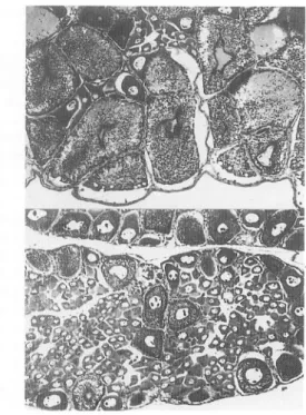
Kig-. 2 Horizontal section of the optic gan
glion. Upper, the arrangement of the
medullae, externa, interna and
terminal-is, and a localization of the cell group of
the medulla externa. Lower, the cell group magnified of the medulla externa. ME, medulla externa; MEC, medulla
externa cell group; MI, medulla interna;
MT, medulla terminalis; MTC, medulla terminalis cells.
Fig. 3 Ovaries of the prawns No. 4 and No. 15 treated with the medulla externa removal
externa and eyestalk removals are presented. The longer rearing than the period of this experiment will yield more developed ovaries, especially for the group of the medulla
externa removal provided that the improved operation is treated suppressing the regenera tion of the exerted tissue. As for the operation effect on the molting, there occurred at least
one molting individually during the 2 weeks rearing. A few of them showed further molting
(No. 1 and 15 in Table 1). There were no tendencies of the molting frequency and gained
weight at the molting among the groups. It needs the repeated experiment for recognition of the effects of the operations on the molting mechanism of the prawn.
References
1 ) J. Panouse (1943) I Influence de l'ablationdu pedoncule oculairesur la croissance de l'ovaire chez la crevette Leander serratus. C. R. Acad. Sci., 217, 553-555.
2 ) J. Panouse (1944) ! L'action de la gland du sinus sur l'ovaire chez la crevette Leander serratus.
C. R. Acad. Sci., 218, 293-294.
3) F.A.Brown and G.B.Jones (1947) ! Hormonal inhibition of ovarian growth in the crayfish,
Cambarus. Anat. Rec., 99, 657.
4 ) F. A. Brown and G. M. Jones (1949) \ Ovarian inhibition by a sinus-gland principle in the fiddler crab. Biol.Bull, 96, 228-232.
5) K. Takewaki and Y. Yamamoto (1950) '. Hormonal regulation of ovarian development in the shrimp, Paratya compressa. Annot. Zool Japon. ,23, 187-190.
6) D.B.Carlisle (1953) : Studies on Lysmata seticaudata RISSO(Crustacea Decapoda).V. The ovarian inhibiting hormone and hormonal inhibition of sex-reversal. Pubbl. Staz. Zool. Napoli, 24, 355-372.
7 ) T. Aoto and H. Nishida (1956) : Effect of removal of the eyestalks on maturation of the oocyte in a
hermaphroditic prawn, Pandalus kessleri. J. Fac. Sci. Hokkaido Univ., Ser. VI, 12, 412-425. 8 ) K. G. Adiyodi and R. G. Adiyodi (1970) I Endocrine control of reproduction in decapod Crustacea.
Biol.Rev., 45, 121-165.
9 ) A. Bomirski and E. Klek (1974) I Action of eyestalks on the ovary in Rhithropanopeus harrisii and
Crangon crangon (Crustacea: Decapoda). Marine Biol., 24, 329-337.
10) G. K. Kulkarni and R. Nagabhushanam (1980) \ Role of ovary-inhibiting hormone from eyestalks
of marine penaeid prawns (Parapenaeopsis hardwickii) during ovarian developmental cycle.
Aquaculture, 19, 13-19.
11) D.E. Bliss and J.H.Welsh (1952) ! The neurosecretory system of brachyuran Crustacea. Biol. Bull, 103, 157-169.