Posted at the Institutional Resources for Unique Collection and Academic Archives at Tokyo Dental College,
Title
A novel model demonstrates the successful repair of
alveolar cleft defects by tooth germ transplants
Author(s)
Kei, Nakajima; Yoshihiko, Akashi; Masami, Sumi;
Takatoshi, Chujo; Katsutoshi, Kokubun; Kenichi,
Matsuzaka
Journal
日本口腔検査学会雑誌, 12(1): 46-52
URL
http://hdl.handle.net/10130/5205
Right
Description
A novel model demonstrates the successful repair of
alveolar cleft defects by tooth germ transplants
Kei Nakajima
1, 2), Yoshihiko Akashi
2), Masami Sumi
2),
Takatoshi Chujo
1, 2), Katsutoshi Kokubun
1, 2), Kenichi Matsuzaka
1, 2)*1)Oral Health Science Center, Tokyo Dental College 2)Department of Clinical Pathophysiology, Tokyo Dental College Abstract
Cleft lip and palate is generally treated by promoting the growth and development of the maxillofacial region, and alveolar cleft defects are often repaired with artificial bones or cancellous bones harvested from the anterior iliac crest. The purpose of this study was to develop a mouse model to investigate the potential application of using tooth germs from wisdom teeth to repair alveolar cleft defects via their ability to form bone tissue. The upper first molars of 3-week-old mice were extracted under deep anesthesia to produce alveolar cleft defects using a dental engine with a round tungsten carbide burr. Tooth germs from embryonic day 16 mice together with collagen sponges were transplanted into alveolar cleft-like bone defects for the experimental group. Collagen sponges alone were transplanted into alveolar cleft-like bone defects for the control group. Hard tissue recoveries were evaluated from micro-CT images immediately after, and 3 and 7 weeks after the transplantations, and hard tissue volumes were measured at 7 weeks after the transplantation. Further, the alveolar cleft defects were observed his-tologically. CT-images of the control group showed no newly formed bone in the alveo-lar defects at any time point, but newly formed bone was observed in the experimental group at 3 and 7 weeks after the transplantations. At 7 weeks in the experimental group, the bone defects were filled with high density hard tissue, and the volume of newly formed bone at 7 weeks was significantly higher than in the control group (P < 0.05). Histological observations revealed that newly formed bone was not observed in al-veolar cleft defects in the control group at any experimental period but was observed in the defects in the experimental group at 3 and 7 weeks after the transplantation. Fur-ther, osteocalcin-positive cells and TRAP-positive cells were observed adjacent to the new bone at 7 weeks after the transplantation. These findings indicate that transplanta-tion of tooth germs with collagen sponge scaffolds is capable of healing large bone de-fects.
Key words: tooth germ, alveolar cleft, transplantation, bone formation Received: December 20th 2019 Accepted: January 29th 2020
*:2︲19︲18, Kandamisaki-cho, Chiyoda-ku, Tokyo 101︲0061, JAPAN TEL:+81︲3︲6380︲9247 FAX:+81︲3︲6380︲9606
JJ S E D P Vol. 12 No. 1:46︲52,2020
Introduction
The jawbone plays important roles in various functions such as mastication, articulation and pronunciation. Jawbone loss can be caused by cleft lip and plate (CLP), peripheral resection of gingival cancers, alveolar bone loss caused by periodontitis, and so on. CLP is a congenital fa-cial defect with an average prevalence of 7.94 per 10,000 live births internationally1), and it
causes dysfunctions such as deglutition and ar-ticulation disorders. Reconstruction of CLP is done via alveolar cleft bone grafting procedures using autologous bone, allogenic and xenogeneic bone grafting materials as well as various tissue-engineered bone replacement materials2︲6). CLP
is generally treated by promoting the growth and development of the maxillofacial region, and alveolar clefts are repaired with artificial bone or cancellous bone harvested from the anterior iliac crest7). However, those graft procedures
have the risk of various complications, including infection, pain, defective bone healing of the lo-cal area and postoperative gait disorder from ili-ac grafting. Although CLP patients usually re-ceive treatment at 7 to 12 years of age, they have normal impacted tooth germs of wisdom teeth in their jaws. In the dental field, stem cells derived from the dental pulp, periodontal ligament and dental follicles have been used for bone regeneration therapy, and third molar teeth or tooth germs are thought to be potential sources of cells and tissues.
There have been a few experimental reports about animal models of alveolar cleft grafting8, 9).
Those reports evaluated tissue-engineered bone grafts in artificially created bone defects, and obtained predictable models for testing of bone grafting materials.
We considered that the tooth germs of im-pacted teeth in CLP patients may be useful to repair their bone defects. So, the purpose of this study was to investigate the potential appli-cation of tooth germs obtained from mice to re-pair alveolar cleft defects and their ability to form bone tissue.
Materials and Methods
Animals
C57BL/6 mice (n
=
24) and embryonic day 16 mice (n=
6) were purchased from SLC Inc. (Shizuoka, Japan). All experimental protocols involving animals were approved by the Tokyo Dental College Animal Care and Use Committee (Approval # : 293204). All surgeries were performed under general anaesthesia using a combination of three types of anesthetics, which were mixtures of 0.75 mg/kg medetomidine hydro-chloride (Nippon Zenyaku Kogyo, Fukushima, Japan), 4.0 mg/kg midazolam (Sandoz, Tokyo, Japan) and 5.0 mg/kg butorphanol tartrate (Meiji Seika Pharma, Tokyo, Japan).Development of the alveolar cleft defect model in mice
The upper first molars of 3-week-old C57BL/6 mice were extracted under deep anesthesia, then left for 4 weeks to promote healing of the bone. Briefly, the oral mucosa at the extraction site was incised with a surgical knife approxi-mately 2.0 mm in length. Alveolar cleft model mice were produced using a dental engine with a round tungsten carbide burr (1.0 mm in diam-eter) (MANI, Utsunomiya, Tochigi) to create bone defects reaching the maxillary sinus and the nasal cavity (Fig. 1a). The oral mucosa was then closed with a 8︲0 nylon suture (Natsume Seisakusyo, Tokyo, Japan). C57BL/6 mice 7 weeks after this surgery were used for the alve-olar cleft model in this study. We confirmed the prepared clefts using micro-CT (R_mCT ; Rigaku, Tokyo, Japan) imaging with an expo-sure at 90 kV and 150 mA. A micro-CT image of the alveolar cleft model is shown in Fig. 1b. Isolation of tooth germs
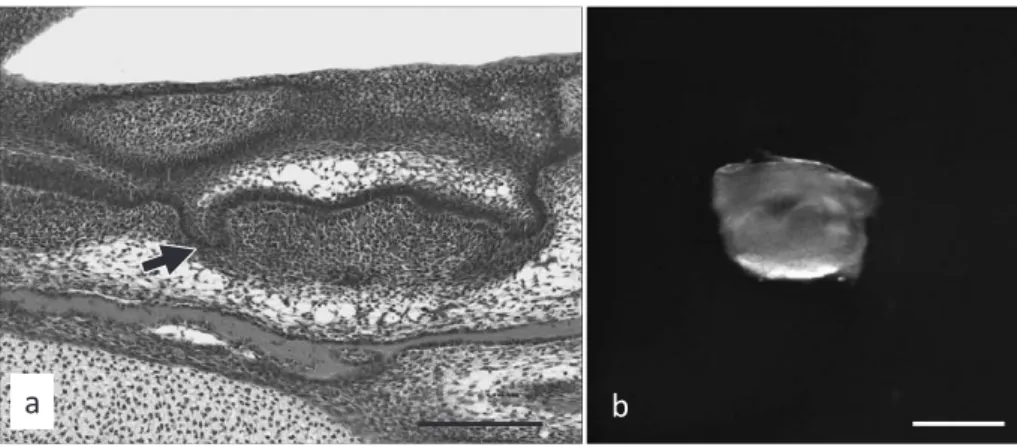
Mice were sacrificed by euthanasia to excise the tooth germs and paraffin sections were cut and stained with H-E (Fig. 2a). Tooth germs were isolated surgically from the mandible of each mouse at embryonic day 16. An example of an isolated tooth germ is shown in Fig. 2b. These procedures were conducted in Dulbecco's
modified Eagle medium (Thermo Fisher Scien-tific, Waltham, MA, USA) supplemented with 10% fetal bovine serum (Biosera, Nuaille, France), 100 U/mL penicillin and 100 µg/mL streptomycin (Thermo Fisher Scientific). Transplantation of tooth germs to the alveolar cleft mouse model
Tooth germs isolated from embryonic day 16 mice together with collagen sponges (Olympus Terumo Biomaterials, Tokyo, Japan) were transplanted into the alveolar cleft-like bone de-fects for the experimental group. Only collagen sponges were transplanted into the alveolar cleft-like bone defects for the control group. Af-ter transplantation, the oral mucosa of each mouse was closed with a 8︲0 nylon suture (Nat-sume Seisakusyo).
Micro-computed tomography (micro-CT) Radiological images were obtained using a mi-cro-CT device (R_mCT) with exposure at 90 kV and 150 mA immediately after, and at 3, 7, 10 and 15 weeks after the transplantation. All mice were placed on the object stage and imag-ing was performed over a full 360-degree rotation with an exposure time of 2 minutes. CT-images were captured using i-view R software (Morita, Kyoto, Japan).
Hard tissue volume measurement
Hard tissue recovery was evaluated from the micro-CT images. The hard tissue volume was measured using TRI/3D-BON software (Ratoc, Osaka, Japan) immediately after and at 7 weeks after the transplantation. The hard tissue volume at 0 weeks was subtracted from the volume at 7 weeks, and was used to determine the volume
Fig. 1 Macroscopic and CT-images
Macroscopic image of the round burr and the oral mucosa (a) and a representative CT-image of a bone defect (3D CT-image of an occlusal view).
Fig. 2 HE staining and a macroscopic image of a tooth germ
HE staining of a tooth germ (arrow) at embryonic day 16 (a, scale bar: 200 µm) and a macroscopic image of a tooth germ (b, scale bar: 500 µm).
JJ S E D P Vol. 12 No. 1:46︲52,2020
of hard tissue recovery.
Histochemical analysis and immunohisto-chemistry
Animals were sacrificed and their upper jaws were excised immediately after or at 7 weeks after producing the alveolar clefts, and at 3 and 7 weeks after the transplantation. These tissues were fixed in 20% neutral buffered formalin for 24 hours and then were decalcified in 10% EDTA for 3 weeks at 4℃. Decalcified tissues were paraffin-embedded, sectioned at
approxi-mately 3 to 5 micrometers thickness and stained with hematoxylin-eosin (H-E). For immunohis-tochemistry, sections were prepared and were incubated with anti-osteocalcin (OCN) antibody (5 µg/mL, rabbit ; Abcam, Cambridge, MA) as the primary antibody. TRAP staining was per-formed using a TRAP/ALP stain kit (FUJIF-ILM Wako, Osaka, Japan).
Statistical analysis
Data are presented as means
±
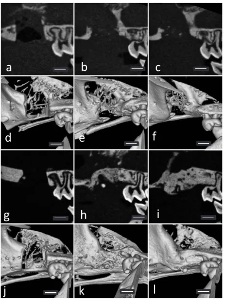
standard de-viation (S.D.). We used the Mann-Whitney UFig. 3 CT-images of the control (a︲f) and experimental (g︲l) groups immediately after and at 3 and 7 weeks after the transplantation
a, g: 2D images immediately after the transplantation. b, h: 2D images at 3 weeks after the transplantation.
c, i: 2D images at 7 weeks after the transplantation. d, j: 3D images immediately after the transplantation. e, k: 3D images at 3 weeks after the transplantation.
test to determine p-values for statistical signifi-cance. Analyses were performed using Graph-Pad Prism 7 (GraphGraph-Pad Software, San Diego, CA, USA).
Results
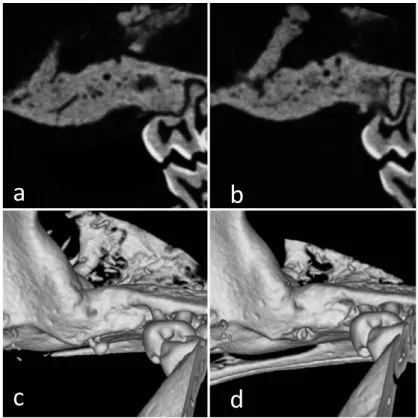
CT images of the control group showed no newly formed bone in the alveolar defects at any evaluation time point but newly formed bone was observed in the experimental group at 3 and 7 weeks after the transplant (Fig. 3). At 7 weeks in the experimental group, the bone defects were filled with high density hard tis-sue. At 10 and 15 weeks in the experimental group, the bone defects had been completely re-placed with radio-opaque hard tissue similar to the surrounding bone (Fig. 4). The volume of newly formed bone in the experimental group at 7 weeks was significantly higher than in the control group (P < 0.05) (Fig. 5).
Histological observations revealed that newly formed bone was not observed in the alveolar
cleft defects in the control group at any experi-mental time point, but was observed in the de-fects in the experimental group at 3 and 7
Fig. 4 CT-images of the experimental group at 10 and 15 weeks after the transplantation
a : 2D image at 10 weeks after the transplantation. b: 2D image at 15 weeks after the transplantation. c : 3D image at 10 weeks after the transplantation. d: 3D image at 15 weeks after the transplantation.
*
Fig. 5 Graph of bone volume
Newly formed bone volume in alveolar cleft defects at 7 weeks after the transplantation (*, P < 0.05)
JJ S E D P Vol. 12 No. 1:46︲52,2020
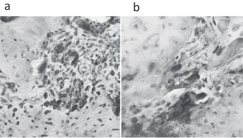
weeks after the transplantation (Fig. 6). Immu-nohistochemically, many osteocalcin-positive cells were observed in osteoblasts located adja-cent to the new bone at 7 weeks after the trans-plantation (Fig. 7a). Some TRAP-positive cells could be seen adjacent to the new bone at 7 weeks after the transplantation (Fig. 7b).
Discussion
Patients with CLP often undergo alveolar bone grafting to stabilize the dental arch and to enable the eruption of permanent teeth into the grafting area10). Our experimental design
uti-lized the tooth germ and a collagen gel scaffold to encourage bone development in the CLP area.
Fig. 6 HE staining
a︲c: Control group at 7 weeks after the transplantation. Fibrous connective tissue invaded the alveolar cleft defects. d︲f: Experimental group at 7 weeks after the transplantation. Newly formed bone filled in the alveolar cleft defects. Boxes in panels a and d indicate areas shown at higher magnification in panels b & c and d & f, respectively. Scale bars in panels a and d: 200 µm, in panels b, c, e and f: 20 µm.
Fig. 7 Immunohistochemical staining of anti-osteocalcin (a) and TRAP staining (b)
Experimental animal models of CLP and cleft alveolar osteoplasty have been reported8, 9).
Ka-mel et al. created alveolar clefts in rabbits fol-lowing the existing anatomy, extracted a central incisor tooth, modified the extraction socket by extending it to the nasal mucosa, and applied a simple bone wax and oxidized cellulose material to help modulate the healing phase in the cleft area and avoid rapid bone generation and filling of the defect8). In our study, alveolar clefts
be-tween the maxillary sinus and nasal cavity were created after wound healing of an extraction socket without any materials to avoid bone re-generation. Therefore, our study model avoids the influence of such artificial materials. Korn et al. attempted to create a cleft model in rats and reported the usefulness of hydroxyapatite-tricalciumphosphate scaffolds9). The innovative
idea of our study was derived from the use of extracted isolated tooth germs into artificially created alveolar clefts instead of bone grafting materials. In our study, we observed newly formed bone in alveolar bone defects in a tooth germ transplantation alveolar cleft model. Further, osteocalcin-positive cells and TRAP-positive cells were identified adjacent to the new bone, which revealed that the new bone remodels af-ter producing new bone.
From this study, we conclude that our alveo-lar cleft mouse model is appropriate because the bone defects did not heal naturally. These find-ings indicate that the transplantation of tooth germs with a collagen sponge scaffold is capable of healing large bone defects. This study shows the potential of a novel approach for the treat-ment of alveolar clefts.
References
1) Tanaka SA, Mahabir RC, Jupiter DC, Menezes JM: Up-dating the epidemiology of cleft lip with or without cleft palate, Plast Reconstr Surg, 129:511e︲518e, 2012 2) Chung VH, Chen AY, Jeng LB, Kwan CC, Cheng SH,
Chang SC: Engineered autologous bone marrow mes-enchymal stem cells: alternative to cleft alveolar bone graft surgery, J Craniofac Surg, 23:1558︲1563, 2012 3) Gładysz D, Hozyasz KK: Stem cell regenerative
thera-py in alveolar cleft reconstruction, Arch Oral Biol, 60: 1517︲1532, 2015
4) Janssen NG, Weijs WL, Koole R, Rosenberg AJ, Meijer GJ: Tissue engineering strategies for alveolar cleft re-construction: a systematic review of the literature, Clin Oral Invest, 18:219︲226, 2014
5) Kawata T, Kohno S, Fujita T, Sugiyama H, Tokimasa C, Kaku M, Tanne K: New biomaterials and methods for craniofacial bone defect: chondroid bone grafts in max-illary alveolar clefts, J Craniofac Genet Dev Biol, 20: 49︲52, 2000
6) Khojasteh A, Kheiri L, Motamedian SR, Nadjmi N: Re-generative medicine in the treatment of alveolar cleft defect: A systematic review of the literature, J Cranio-maxillofac Surg, 43:1608︲1613, 2015
7) Saha A, Shah S, Waknis P, Bhujbal P, Aher S, Vaswani V : Comparison of minimally invasive versus conven-tional open harvesting technique for iliac bone graft in secondary alveolar bone grafting in cleft palate pa-tients: a systematic review, J Korean Assoc Oral Maxil-lofac Surg, 45:241︲253, 2019
8) Kamal M, Andersson L, Tolba R, Bartella A, Gremse F, Hölzle F, Kessler P, Lethaus B: A rabbit model for ex-perimental alveolar cleft grafting, J Transl Med, 15:50, 2017
9) Korn P, Schulz MC, Range U, Lauer G, Pradel W: Effi-cacy of tissue engineered bone grafts containing mes-enchymal stromal cells for cleft alveolar osteoplasty in a rat model, J Craniomaxillofac Surg, 42:1277︲1285, 2014
10) Benlidayi ME, Tatli U, Kurkcu M, Uzel A, Oztunc H: Comparison of bovine-derived hydroxyapatite and au-togenous bone for secondary alveolar bone grafting in patients with alveolar clefts, J Oral Maxillofac Surg, 70:e95︲e102, 2012