Title
[症例報告]Enema-Induced Colonic Perforation in a
Psychiatric Patient being treated with Antipsychotic Drugs and
Hemodialysis for Renal Failure : A Case Report
Author(s)
Kinjo, Saneo; Hiroyasu, Shungo; Samura, Hironori; Shiraishi,
Masayuki; Muto, Yoshihiro; Nomura, Ken
Citation
琉球医学会誌 = Ryukyu Medical Journal, 20(2): 89-92
Issue Date
2001
URL
http://hdl.handle.net/20.500.12001/3474
Enema-Induced Colonic Perforation in a Psychiatric Patient being treated with
Antipsychotic Drugs and Hemodialysis for Renal Failure. A Case Report
Saneo Kinjo", Shungo Hiroyasull, Hironori Samura", Masayuki Shiraishil
Yoshihiro Mutoll and Ken Nomura
l The First Department of Surgery, Faculty of Medicine, University of the Ryuわ・us, and
21 Kitagami Chuo Hospital, Okinawa, Japan
(Received on August 8, 2000, accepted on November 28, 2000)
AB STRACT
A 44-year-old male psychiatric patient (suffering from schizophrenia) with renal failure was referred to the University Hospital because of a massive rectal bleeding. A few days after undergoing retrograde irrigation enemas performed by the health care staff, the patient devel-oped rectal bleeding. In his past medical history, the patient had been diagnosed to have schizo-phrenia at 14 years of age and renal failure (etiology unknown) requiring hemodialysis at 34 years of age. Colonoscopy revealed a large perforation occupying half of the luminal circumfer-ence of the sigmoid colon. Immediately after performing blood transfusion and hemodialysis, the patient underwent Hartman's procedure. The resected sigmoid colon revealed a large, longi-tudinal perforation measuring 4 cm in length on the mucosal surface and a ruptured configu-ration on the serosal surface. There were no diverticuli either at the site of or in the vicinity of the perforation. The perforated lesion showed mild inflammatory changes, a normal-appearing mucosa and tearing of the muscle fibers in the margins of the perforation. The patient recovered
without any postoperative complications. Ryukyu Med. J. , 20(2)89--92, 2001
Key words: chrome constipation, retrograde irrigation enema, colonic perforation, psychiatric patient, hemodialysis for renal failure
I NTRODUCTIO N
The frequently reported adverse effects of antipsychotic drugs in the treatment of patients with schizophrenia such as chronic constipationト and abdominal pain have al-ways been a major concern for general clinicians. Enemas are commonly used in the treatment of chronic constipation, especially in elderly, bedridden and psychiatric patients6- BI The habitual use of laxatives also increases with age and the use of retrograde irrigation enemas in long-term cases is also increasing. As a result of the common use of rectal tubes or similar devices for retrograde irrigation enemas, a perforation of the sigmoid colon and rectum can occur7', but the incidence of enema-induced colonic perforations is rare .
To our knowledge, a perforation of the sigmoid colon caused by cleansing enemas used by chronically consti-pated patients has up to now only rarely been described.
We herein describe a case of an enema-induced perforation
of the sigmoid colon in a chronically constipated schizo-phrenia patient who was also undergoing hemodialysis for renal failure.CASE REPORT
A 44-year-old male psychiatric patient (suffering from schizophrenia) was referred to Ryukyu University Hospital because of a massive rectal bleeding on May 24, 2000. The patient presented with abdominal pain due to chronic constipation. A laxative enema and an irrigation enema through a rectal tube had been performed by the health care staff. Thereafter, he developed rectal bleeding, and immediately was referred to our hospital for further examination and treatment. His past medical history re-vealed that the patient had been diagnosed to have schizo-phrenia at 14 years of age and renal failure ( etiology unknown) was later identified at 34 years of age. The pa-tient had been treated with anti-psychotic drug [Dogmatil (sulpiride) 600 mg, Lodopin (zotepine 75 mg and Artane (trihexyphenidyl HCl) 6 mg], and hemodialysis for renal failure every other day. On admission, the patient ap-peared to be apathetic and anemic. A physical examination revealed the abdomen to be moderately distended and ten-derness and muscular guarding were noted over the lower abdomen. No papable mass was observed in the abdomen. A rectal examination showed a bloodstain on the doctor s
90 Enema-Induced Colonic Perforation
Fig. 1 An abdominal CT scan demonstrating a small mass-like lesion and the probable presenceof entrapped air (arrow) in the lower abdominal cavity (left) and colonoscopy showing a linear perforation with oozing (right).
glove, but no lesion could be identified in the rectum. The blood pressure was 160 mmHg systolic and lOOmm Hg dia-stohc. The pulse rate was 120/minute and regular. Rou-tine laboratory findings revealed the Hgb to be 6.3 mg/dl, Hct 18.2%, total protein 4.8 g/dl, albumin 2.2 g/dl, BUN 32 mg/dl and creatinine 7.5 mg/dl.
Plain radiographs and an abdominal CT scan re-vealed neither any definite free air nor a mass lesion. Colonoscopy revealed a large perforation with oozing oc-cupying about half of the luminal circumference of the sigmoid colon (Fig. 1 ). Immediately after the patient became stable following a blood transfusion of 4 units and hemodialysis for renal failure, the patient underwent an exploratory laparotomy. A large perforation covered with the omental tissue was observed at the anterior wall of the sigmoid colon with no evidence of gross fecal contami-nation. The patient was treated by a resection of the per-forated segment, a colostomy with the proximal sigmoid colon, and a closure of the rectal stump by a linear sta-pling device (Hartmann's procedure).
Grossly, the resected colon showed a large perforation measuring4 × 4cm in size at the anterior wall of the sigmoid colon with no evidence of diverticuli. A mucosal prolapse was noted from the margin of the perforation. No fecalomas, fecal contamination, ischemic necrosis of the margins of the perforation or abscess were observed (Fig. 2 ). Histologically, the perforated lesion revealed mild acute inflammatory changes, an almost normal-appearing mucosa and tearing of the muscle fibers in the margins of the perforation. No ischemic necrosis of the colonic wall, diverticuli or amyloid deposits in the muscle layers and blood vessels were identified (Fig. 3 ). Accord-ingly, the gross findings at surgery and the histologic ex-animation of the specimen ruled out any other cause of perforation, such as either cancer, diverticuli at the perfo-ration site or ischemia. In this case the observed tearing of the muscle fibers in the muscle layers also exclusively sup ported an enema-induced perforation. A history of repeated use of retrograde irrigation enemas was also confirmed by the health care staff. The patient recovered with no post-operative complications and was discharged after 4 weeks
Fig. 2 Macrophotographs of the resected sigmoid colon showing a rather ruptured configuration on the serosal surface (top) and a linear perforation on the mucosal sur-face (bottom).
of hospitahzation.
DISCUSSION
Perforations of the colon and rectum have been well described in the literature as complications of colonoscopy and barium enemas", but they only rarely show serious complications. The reported incidence of colorectal perfo-ration ranges from 0.1% to 0.9% and 0.01% to 0.04%
fol-lowing colonoscopy and barium enemas, respectively9 Nevertheless, the true incidence of retrograde irrigation enemas-induced perforation of the colon and rectum is unknown7'. To our knowledge, no case of enema-induced-colomc perforation in a chronically constipated, psychiat-ric patient undergoing hemodialysis treament for renal failure every other day has previously been reported.
In comparison to such latrogenic colonic perforations mentioned above, non-iatrogenic or spontaneous colonic perforations are classified as either "stercoral" or "idiopathic" perforations based on the etiopathogenetical causes of the lesions'31. Anatopathologically, stercoral and idiopathic perforations present different characteristics. Grossly, stercoral perforations originate from an ulcerative lesion often situated on the sigmoid colon or rectum. The histological characteristics are represented by the superfi-cial ischemic necrosis of the mucosa (caused by fecalomas)
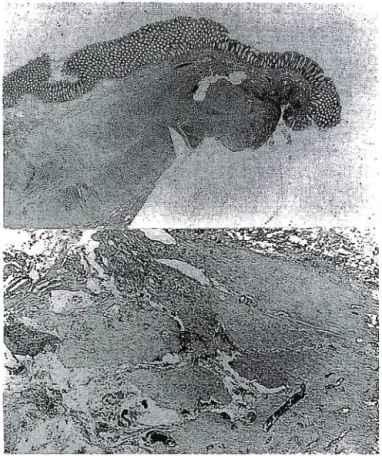
Fig. 3 Microphotographs of the perforated lesion reveal-ing a normal-appearreveal-ing mucosa and mild inflammation (top right ; HE, ×10), and a tearing of the muscle fibers (top right; HE,×25).
followed by an extension to the submucosa and muscle lay-ers of the colonic wall. On the other hand, idiopathic perforations,which are frequently located in the sigmoid colon, are linear lacerations on the anti-mesenteric side of the colon without any pathologic modification of the colon.
Morphologically, the colonic perforation in our case did
not reveal either any ischemic necrosis of the colonic wall or a linear laceration on the antimesenteric side of the colon. Therefore, the lesion of the colonic perforation in our case was comparable to that of an iatrogenic colonic perforation. As aforementioned, the perforated lesion showed characteristical tears of the muscle fibers, thus indi-eating either a rectal tube tip injury or excessive intracolonic pressure during a retrograde irrigation enema.
The clinical presentation of patients with a colonic perforation varies and depends on the size, site and mecha-nism of perforation, the underlying pathology, the degree of peritoneal contamination, and the condition of the pa-tient. Our patient presented with abdominal pain a few days after undergoing retrograde irrigation enemas and
developed rectal bleeding prior to this admission.
Mean-while, plain radiographs did not demonstrate any free air in the abdominal cavity, but an abdominal CT scan did show localized air in the pelvic cavity which was con-firmed to be covered with omental tissue at surgery. Plain radiographs and CT scan usually demonstrate free air, but
if free air can not be confirmed by both examinations, then colonoscopy may be useful for determining the pres-ence of a perforation of the colon.
Our patient underwent Hartmann's procedure. The decision to perform surgery has been based on the immediate recognition of the injury and also the clinical symptoms and signs of peritonitis. There are three types of surgical proce-dures (primary repair, resection and primary anastomosis, Hartmann's procedure) depending on the degree of perfora-tion and gross fecal contaminaperfora-tion71. A primary repair has been advocated for a traumatic perforation of the colon and rectum without any gross contamination. Hartmann s proce-dure should be performed for patients with much more exten-sive colonic damage and!or severe contamination. Our patient underwent Hartmann's procedure because of an exten sive colonic perforation and a delayed presentation. It is generally accepted that the complications related to the operative treatment of patients with colonic perforation closely correlated to the length of the surgery as well as to the surgical techniques used8'.
In conclusion, an awareness of the possibility of in-ducing injury should be stressed to the medical staff of all institutions for chronically constipated patients using colonic irrigation. As a result, an early diagnosis and timely surgical treatment should result in a good progno-sis, regardless of the patient's age and previous medical conditions.
ACKNOWLEDGEMENTS
We thank Mr. Brian Quinn for his comments on this
manuscript.
REFERENCES
1 ) Whitworth A.B. and Fleischhacker W.W∴ Adverse effects of antipsychotic drugs. Int. Clin. Psychopharmacol. 9 (Suppl 5): 21-27, 1995.
2 ) Kopala L.C.: Clinical experience in developing regi-mens with the novel antipsychotic rispendone. Int. Clin. Psychopharmachol. 12 (Suppl 4): S ll-18, 1997. 3 ) Bever K.A. and Perrry P.J.: Olanzapine: a
serotonin-dopamine-receptor antagonist for antipsychotic ther-apy. Am. J. Health Syst. Pharm. 55: 1003-1016, 1998. 4 ) Murasaki M., Yamauchi T., Yagi G., Nakajima T.,
一Nffka三ne Y. and Kudo Y.: Early phase II study of
quetiapine fumarate on schizophrenia. Ninon Shinkei Seishin Yakurigaku Zasshi 19: 53-66, 1999.
5 ) Harari D., Gurwitz J.H., Avorn J., Choodnovskiy I. and Minaker K.L.: Constipation: assessment and
management in an institutionalized elderly popula-tion. J. Am. Geriatr. Soc. 42: 947-952, 1994.
6) Harari D., Gurwitz J. H. and Minaker K. L∴ Consti-pation in the e】iderly. J. Am. Geriatr. Soc. 41: 1130-1140, 1993.
92 Enema-Induced Colonic Perforation
Kahsai D. and Hunter G.C.: Clinical presentation and management of latrogenic colon perforation. Am. J. Surg. 172: 454-458, 1996.
8) Paran H., Butnaru G., Neufeld D‥ Magen A. and
Freund U.: Enema-induced perforation of the rectum in chronically constipated patients. Dis. Colon
Rec-turn 42: 1609-1612, 1999.
9 ) Hakim N. S., SarrM. G., BenderC. E. andNivatvongs
S∴ Management of barium enema-induced colorectalperforation. Am. Surg. 58: 673-676, 1992.
10) Hall C, Dorricott N.J., Donavan I.A. and Neoptolemos
J.P∴ Colon perforation during colonoscopy: surgicalversus conservative management. Br. J. Surg. 78: 542-544, 1991.
ll) Macrae F.A., Tan K.G. and Williams C.B.: Towards
safer colonoscopy: a report on the complications of 5000 diagnostic or therapeutic colonoscopies. Gut 24: 376-383, 1983.
12) Woodhall J. P. and Oshner A. : The management of
perforating injuries of the colon and rectum in civilian practice. Surgery 29: 305-320, 1951.
13) Basile M., Montini F., Cipollone G. and Errichi B. M∴ Spontaneous perforations of the large intestine. Ann. Ital. Chir. 63: 625-629, 1992.