Effect of Dose and Pressure on the Transformation from
Neutron-Irradiated Graphite to Amorphous Diamond
K Niwase1, T Atou2, K G Nakamura2, and T. Iwata3
1
Hyogo University of Teacher Education, Kato-shi, Hyogo 673-1494, Japan 2
Materials and Structures Laboratory, Tokyo Institute of Technology, 4259 Nagatsuta, Yokohama, 226-8503, Japan
3
Nuclear Professional School, School of Engineering, University of Tokyo, Tokai, Ibaraki 319-1188, Japan
E-mail: [email protected]
Abstract. We previously discovered a novel pathway for the transformation from highly orientated pyrolytic graphite foils irradiated with neutrons into amorphous diamond platelets. Here, we utilized graphite foils irradiated with a neutron dose of about a half of the previous study, and confirmed the pathway for the transformation. Raman spectrum of the irradiated graphite exhibits a broad amorphous one but still possessed weak G and D peaks, appearing in a less disordered state. The irradiated graphite foils were shock compressed at 46, 51 and 54 GPa. At 46 GPa, Raman spectra clearly show the G and D peaks but a weak photoluminescence appears. At 51 GPa, we can find transparent platelets with intense photoluminescence but no diamond Raman peak, implying the transformation to amorphous diamond. At 54 GPa, we obtained transparent platelets with intense photoluminescence and a small and broad diamond peak, suggesting the formation of nano-diamond.
1. Introduction
The transformation from graphite to diamond is a long time issue and polycrystalline diamond has been synthesized by direct conversion of graphite under static high pressure and temperature [1]. The ultimate smallest crystalline size of diamond was synthesized from C60 fullerene by shock compression and rapid quenching (SCARQ) technique [2]. The obtained material is transparent and comprised wholly of sp3 carbons, suggesting diamond structure, but the diamond crystalline size is within the range of unit cell. Then, it was labeled “amorphous diamond” as it is amorphous in the long-range order and diamond in the short-range order [3]. Graphite, on the other hand, has not been found to transform to amorphous diamond by shock compression but to crystalline diamond by the martensitic mechanism [4], if shock pressure is applied parallel to the c axis.
Recently, we have discovered a novel pathway for the transformation from highly oriented pyrolitic graphite (HOPG) into amorphous diamond [5]. The pathway consists of neutron irradiation, shock compression, and rapid quenching. Defects produced by irradiation (Wigner defects) are considered to work as the nucleation sites for diamond. In the pathway, we can control the introduction of Wigner defects by changing the conditions of irradiation and shock compression.
Here, we aimed to investigate the dose and pressure dependencies on the novel pathway by utilizing HOPG specimen irradiated with neutron to a dose which is about half of the last one and by giving shock compression in three different conditions.
2. Experimantal procedure
We utilized highly oriented pyrolytic graphite (HOPG-ZYA) from Union Carbide as a starting material, which was irradiated with fast neutrons at about 333 K to a dose of 1.4 x 1024 n m-2 (E> 1 MeV) in JAERI JRR-2 nuclear reactor. The dose is about half of the previous study, in which the specimen was irradiated to a dose of 2.6x1024 n m-2 [5]. Thin films of about 8 μm in thickness were prepared by cleavage with a razor. The sample was sandwiched by gold foils, which work for heat sinks. The tungsten flyer was accelerated by a powder gun and was impacted to the front side of the iron capsule. The shock pressures determined by the flyer velocity were 46, 51 and 54 GPa. The samples after the shock compression were observed by using a scanning electron microscope (SEM). Raman measurements were done with a micro-Raman spectroscope (SPEX Raman-500) using an Ar (488 nm) laser.
3. Results and discussion
Figure 1 shows Raman spectra for the unirradiated and neutron-irradiated HOPG specimens. The Raman measurements were done in the same condition to compare the relative intensity. The unirradiated HOPG specimen shows the first-order peak near 1580 cm-1 (G band) and the second-order Raman band near 2700 cm-1. After the neutron irradiation, a broad band ranging from 800 to 1700 cm-1 appeared. However, there still remains small G band and a defect peak near 1360 cm-1 (D band) appears. This means that the neutron irradiated specimen is in a less disordered state, compared to the specimen used in the previous study [5].
Figure 1. Raman spectra of an unirradiated HOPG specimen as a starting material and a HOPG specimen irradiated with fast neutrons at about 333 K to a dose of 1.4 x 1024 n m-2 (E> 1 MeV) in JAERI JRR-2 nuclear reactor. Spectrum of the neutron-irradiated HOPG shows a broad band in a range from 800-1700 cm-1. Peaks of the G and D bands still remain in the spectrum.
Figure 2 shows an optical micrograph of neutron-irradiated HOPG recovered after the shock compression. The shock pressure determined by the flyer velocity is 46 GPa. We observed an occurrence of fracture of the graphite foils into platelets, similar to the previous studies [5,6]. A platelet is seen in the center of Fig. 2. A bright square area beside the platelet is gold foil, from which a platelet has fallen off. The colour of the lower part of the platelet is seen to be brighter than that of the upper part.
Figure 2. Optical micrograph of neutron-irradiated HOPG recovered after the shock compression at 46 GPa. A platelet can be seen in the center of the figure. The colour of the lower part of the platelet is brighter than that of the upper part, suggesting an occurrence of some transparency in the lower area. A bright square area in the left hand side of the platelet corresponds to gold foil, from which a platelet has fallen off.
Figure 3 shows Raman spectra for the neutron-irradiated HOPG specimen and for the platelet shown in Fig. 2, which is obtained after the shock compression at 46 GPa. The Raman measurements were done in the same condition to compare the relative intensity. The Raman intensity is seen to increase after the shock compression due to photoluminescence, of which intensity in the lower part of the platelet is higher than that in the upper part, suggesting some fluctuation on the transformation. The G and D bands are seen to remain after the shock compression.
Figure 3. (a) Raman spectrum for the neutron-irradiated HOPG specimen. (b) Raman spectrum, obtained after the shock compression at 46 GPa, at upper part of the platelet in Fig. 2. (c), (d) Raman spectra obtained at lower part of the platelet in Fig. 2.
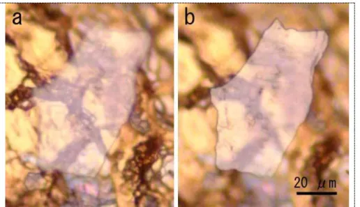
Figure 4 shows optical micrographs of a platelet of 100 μm in length, which was obtained after the shock compression at 54 GPa. Photos (a) and (b) are respectively focused on the surface of gold foil and the recovered specimen. The platelet is seen to be transparent.
Figure 4. Optical micrographs of neutron-irradiated HOPG recovered after the shock compression at 54 GPa. (a) Focused on the gold foil. (b) Focused on a platelet of the recovered specimen.
Figure 5 shows Raman spectra obtained after the shock compression at 54 GPa. Raman spectrum for the neutron-irradiated HOPG specimen is shown at the bottom of the figure for comparison. Intensity of the Raman spectra is seen to remarkably increase after the shock compression. The increase of the background intensity has been seen for the amorphous diamond synthesized from C60 fullerene and neutron-irradiated graphite by shock compression, and is due to photoluminescence, probably originating in an existence of defective sp3 bondings [6]. A small and broad peak denoted by arrows can be seen at about 1330 cm-1, implying the formation of nano-diamond. We did another shock compression at 51 GPa and found transparent platelets with remarkable photoluminescence. There are slight G and D peaks but no diamond peak, implying the formation of amorphous diamond.
Figure 5. (a) Raman spectrum for the neutron-irradiated HOPG specimen. (b), (c) Raman spectra, obtained after the shock compression at 54 GPa, from the platelet shown in Fig. 2. Intensity of the Raman spectra significantly increases after the shock compression due to photoluminescence. A broad peak can be seen at about 1330 cm-1, suggesting the formation of nano-diamond.
The neutron irradiated specimen utilized in the present study has been proved to be in a disordered state by means of ray diffraction and high resolution transmission electron microscopy [7]. The X-ray profile was highly asymmetrical and the TEM analysis showed breaking and bending of the 0002
lattice fringes which were found in other irradiation studies [8-10]. The result approves the present result of broad Raman spectra shown in fig. 1 and the dislocation accumulation model [11] which attributed the irradiation-induced amorphization to the accumulation of dislocation dipoles, inducing the buckling and the rotation of basal planes. We have proposed a tentative scenario on the synthesis of amorphous diamond platelets from the neutron irradiated HOPG under shock compression [5] as follows. The disordered layered sequences induced by irradiation prevent the martensitic transformation from graphite to diamond under shock compression. Defective sites relating to Wigner defects make a high density of diamond nucleation sites, leading to the synthesis of the amorphous diamond platelets. There remains a question on the kind of defect, relating to the transformation. To clarify the effect of defects on the transformation, it may be interesting to utilize lower damaged graphite specimens before the onset of irradiation-induced amorphization, which possess the layered sequence of the basal planes.
4. Conclusions
A novel pathway for the transformation from thin foils of neutron irradiated graphite into amorphous diamond platelet was confirmed with a neutron dose of about a half of the previous study. The Raman spectrum of the neutron irradiated graphite shows a broad amorphous one but is in a less disordered state, compared to the specimen used in the previous study. The graphite foils were shock compressed at 46, 51 and 54 GPa. At 46 GPa, we find the G and D peaks, and additional photoluminescence. At 51 GPa, we obtained transparent platelets with a strong photoluminescence but no diamond peak, suggesting the transformation to amorphous diamond. At 54 GPa, we find transparent platelets with strong photoluminescence. Also, a weak peak at about 1330 cm-1 appeared, suggesting the formation of nano-crystallized diamond. Thus, the combined method of irradiation and shock compression should be a promising way to synthesize a various state of diamond such as amorphous diamond and nanodiamond.
Acknowledgements
This research was partly supported by Collaborative Research Project of Materials and Structures Laboratory, Tokyo Institute of Technology and by JSPS Grant-in Aid (No. 23560787).
References
[1] Irifune T, Kurio A, Sakamoto S, Inoue T and Sumiya H 2003 Nature 421 599 [2] Hirai H, Kondo K, Yoshizawa N and Shiraishi M 1994 Appl. Phys. Lett. 64 1797 [3] Hirai H, Tabira Y, Kondo K, Oikawa T and Ishizawa N 1995 Phys. Rev. B 52 6162 [4] Erskine D J and Nellis W J 1991 Nature (London) 349 317
[5] Niwase K, Nakamura K G, Yokoo M, Kondo K and Iwata T, 2009 Phys. Rev. Lett. 102 116803 [6] Niwase K, Homae T, Nakamura K G and Kondo K 2006 Physica (Amsterdam) 376B–377B 280 [7] Asthana A, Matsui Y, Yasuda M, Kimoto K, Iwata T and Ohshima K, 2005 J. Appl. Crystallogr.
38 361. The neutron dose for JR and ICM19 samples should be revised to 3×10¹⁹ n/cm2 and 1.4×10²⁰ n/cm2, respectively.
[8] Tanabe T, Muto S and Niwase K 1992 Appl. Phys. Lett. 61 1638 [9] Koike J and Pedraza D F, 1994 J. Mater. Res. 9 1899
[10] Karthik C, Kane J, Butt D P, Windes W E and Ubic R 2011 J. Nucl. Mater. 412 321 [11] Niwase K, Philos. Mag. Lett. 2002 82 401; Mater. Sci. Eng. A 2005 400–401 101