A Simple Technique Using Tape to Expose
the Subcarinal Lymph Node Station :
M ove the Field
Mitsuhiro Kamiyoshihara,
Takashi Ibe
and Izumi Takeyoshi
In systemic lymph node dissection for lung cancer surgery,the subcarinal lymph node(#7)is situated far from any access incision. Therefore,it is difficult to identify#7 through the access wound used for video-assisted thoracic surgery lobectomy. This is also true in the case of a left-sided approach because of the descending aorta. Here,we propose a simple technique using tape to expose#7. This saves time and benefits the patient.(Kitakanto Med J 2008;58:403∼404)
Key Words: primary lung cancer, subcarinal lymph node dissection
Introduction
Radical surgery for primary lung cancer requires systemic lymph node dissection, which routinely includes dissection of the subcarinal lymph node(#7; station 7). Of the lymph node stations,#7 is situated far from any access wound. Therefore,it is difficult to identify#7 through the access wound used for video-assisted thoracic surgery(VATS) lobectomy. This is also true in the case of a left-sided approach because of the descending aorta. Here, we propose a simple technique using tape to expose #7. This saves time and benefits the patient.
Technique
We use the lymph node nomenclature used in the original lymph node map for lung cancer reported by Naruke et al.
As a working port,we routinely make a 5-to 7-cm anterior or lateral incision in the fourth or fifth inter-costal space. Additional 0.5-to 1.5-cm thoracic ports are made in the seventh intercostal space in the anterior axillary line or the ninth intercostal space in the posterior axillary line. The working port is protected with a polyurethane Alexis wound retractor (Applied Medical,CA,USA). A rib spreader is not used. We use nearly direct visualization through the working port.
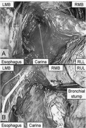
When performing a lower lobectomy, we divide the pulmonary veins and arteries,and dissect the hilar, interlobar, and lobar lymph nodes around the lower bronchus. Next, we loop non-stretch cotton tape around the bronchus (Fig. 1A), pull the tape forward through one of the thoracic ports, and elevate the carina (Fig.1B). Then,the subcarinal lymph node is dissected (Fig.1C). This provides very good exposure of the subcarinal region (Fig. 2). Care should be taken to dissect#7 before dividing the lower bronchus.
Comment
Naruke et al. studied the incidence of lymph node metastasis in each lobe in primary lung cancer. The extent of lymph node metastasis was lobe specific. The usual site of metastasis to the mediastinal lymph nodes for both lower lobes was #7. Therefore, a thorough dissection that includes#7 is very important for lower lobe tumors.
In an upper lobectomy, the same view cannot be obtained because the tissue around the lower lobe bronchus is not exposed. However, a subcarinal lymphadenectomy is not always necessary for tumors of the right upper lobe or left upper segment. Never-theless, when a subcarinal lymphadenectomy is essen-tial for tumors of the middle lobe and lingular seg-ment,a similar view can be obtained with difficulty by holding the lung parenchyma of the superior segment
403 Kitakanto Med J
2008;58:403∼404
1 Department of General Thoracic Surgery, Maebashi Red Cross Hospital, Maebashi, Gunma, Japan. 2 Division of Thoracic and Visceral Organ Surgery, Gunma University Graduate School of Medicine, Maebashi, Gunma, Japan.
Received : August 24, 2008
Address: MITSUHIRO KAMIYOSHIHARA Department of General Thoracic Surgery, Maebashi Red Cross Hospital, 3-21-36 Asahi-Cho, Maebashi, Gunma 371-0014, Japan
of the lower lobe with lung-grasping forceps.
Basically, we perform a sharp dissection for lymph nodes, using scissors and vascular clips instead of an electric scalpel. In our experience,the use of an electric scalpel results in bronchial fistulas and perfora-tion. Moreover,the electric scalpel has caused greater tissue damage in animal experiments. Care should be taken to avoid injuring the bronchial arteries near the carina. Some surgeons use an especially long retractor to expose#7,but our technique does not use a special device.
References
1. Naruke T,Suemasu K,Ishikawa S. Lymph node mapping and curability at various levels of metastasis in resected lung cancer. J Thorac Cardiovasc Surg 1978; 76: 832-839. 2. Naruke T, Tsuchiya R, Kondo H, et al. Lymph node
sampling in lung cancer: how should it be done? Eur J Cardiovasc Surg 1999 ; 16: S17-24.
3. Turna A, Solak O, Kilicgun A, et al. Is lobe-specific lymph node dissection appropriate in lung cancer patients undergoing routine mediastinoscopy? Thorac Cardiovasc Surg 2007; 55: 112-119.
4. Asamura H, Nakayama H, Kondo H, et al. Lobe-specific extent of systematic lymph node dissection for non-small cell lung carcinomas according to a retrospective study of metastasis and prognosis. J Thorac Cardiovasc Surg 1999 ; 117: 1102-1111.
5. Kamiyoshihara M, Ibe T. Does the electric scalpel facili-tate bronchial tissue damage? Eur J Cardiothorac Surg 2008; 34: 223.
6. Meyer F,Ioshii SO,Chin EW,et al. Laparoscopic partial nephrectomy in rats. Acta Cir Bras 2007; 22: 152-156. Technique for exposing subcarinal lymph node
Fig. 1 A : Non-stretch cotton tape is looped around the bron-chus
B: The tape is pulled forward to elevate the carina C : The subcarinal lymph node(#7) is dissected. Note
the relative position of the carina and vertebra.
Fig. 2 A : The subcarinal region is very well exposed
B: A close-up picture of the carina after a lower lobectomy.
Abbreviations: RUL : right upper lobe; RML : right middle lobe; RLL : right lower lobe; RMB: right main bronchus; LMB: left main bronchus