Ext r ac or por eal s hoc k w
ave t her apy f or avul s i on
f r ac t ur es of t he s ubl i m
e t uber c l e of t he ul na
i n hi gh s c hool bas ebal l pl ayer s : A r epor t of
t w
o c as es
著者
Tanaka Kent a, Kanam
or i Aki hi r o, Yam
am
ot o Yuki ,
H
ar a Yuki , N
i s hi ur a Yas um
as a, N
i s hi no
Tom
of um
i , Yam
az aki M
as as hi , M
i yakaw
a Shum
pei
j our nal or
publ i c at i on t i t l e
As i a- Pac i f i c J our nal of Spor t s M
edi c i ne,
Ar t hr os c opy, Rehabi l i t at i on and Tec hnol ogy
vol um
e
10
page r ange
1- 3
year
2017
権利
( C) 2017 As i a Pac i f i c Knee, Ar t hr os c opy and
Spor t s M
edi c i ne Soc i et y. Publ i s hed by El s evi er
( Si ngapor e) Pt e Lt d. Thi s i s an open ac c es s
ar t i c l e under t he CC BY- N
C- N
D
l i c ens e (
ht t p: / / c r eat i vec om
m
ons . or g/ l i c ens es / by- nc - nd/ 4
. 0/ ) .
U
RL
ht t p: / / hdl . handl e. net / 2241/ 00150618
doi: 10.1016/j.asmart.2017.05.298
Extracorporeal shock wave therapy for avulsion fractures of the
sublime tubercle of the ulna in high school baseball players: A report
of two cases
Kenta Tanaka, M.D.
a,*, Akihiro Kanamori, M.D., Ph.D.
a, Yuki Yamamoto, M.S.
b,
Yuki Hara, M.D., Ph.D.
a, Yasumasa Nishiura, M.D., Ph.D.
c, Tomofumi Nishino, M.D., Ph.D.
a,
Masashi Yamazaki, M.D., Ph.D.
a, Shumpei Miyakawa
daDepartment of Orthopaedic Surgery, Faculty of Medicine, University of Tsukuba, Japan bSports Research&Development Core, University of Tsukuba, Japan
cTsuchiura Clinical Education and Training Center, Tsukuba University Hospital, Japan dFaculty of Health and Sport Sciences, University of Tsukuba, Japan
a r t i c l e
i n f o
Article history:
Received 16 December 2016 Received in revised form 7 May 2017
Accepted 26 May 2017 Available online 7 June 2017
Introduction
Injury to the structures of the medial elbow is common among baseball players. The injury can result from repeated valgus stress of the elbow. Several types of injury to the structures of the medial elbow have been reported.1These injuries cause valgus instability and pain of medial aspect of the elbow during pitch.2e4Avulsion
fractures of the sublime tubercle of the ulna are rare and generally develop after bone maturation in baseball players.5To the best of our knowledge, there are only three reports in the English ortho-pedic literature regarding this type of fracture.5e7 According to
these reports, conservative treatment is performed initially. As a secondary treatment, bone fixation or ulnar collateral ligament reconstruction is performed when bone union cannot be ach-ieved.6,7Additionally, avulsion fractures of the sublime tubercle of the ulna are often overlooked, and therefore, are liable to involve non-union. Ulnar collateral ligament reconstruction results in a high rate of successful return to baseball, as reported in a system-atic review,8 but no reports have exclusively studied avulsion fractures of the sublime tubercle of the ulna. If patients receive the surgery for an avulsion fracture of the sublime tubercle of the ulna,
they should inevitably receive post-operative management and long-term rehabilitation for at least 6 months.8,9 Thus, a novel treatment for avulsion fractures of the sublime tubercle of the ulna is needed. Extracorporeal shock wave therapy (ESWT) is performed for stress fractures and non-union cases, and good results have been obtained in recent years.10e14ESWT can be a novel treatment
for avulsion fractures of the sublime tubercle of the ulna; however, no reports of its use have been published. This is thefirst report describing ESWT for delayed-union avulsion fractures of the sub-lime tubercle of the ulna.
Methods
The present study was approved by the Ethics Committee of our institution (H24-144). We obtained written informed consent from the patients. As per our criteria, patients who did not develop union over the 3 months, and who were not administered pain relief medication were included in the study, while those who had acute fractures were excluded.
ESWT protocol
The focused shock wave generator was used (Epos Ultra® ; Dornier MedTech GmbH, Wessling, Germany). Patients received 3500 impulses at 0.36 mJ/mm2without anesthesia, and the energy was gradually increased (total, 1300 mJ/mm2).
Irradiation and follow up
ESWT was performed by the same orthopedic surgeon in both cases. Patients were placed in the supine position with a 90 shoulder abduction and a 0 elbow extension. At the start of treatment, the most painful point of the medial elbow was confirmed to be the sublime tubercle by ultrasound. The point was *Corresponding author. 1-1-1, Tennodai, Tsukuba city, Ibaraki 305-8575, Japan.
E-mail address:kenattanaka@tsukuba-seikei.jp(K. Tanaka).
Contents lists available atScienceDirect
Asia-Paci
fi
c Journal of Sports Medicine, Arthroscopy,
Rehabilitation and Technology
j o u r n a l h o m e p a g e :w w w . a p - s m a r t . c o m
http://dx.doi.org/10.1016/j.asmart.2017.05.298
2214-6873/©2017 Asia Pacific Knee, Arthroscopy and Sports Medicine Society. Published by Elsevier (Singapore) Pte Ltd. This is an open access article under the CC BY-NC-ND license (http://creativecommons.org/licenses/by-nc-nd/4.0/).
highlighted with a waterproof marker. The shock waves were delivered to the highlighted site (Fig. 1). ESWT was performed once on each patient. Bone union was checked every 4 weeks by plain X-ray or computed tomography (CT).
Case reports
Case 1: A 15-year-old male high school baseball pitcher expe-rienced pain in the medial aspect of the elbow of his dominant hand during pitching. The patient initially rested from pitching and did not seek medical attention, but the pain in his elbow did not improve. He was examined at our orthopedic clinic after a 3-month break from pitching and upper limb training. Local compression pain in the medial aspect of the elbow and pain causing valgus stress were detected. No limitation of the range of motion was described. CT showed a bone fragment in the sublime tubercle of the ulna. Bone sclerosis was noted in the fracture edge, and, therefore, he was diagnosed with a stress fracture (Fig. 1). After 1 month of fulltime immobilization of the elbow, the pain was not relieved. ESWT was performed 4 months after thefirst incident of pain. The elbow was immobilized fulltime with a splint for 4 weeks after ESWT. The compression pain was decreased but was still present. Range of motion exercises were allowed 4 weeks after ESWT. Eight weeks after ESWT, no elbow pain was reported, and bone union was identified on CT (Fig. 2). He was allowed to play a game of catch 8 weeks after ESWT and to pitch at full effort 3 months after ESWT. No elbow pain during pitching had been re-ported at 6 months after ESWT.
Case 2: A 16-year-old male high school baseball pitcher expe-rienced pain in the medial aspect of the elbow of his dominant hand during pitching. He had experienced pain in the same area and the pain had been improved with rest from pitching when he was in junior high school. Therefore, the patient initially rested from pitching and did not seek medical attention, but the pain in his elbow did not improve. He was examined at the orthopedic clinic after a 1-month break from pitching, Local compression pain in the medial aspect of the elbow and pain causing valgus stress was detected. The range of motion was limited from 17to 135. Anteroposterior radiography and CT showed a bone fragment in the sublime tubercle of the ulna (Fig. 3). After 1 month of immobili-zation, ESWT was performed, because the pain did not subside. The elbow was immobilized fulltime with a splint for 4 weeks after ESWT before range of motion exercises were allowed. Six weeks after ESWT, no local compression pain of the medial aspect of the
elbow was reported. Bone union was identified on performing anteroposterior radiography 14 weeks after ESWT, and the patient could pitch with full effort (Fig. 4). The range of motion was
Fig. 1. Extracorporeal shock wave therapy being performed. The sublime tubercle was confirmed to be the point of irradiation by ultrasound.
Fig. 2.Case 1. Computed tomography scan before extracorporeal shock wave therapy (left). Note the bone fragment at the sublime tubercle (arrow, left). Computed to-mography scan 8 weeks after extracorporeal shock wave therapy (right) showed union of the fractured bone of the sublime tubercle (arrow, right).
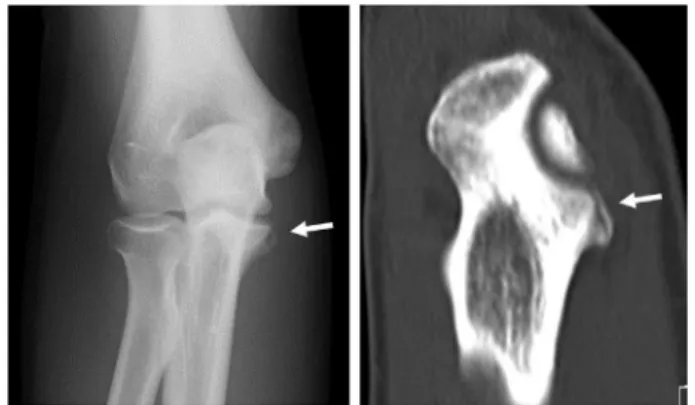
Fig. 3.Case 2. Anteroposterior radiograph (left) and computed tomography (right) of the right elbow joint. Note the bone fragment at the sublime tubercle (arrow).
Fig. 4.Case 2. Anteroposterior radiograph 14 weeks after treatment. Note the union of the fractured bone of the sublime tubercle (arrow).
K. Tanaka et al. / Asia-Pacific Journal of Sports Medicine, Arthroscopy, Rehabilitation and Technology 10 (2017) 1e3
improved from 10 to 142. No elbow pain during pitching had been reported at 6 months after ESWT.
Discussion
In the present cases, stress fractures of the sublime tubercle of the ulna that did not heal after resting from pitching were suc-cessfully treated with ESWT. In the previous study, six of eight patients with sublime tubercle fractures did not respond to non-operative treatment consisting of immobilization with a range of motion brace for 8 weeks, followed by full-effort pitching at 12 weeks. Two of these patients ultimately underwent ulnar collat-eral ligament reconstruction, and four required bone fixations using bioabsorbable anchors. Finally, these patients returned to pitching 4e6 months after bonefixation and 9e12 months after ulnar collateral ligament reconstruction.3 Screw fixation for an avulsion fracture of the sublime tubercle was reported in a case report, and the patient returned to pitch 6 months after surgery.6 In each reported case, patients inevitably received operative management and long-term rehabilitation. In the present two cases, patients were able to avoid surgery, and through ESWT returned to pitch 3 months after treatment without any reported re-fractures, which is faster than any other reported case. Addi-tionally, ESWT is believed to activate osteoblasts surrounding the fracture, which aid in bone union. Shockwaves activate extracel-lular signal-regulated kinase and p38 kinase, which are important factors for bone formation, promote bone marrow stromal cell growth and differentiation toward osteoprogenitors associated with TGF-b1 and VEGF induction.15,16ESWT is clinically performed for stress fractures and non-union cases with good results.10e14
ESWT can be a novel treatment for avulsion fractures of the sub-lime tubercle of the ulna, but no reports of its use have been published. This is thefirst report describing ESWT for delayed-union avulsion fractures of the sublime tubercle of the ulna. In the present cases, no major complications were detected. ESWT is a known treatment with few complications.12 Local pain during irradiation is the most frequent complication, and some patients experience local swelling and perceptual disorder.17Rupture of the Achilles tendon after ESWT has been previously reported in a case report, but this case had undergone injection of steriods.18To the best of our knowledge, there are no reports describing major complications such as refractures, continuous pain, or local dysfunction associated with ESWT for musculoskeletal tissue. However, no previous reports have described irradiation of the medial aspect of the elbow. For this reason, long-term follow up is needed.
This report has some limitations. Both subjects were treated by the same two methods; ESWT and immobilization. A further limi-tation was that case 2 did not undergo a CT scan after ESWT.
Conclusion
ESWT appears to be a successful non-invasive therapy to achieve bone union for stress fractures of the sublime tubercle of the ulna, allowing the patient to return to pitching.
Conflicts of interest and source of funding
None declared.
References
1. Chen FS, Rokito AS, Jobe FW. Medial elbow problems in the overhead-throwing athlete.J Am Acad Orthop Surg. 2001;9(2):99e113.
2. Ciccotti MG, Atanda Jr A, Nazarian LN, et al. Stress sonography of the ulnar collateral ligament of the elbow in professional baseball pitchers: a 10-year study.Am J Sports Med. 2014;42(3):544e551.
3. Jobe FW, Stark H, Lombardo SJ. Reconstruction of the ulnar collateral ligament in athletes.J Bone Joint Surg Am Vol. 1986;68(8):1158e1163.
4. Morrey BF, An KN. Articular and ligamentous contributions to the stability of the elbow joint.Am J Sports Med. 1983;11(5):315e319.
5. Glajchen N, Schwartz ML, Andrews JR, et al. Avulsion fracture of the sublime tubercle of the ulna: a newly recognized injury in the throwing athlete.Am J Roentgenol. 1998;170(3):627e628.
6. Akagi M, Ito T, Ikeda N, et al. Total avulsion fracture of the coronoid tubercle caused by baseball pitching. A case report. Am J Sports Med. 2000;28(4): 580e582.
7. Salvo JP, Rizio 3rd L, Zvijac JE, et al. Avulsion fracture of the ulnar sublime tubercle in overhead throwing athletes.Am J Sports Med. 2002;30(3):426e431. 8. Erickson BJ, Chalmers PN, Bush-Joseph CA, et al. Ulnar collateral ligament reconstruction of the elbow: a systematic review of the literature.Orthop J Sports Med. 2015;3(12), 2325967115618914.
9. Erickson BJ, Bach Jr BR, Verma NN, et al. Treatment of ulnar collateral ligament tears of the elbow: is repair a viable option?Orthop J Sports Med. 2017;5(1), 2325967116682211.
10. Alvarez RG, Cincere B, Channappa C, et al. Extracorporeal shock wave treat-ment of non- or delayed union of proximal metatarsal fractures.Foot Ankle Int. 2011;32(8):746e754.
11. Furia JP, Juliano PJ, Wade AM, et al. Shock wave therapy compared with intramedullary screw fixation for nonunion of proximal fifth metatarsal metaphyseal-diaphyseal fractures. J Bone Joint Surg Am Vol. 2010;92(4): 846e854.
12. Leal C, D'Agostino C, Gomez Garcia S, et al. Current concepts of shockwave therapy in stress fractures.Int J Surg. 2015;24(Pt B):195e200.
13. Taki M, Iwata O, Shiono M, et al. Extracorporeal shock wave therapy for resistant stress fracture in athletes: a report of 5 cases.Am J Sports Med. 2007;35(7):1188e1192.
14. Moretti B, Notarnicola A, Garofalo R, et al. Shock waves in the treatment of stress fractures.Ultrasound Med Biol. 2009;35(6):1042e1049.
15. Chen YJ, Kuo YR, Yang KD, et al. Activation of extracellular signal-regulated kinase (ERK) and p38 kinase in shock wave-promoted bone formation of segmental defect in rats.Bone. 2004;34(3):466e477.
16. Wang FS, Yang KD, Chen RF, et al. Extracorporeal shock wave promotes growth and differentiation of bone-marrow stromal cells towards osteoprogenitors associated with induction of TGF-beta1.J Bone Joint Surg Br Vol. 2002;84(3): 457e461.
17. Kudo P, Dainty K, Clarfield M, et al. Randomized, placebo-controlled, double-blind clinical trial evaluating the treatment of plantar fasciitis with an extracoporeal shockwave therapy (ESWT) device: a North American confi r-matory study.J Orthop Res. 2006;24(2):115e123.