This is an unedited version of the accepted manuscript for the final version please refer to : http://www.sciencedirect.com/science/article/pii/S0022098115300629
Mitochondrial electron transport activity and metabolism of
experimentally bleached hermatypic corals
Sylvain Agostini1*, Hiroyuki Fujimura2. Hiroyuki Hayashi2, Kazuhiko Fujita3
1Shimoda Marine Research Center, University of Tsukuba, 5-10-1 Shimoda, Shizuoka, Japan
2 Department of Chemistry, Biology and Marine Science, University of the Ryukyu, 1 Senbaru,
Nishihara-cho, Okinawa 903-0213, Japan
3 Department of Physics and Earth Science, University of the Ryukyu, 1 Senbaru, Nishihara-cho,
Okinawa 903-0213, Japan
citation:
Agostini, S., Fujimura, H., Hayashi, H., Fujita, K., 2016. Mitochondrial electron transport activity
and metabolism of experimentally bleached hermatypic corals. Journal of Experimental Marine
Abstract
Bleached corals (Porites cylindrica and Galaxea fascicularis) were obtained through extended
incubation (over 45 days) under light depletion and privation: low light and dark conditions, and
heat stress (32 °C). The colonies in the different treatments became bleached and had reduced
metabolic rates, photosynthesis, calcification and respiration; reduced biomass, zooxanthellae
density, and chlorophyll a concentrations; and reduced mitochondrial electron transport system
activity, which represent potential respiration rates. The most important reduction in mitochondrial
electron transport activity was shown when the activities were normalized by the unit of surface and
not by the unit of host protein. This result indicates that the reduction in activity could be mainly
explained by the reduction of biomass and tissue thickness. However, increased Manganese
Superoxide dismutase (MnSOD) activity, a mitochondrial SOD, suggests that ROS production
occurs in the mitochondria under heat stress with the consequence of potentially damaging the
electron transport system. The observed reduced calcification rates observed are hypothesized to be
the results of a decrease in the energy available for calcification due to the reduced photosynthetic
rates, limiting the availability of substrates for respiration and therefore the energy production, and
the decreased in the number of active mitochondrial electron transport system. Electron transport
system activity associated with respiration is the basis of all metabolic processes and is not biased
by incubation like traditional measurements of respiration in an aquarium. Therefore, ETSA could
be used as an overall indicator of coral health, especially for host animal health.
Keywords
Coral, Bleaching, Mitochondrial activities, Oxidative Stress, Calcification
Highlights
Bleached corals, through light depletion and heat stress showed reduced metabolism and
mitochondrial electron transport system.
Reduced respiration and mitochondrial electron transport system activities were associated
with reduced calcification rates.
Reduction in coral biomass and/or direct damage due to reactive oxygen species lead to the
Introduction
Respiration is the basis of metabolism in animals, including corals. It provides the energy required
for all of the subsequent metabolic processes in the form of ATP. Aerobic respiration is divided in
two phases: the oxidation of an organic substrate through glycolysis and the citric acid cycle and the
reduction of the terminal electron acceptor, oxygen. The transfer of electrons from the reduced
co-factors, NADH and FADH2 produced during the oxidation phase is accomplished through a chain
of enzymatic transporters embedded in the internal membrane of the mitochondria. Impairment of
the mitochondrial machinery, especially the electron transport chain, will have strong repercussions
on the respiration process and, subsequently, the metabolism of the organism.
Elevated temperature and other environmental factors can cause coral bleaching: the loss of their
photosynthetic symbionts or their pigments (Glynn et al., 1992; Hoeghguldberg and Smith, 1989).
The most accepted model for the mechanism of bleaching proposes that it starts with the
impairment of the zooxanthellae photosystems (Warner et al., 1999), which leads to the production
of reactive oxygen species (ROS) (Higuchi et al., 2010; Jones et al., 1998). However, mitochondria
and their electron transport system are also an important source of ROS in animals, and the
mitochondrial production of ROS may not be negligible in corals (Downs et al., 2002). This
suggests that the importance of the host in the bleaching process may have been underestimated.
Recently, Dunn et al. (2012) showed degradation of the host mitochondria in anemone under heat
stress. This degradation was associated with the decreased expression of the gene coding for
cytochrome c and complex IV, both of which are proteins that are important parts of the
mitochondrial electron transport system. Corals incubated in the dark for a long period have also
been shown to bleach (Hoegh-Guldberg and Smith, 1989; Tolleter et al., 2013; Yonge et al., 1930).
It has been shown that bleached corals have lower calcification rates independently of the cause of
bleaching, whether due to heat stress, bacterial factors (Higuchi et al., 2013) or depletion of light.
Corals incubated in total darkness for a long period have highly reduced calcification rates with
only 10% of the light calcification rate after 7 h (Al-Horani et al., 2007).
Photosynthesis and calcification show an intimate relationship at both the community level (Barnes
and Chalker, 1990; Gattuso et al., 1996; Kinsey, 1985) and organism level (Allemand et al., 2004;
Furla et al., 2000; Moya et al., 2006). The increase in the calcification rates under light for
photosynthetic organisms is called light-enhanced calcification. On average, corals show three
times higher calcification rates in the light than in the dark. Several hypotheses have been raised to
is that OH- resulting from photosynthesis titrate H+ that is formed during the calcification process. For corals, photosynthesis is generally the main source of reduced carbon and is used as
combustible for respiration and therefore for the production of the energy required for calcification,
which could represent 30% of the coral’s total energy budget (Cohen and Holcomb, 2009).
Inhibition of respiration (oxidative phosphorylation) has been shown to inhibit calcification in the
light, which then falls to the same level as calcification in the dark (Chalker and Taylor, 1975). In a
recent paper (Agostini et al., 2013), the respiration limited calcification model, which support the
models by Chalker and Taylor (1975) and, more generally, the Trans-calcification model of
McConnaughey (1997) was introduced. In this model the hypothesis made is that calcification in
symbiotic corals rates are limited by the energy production through host respiration, itself limited
by the ETSA and the amount of substrate for respiration produced by the photosynthetic activity of
the symbiont.
Many tools are available to assess the state of the symbionts in corals, such as PAM fluorescence,
zooxanthellae density, and pigment concentration. However, only a few are available for the host
and are rather general, such as the lipid or protein content, or require incubation, such as respiration
and calcification. Moreover, traditional respiration measurements also include symbiont respiration,
which may bias the interpretation of the results. Therefore, coral biology is in need of tools to assess
host health, and ETSA could be one of these tools.
The long-term effect of the decrease in photosynthetic activity on the respiratory activity and
calcification rates was tested on two different coral species: Galaxea fascicularis and Porites
cylindrica. Reduced photosynthetic rates were obtained in two ways: corals were maintained under
reduced light or exposed to a gradual elevation of temperature. The aim of this experiment is to first
better understand the relationship of photosynthesis, respiration and calcification in corals, and
second, to investigate the possibility of a direct effect of temperature on mitochondrial ETS, which
leads to decreased growth rates. Two different hypothesis were made regarding the mechanisms that
lead to a decrease in the respiration potential (ETSA) which both lead to reduced host metabolism
(calcification and respiration). The first mechanism would be a decrease in the biomass based on
tissue per unit of surface and tissue thickness, resulting in a reduction in the number of active ETS
(expressed per unit of surface). The second, caused by damage due to heat stress, would be a
decrease in the number of active ETS (expressed in per mg protein, representing the host biomass).
Materials and Methods
Coral specimens
Colonies of Galaxea fascicularis and Porites cylindrica were collected from a coastal region off
Okinawa Island, Japan, with permission from the Okinawa Prefecture government (No. 23-7). The
P. cylindrica colonies were fractionated to obtain small fragments, and the colony of G. fascicularis
was fractionated into single polyps. The fragments were suspended on a nylon thread and
maintained for several months in an outdoor aquarium with running seawater at the Sesoko Station,
Tropical Biosphere Research Center, University of the Ryukyus, Okinawa, Japan. Micro-colonies,
of which the skeleton was entirely covered by tissue, were selected for the experiments.
Experimental design
Colonies were moved into indoor aquariums with running seawater and maintained under controlled
conditions for a minimum of 45 days. Four different conditions were tested, and the different
treatments were applied after a minimum of one week to let the corals acclimatize to the indoor
conditions. The control treatments corresponded to the natural seawater temperature (24 to 27 °C
for P. cylindrica and 22 to 26 °C for G. fascicularis), and an illumination of 300 μmoles of photon s -1 m-2 (measured with a 2pi quantum sensor, JFE Advantech, Japan) provided a 12 h cycle by metal
halide lamps. The heat treatment (high temperature stress) was conducted under the same
illumination as the control treatment, and the temperature was gradually elevated to reach 32 °C in
the last week of the experiment over the total incubation period at a maximum rate of 1 °C per
week. In the dim treatment (dimmed light), the colonies were incubated under ambient light with a
maximum recorded light of 50 μmoles photon s-1 m-2 and an average of 2.5 μmoles photon s-1 m-2 in the daytime, and the temperature was the natural seawater temperature (the same as the control).
The dark treatment was obtained by covering the aquarium with dark cloth; no measurable light
occurred, and the temperature was same as the control. Four micro-colonies of each species were
used for each treatment and suspended on a nylon thread. Aeration was continuously provided in all
treatments.
Metabolism measurement
The colonies were enclosed in individual 300-ml vessels directly in their respective aquariums to
avoid stress due to manipulation and light regime changes. The water was continuously stirred in
the vessels. The incubation was conducted under the appropriate treatment light levels and
for 2 h. For the dark treatment, only a dark incubation was conducted. The dissolved oxygen was
measured at the beginning and end of each incubation using an Orion 4-Star pH-DO sensor
equipped with an RDO probe (Thermo Scientific). Sub-samples of the incubation water were
sampled at the beginning and end of each incubation period and filtered through a 0.45-μm
membrane filter to measure the total alkalinity. The total alkalinity was determined via titration with
HCl at 0.1 mol l-1 with a Metrohm titrator (785 DMP titrino). The calcification rates were calculated using the alkalinity anomaly method (Gattuso et al., 1996), and net photosynthesis and respiration
were calculated based on the variation in the dissolved oxygen during the light and dark incubation,
respectively.
Separation of the host fraction
The coral tissues were removed using an airbrush with phosphate buffer saline and then
homogenized using a Teflon potter homogenizer to break the host cells and release the
zooxanthellae. The host and zooxanthellae fraction were separated by centrifugation at 1500 rpm
for 20 min.
ETSA measurement
Electron transport system acitivity was measure as described in Agostini et al. (2013). Five
milliliters of the supernatant (the host fraction) was collected and homogenized using a sonicator
(Smurt 155 NR-50 M, Microtec Co., Ltd, Funabashi, Japan) at 25% for 5 min in the presence of
polyvinylpirrolidone k30 at 1.5 mg l-1, MgSO4 at 75 μmol l-1, Triton X-100 at 0.20% and EDTA 2Na at 10 mmol l-1. The extract was then cleared by centrifugation at 10,000 rpm for 5 min. All the steps were conducted on ice or in a cooling centrifuge at 3 °C; 300 μl of the cell free extract was
incubated in the presence of NADH (1.5 mmol l-1), NADPH (0.15 mmol l-1) and tetrazolium salt 2-(p-iodophenyl)-3-(p-nitrophenyl)-5-phenyl tetrazolium chloride (INT) (0.4 mg l-1). The reaction was
stopped using 50% formalin after 20 min, and the absorbance was immediately read at 490 nm. The
absorbances were corrected against a turbidity blank (containing 300 μl of the samples, but the
reagents were replaced by phosphate buffer saline) and a chemical blank (containing all the reagents
but the sample was replaced with phosphate buffer saline). The ETSA were expressed in mg O2 h-1 cm-2 using the following formula, where A is the absorbance at 490 nm, Vs the volume of the sample (in this case 0.3 ml), S is the surface of the colony, Vt is the total volume of the tissue slurry
and 7.85 is a factor to convert the increase in absorbance in 20 min to mg of O2 h-1. Alternatively, a similar formula was used where the surface of the skeleton was replaced by the total amount of host
SOD measurements
The host cells were lysed by sonication in phosphate buffer with 0.05% Triton X-100. The
Cu/Zn SOD activity was assayed spectrophotometrically as described by Elstner and Heupel (1976)
and Ōyanagui (1984). The standards for activity were prepared using bovine erythrocytic SOD
(Sigma) for calibration of the Cu/Zn SOD measurement and using the Mn SOD extracted from
buttermilk (Sigma) for the Mn SOD measurement. The Mn SOD was measured by additions of 100
μmol l-1 of KCN in the assay (Beauchamp and Fridovich, 1971). The activities, expressed in nitrate units (NU), were calculated using a logit plot (Kobayashi et al., 1978) and normalized by the units
of the host protein.
Zooxanthellae and chlorophyll contents
The zooxanthellae density was determined by counting cells in the tissue slurry using a
hemocytometer (Neubauer modified). A total of 2 to 3 ml of the tissue slurry was filtered on GF/F
filters, excess of water removed and placed in 3 ml of 100% analytical grade methanol. The filters
were left to extract in a freezer in the dark for 24 h and sonicated to ensure complete extraction. The
extracts were cleared by filtration using a 0.50-μm filter (13JP050AN, Advantec, Japan), and the
absorbance was read at 665 nm and 652 nm. The chlorophyll a concentrations were calculated using
the following formula:Chl a
(
μg l−1)
=16. 29×A665−8. 54×A652 (Ritchie, 2006). Thezooxanthellae density and chlorophyll contents were then normalized by the surface area.
Protein and coral surface measurements
An aliquot from the host fraction obtained after centrifugation at 1500 rpm for 20 min was
sonicated and kept in a freezer until measurement. The protein contents were determined by the
Bradford assay (Bradford, 1976). The surface of the colonies was determined by the aluminum
method (Marsh, 1970) for P. cylindrica and the wax method (Johannes and Wiebe, 1970) for G.
fascicularis.
Statistical treatment of the results
The metabolic rates were normalized by the unit of the surface of the coral skeleton. Enzyme
activities, SODs and ETSA were normalized by the host protein contents. The ETSA was also
normalized by the unit of the surface for comparison. The amount of chlorophyll a was expressed
per unit of surface. All the results were analyzed using the non-parametric Kruskal Wallis test and
multiple comparisons adjusted based on the Hochberg method (Hochberg, 1988). Square of the
Pearson correlation coefficient among ETSA and the physiological variables and their significance
were calculated. All the statistical analyses were conducted with R (Team, 2011) using the agricolae
library except for the linear regressions, which were performed with the plotting software qtiplot
Results
Coral observations
All of the corals survived the different treatments. Severe bleaching was obtained for all of the
corals in the dark treatment, with P. cylindrica showing a visibly reduced tissue thickness and few
expanded polyps. In the dim and heat treatment, more or less severe paling of the corals was
observed. Corals in the control treatment showed expanded polyps and bright colors.
Metabolic rates
Net photosynthesis (Figure 1 A) was significantly affected by treatments in both species (Kruskal
Wallis, n = 4, DF = 3, P. cylindrica: p < 0.05 and G. fascicularis: p = 0.01). For P. cylindrica
respiration rates in the dim treatments was significantly lower than control (Kruskal Wallis mc,
n = 4, p = 0.03) but the decreased observed under heat treatments was not significant compare to
control (Kruskal Wallis mc, n = 4, p = 0.18). For G. fascicularis, a significant reduction in the net
photosynthesis rates was found in both treatments compared to the control ((Kruskal Wallis mc,
n = 4, p = 0.180).
The treatments significantly decreased the respiration rates (Figure 1 B) compared to the control for
both species (Kruskal Wallis, n = 4, DF = 3, p < 0.01 and p < 0.01 for P. cylindrica and G.
fascicularis, respectively). The most important decrease was observed for the dark and dim
treatments. Respiration rates in all treatments were significantly lower than in the control in both
species (Kruskal Wallis mc, n = 4, p < 0.001 compared to the control)
Light calcification (Kruskal Wallis, n = 4, DF = 3, P.c.: p = 0.01, G.f.: p< 0.01) (Figure 1 C) and
dark calcification (Kruskal Wallis, n=4, DF=3, P.c.: p = 0.02, G.f.: p < 0.01) (Figure 1 D) were
significantly affected by treatments in both species and the reduction in the treatments was
significant compare to control (Kruskal Wallis mc, n = 4, p < 0.05) except for the dark calcification
rates in the heat treatment which was not different from control. Light calcification was greatly
reduced in the dim treatment for both species, and among these specimens, two showed net
dissolution for P. cylindrica and one for G. fascicularis. In both the dim and dark treatments, three
of the four specimens for each treatment showed a net dissolution in the dark for P. cylindrica.
Among all the specimens in the heat treatment, two colonies of P. cylindrica showed net dissolution
in the dark, but the average response was not significantly reduced compared to the control. For G.
fascicularis, the dark calcification rates were low in both the control and the different treatments.
treatments.
Physiological variables
The zooxanthellae density (Kruskal Wallis, n = 4, DF = 3, P.c.: p = 0.01, G.f.: p < 0.01) and
chlorophyll a concentration (Kruskal Wallis, n = 4, DF = 3, P.c.: p < 0.01, G.f.: p < 0.01) were
significantly decreased compared to the control in both species and in all the treatments compared
to the control (Kruskal Wallis mc, n = 4, p < 0.01 compared to the control) (Figure 2). The lowest
zooxanthellae density for P. cylindrica was observed for the dark treatment. In the heat treatment,
the zooxanthellae densities observed were higher but still significantly lower than the control. The
chlorophyll a concentration normalized per surface of corals showed a similar pattern. For G.
fascicularis, the patterns observed were different with the heat treatment, showing average values
between the Dim and Dark values for the chlorophyll a but the lowest zooxanthellae density.
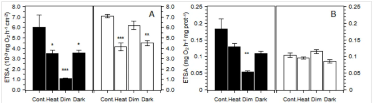
The mitochondria electron transport system activity was measured and was normalized by the unit
of the surface and the amount of host protein (Figure 3). The interpretation of the results differs and
is discussed later. When normalized by the amount of surface, the activities obtained were
significantly reduced by treatments for both P. cylindrica (Kruskal Wallis, n = 4, DF = 3, p < 0.01)
and G. fascicularis (p < 0.01). For P. cylindrica, ETSA measured in all three treatments were
significantly lower than control (Kruskal Wallis mc, n = 4, p < 0.05) with the minimum value
observed for the dim treatment. In G. fascicularis, posthoc tests revealed that the activities in the
dark and heat treatments were significantly reduced compared to the control (Kruskal Wallis mc,
n = 4, p < 0.01 and p < 0.01, respectively) but that the activities in the dim treatment (87%) were
not significantly different from that of the control (Kruskal Wallis mc, n = 4, p = 0.31). ETSA,
expressed by the amount of host protein, showed that the treatments significantly affected the
activities (Kruskal Wallis, n = 4, DF = 3, p = 0.01 for P. cylindrica and p = 0.03 for G. fascicularis).
Significant differences with the control, as indicated by posthoc tests, were only found for the dim
treatment for P. cylindrica. The observed reduction in the averaged activities of the specimen in the
heat treatment of G. fascicularis (91% of the control) and of P. cylindrica (72% of control) were not
significant at the 95% confidence level (Kruskal Wallis mc, n = 4, p = 0.25 and p = 0.07,
respectively). In addition, the activities in the other treatments did not show a significant difference
from the control (Kruskal Wallis mc, n = 4, p > 0.1). In G. fascicularis, the only significant
difference among treatments for the ETSA normalized by the amount of protein was observed
between the Dim and Dark treatments (Kruskal Wallis mc, n = 4, p < 0.01).
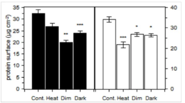
amount of protein per surface (Figure 4). In both species, the maximum values were observed for
the control treatment and a significant effect of the treatments was observed (Kruskal Wallis, n = 4,
DF = 3, p < 0.01 for P. cylindrica and p = 0.01 for G. fascicularis). In P. cylindrica, the minimum
value was obtained for the Dim treatment and a similar value was obtained for the dark. The amount
of protein in the heat treatment was not significantly reduced compared to the control at the 95%
confidence level (Kruskal Wallis mc, n = 4, p = 0.06). For G. fascicularis, the minimum value was
observed for the heat treatment, and the protein contents in the dim and dark treatment were
significantly lower than the control.
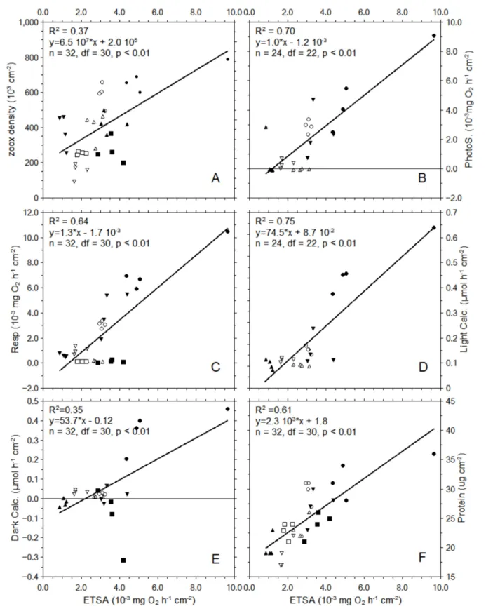
Mitochondrial electron Transport system activity, normalized by the amount of surface, show a
positive correlation with the different metabolic rates and physiological variable (Figure 5). The
correlations were all significant (n = 32 or 24, df = 30 or 22, p < 0.01) with the strongest observed
between light calcification and ETSA, r2 = 0.75. A positive correlation of ETSA with the amount of protein was observed with coefficient of correlation of 0.61.
Two different superoxide dismutase activities were measured (Figure 6): Manganese Superoxide
dismutase (Mn SOD) and Copper Zinc Superoxide dismutase (Cu/Zn SOD). The Mn SOD activities
were minimal in the control treatment for both species. They significantly increased in all other
treatments, except for the dark treatment (Kruskal Wallis, n = 4, DF = 3, p < 0.01), compared to the
control, with a maximum occurring for the heat treatment. No significant effect of the treatments
was observed for Cu/Zn SOD for G. fascicularis (Kruskal Wallis, n = 4, DF = 3, p = 0.24) which
show generally lower activities than P. cylindrica. For P. cylindrica, the activity of Cu/Zn SOD was
significantly affected by the treatments (Kruskal Wallis, n = 4, DF = 3, p = 0.02), with significantly
higher values than the control in the Dim treatments.
Discussion
Both methods, light depletion and heat stress caused bleaching in the two species studied, as shown
by the significant decrease in zooxanthellae densities and chlorophyll contents, which are
parameters that are typically used to assess bleaching (Hoegh-Guldberg and Smith, 1989).
Simultaneous to bleaching, reduced photosynthesis and calcification rates were observed, as is
commonly observed for bleached corals (Abramovitch-Gottlib et al., 2002; Colombo-Pallotta et al.,
2010; Foster et al., 2014; Marshall, 1996). All metabolic rates: net photosynthesis, dark respiration,
and light and dark calcification, as expressed by the unit of the surface, were significantly affected
by the different treatments in both species. Overall, the most visible effects were caused by the dark
realistic in nature and only provide elements to understand the physiology of corals. More variable
responses were obtained for the heat and dim treatments. Low light level (Dim treatment)
incubation of corals for over one month is also an extreme condition, but not unrealistic. It led to an
important decrease in metabolic rates, including photosynthesis and calcification. Dark calcification
was greatly reduced in the light depleted treatments, with net dissolution often observed. For the
respiration rates, the observed decrease in all treatments compared to the control may have two
different explanations that are not exclusive: respiration is limited by the availability of the substrate
to oxidize and the number of working electron transport systems and enzymes required is
decreased. It is interesting to note that for G. fascicularis, while the zooxanthellae density was
lower in the heat treatment than in the dark and dim treatments, the metabolic rates of the corals in
the heat treatment were still higher, which indicated a strong limitation of zooxanthellae by the
energy light available and therefore a reduction in the production of available photosynthetate. The
type of zooxanthellae typically found in G. fascicularis and P. cylindrica is known to differ possibly
explaining the difference observed in the effect of the treatments. On the one hand, G. fascicularis
has been shown to be associated with C1 or C3 zooxanthellae, C1 being known to be adapted to low
light level (Oppen et al, 2005) but sensitive to thermal stress (Baird et al., 2010). Contrasting with
P. cylindrica which could, as other species of the genus Porites in the Asia-Pacific region, be
associated with the thermo-tolerant zooxanthellae from the clade C15 (D'Angelo et al., 2015).
The relationship between photosynthesis and calcification and the enhancement of calcification in
the light has long been known (Goreau, 1959) and widely reviewed (Gattuso et al., 1996; Kinsey,
1978); however, the exact mechanism of the relation has not been determined (Allemand et al.,
2004). The respiration rates also show a strong relation with photosynthesis. In the experiment
reported here, the decrease in photosynthesis was always associated with decreased, sometimes
severely decreased, as in the light depletion treatment, respiration rates. In a previous paper, the
respiration limited calcification model was introduced. This model is similar to the
trans-calcification model (McConnaughey, 1997), but with a stronger focus on the role of respiration.
This model implies that photosynthesis provides the substrates required for respiration, which then
provide the energy required for calcification (Agostini et al., 2013). In this study, the use of
artificially bleached corals, allowed testing whether the model is still valid for a wider range of
respiration rates. In the most extreme case (dark treatment), respiration was strongly limited by the
availability of substrate and calcification was minimal or even resulted in net dissolution, which
suggests that respiration may have limited calcification. The effects of the treatments on the light
to the extremely low dark calcification rates. In G. fascicularis, the ratio decrease from 3 in the
control treatment to less than 1 in the dim and heat treatments showed that the enhancement of
calcification by light was removed due to the lack of photosynthesis and therefore supports the
hypothesis that this enhancement is due to the photosynthetic activity.
A generally accepted model for bleaching through heat stress involves the production of reactive
oxygen species (ROS) (Downs et al., 2002). Typically, zooxanthellae and their photosynthetic
apparatus are associated with the production of ROS; however, mitochondria and their electron
transport systems are known to produce ROS under heat stress, at least in plants (Taylor et al.,
2004), and more damaged mitochondria were observed in heated stressed cnidarian, damages that
may have been caused by ROS (Dunn et al., 2012). In this paper, two different superoxide
dismutase activities in the host tissues were measured: The copper/Zinc superoxide dismutase
(Cu/Zn SOD) which is a cytoplasmic SOD and the Manganese superoxide dismutase (Mn SOD)
which is found strictly in the mitochondria. The following hypothesis were made to assess the
origin of the ROS produced under stress. In the case ROS mainly originates from the zooxanthellae
due to the damage to their photosystems, the host Cu/Zn SOD should increase the most, and in the
case ROS are mainly produced in the host mitochondria, then the Mn SOD should increase the
most. In the last case, where mitochondria is the main source of ROS for coral host under stress, the
ROS could result in damage to the host mitochondrial electron transport systems. The Mn SOD
activity was found to significantly increase in the heat treatment in both species. The Cu/Zn SOD
activity increased in both the dim and heat treatments in P. cylindrica, but was not increased in G.
fascicularis. These results suggest that ROS were produced in the host mitochondria and that
damage to the respiratory apparatus could have occurred. In the light depletion specimens, the
Mn SOD and, in some cases, Cu/Zn SOD activities increased, but not as much as in the heat treated
specimens. The mechanism behind the increase of SOD under depletion of light, and therefore
possible ROS production in the host, remains unknown. It is usually considered that light is
required for the production of ROS in zooxanthellae (Lesser, 1996). Thus, zooxanthellae may not be
the source of ROS under light depletion for some coral species, but the mitochondria may be.
Because the ROS production increase may have been higher under heat stress than in the light
depletion treatment, it is assumed that the decrease in the respiration rates observed is due to both
damage to the respiratory system and substrate limitation in the heat treatment, but mainly by
substrate limitation in the light depletion treatments. To investigate this, the activity of the electron
transport system (ETSA) using the INT methods was measured (Agostini et al., 2013; Packard,
used. To show a decrease in ETSA due to ROS damage, the activities normalized by the amount of
host protein could be used with the assumption that they represent the host biomass and,
consequently, the total number of ETS. While the treatment significantly affected the ETSA
normalized by protein for both species, only P. cylindrica under the dim treatment was significant
decreased compared to the control. For G. fascicularis only the activities in the dim and dark
treatment were significantly different. This result indicates that while possible direct damage to the
mitochondrial electron transport system using the INT methods could be observed, the experiment
could not sufficiently detect the decrease in ETSA for multiple comparison. Moreover, a more
severe decrease was expected in the heat stress treatment than in the light depletion treatment, as the
Mn SOD activities were higher in the heat treatment, but that was not always observed. One
explanation may be that the production of ROS in the mitochondria damages part of the
enzymes/transporters lower in the transporter chain. The tetrazolium salt INT is reduced to
formazan by the cytochrome b - ubiquinone complex and before the complex III, which means that
damage further down the chain may not always lead in decreased ETSA.
Electron transport system activity normalized by the surface of the host can be used as an easy to
measure overall health (metabolism) indicator for the host. It has already been used for this purpose
with other organisms (Finlay et al., 1983; Simcic, 2004) and is typically used to estimate the
respiration rates of plankton (Arístegui and Montero, 1995; Bamstedt, 1980; Packard, 1971) or the
overall metabolism of meiofauna (Cammen and Corwin, 1990; Olanczukneyman and Vosjan, 1977;
Wada et al., 2012). Decreased ETSA in the different treatments and a strong correlation with
metabolic rates was observed, confirming the previous results obtained with the same methods
(Agostini et al., 2013). The experiment conducted consisted of long-term exposure to the
treatments. This led to a change in tissue biomass, which was shown by a reduction in the amount
of protein per surface for the different treatments. Decreased tissue biomass: essential constituent of
coral tissue and energetic reserve such as proteins, and lipids, is typical for bleached coral (Fitt et
al., 2000; Rodrigues and Grottoli, 2007; Porter et al., 1989; Szmant and Gassman, 1990). However,
more than just a decrease in the reserve available to the coral host, this result show that the observed
decrease in biomass is associated with a decrease in ETSA. The reduction in ETSA could be due to
a decrease in the number of mitochondria and therefore a decrease in the number of ETS. Therefore,
ETSA can incorporate both the change in biomass or tissue thickness and the direct effect of stress
on the mitochondrial electron transport system.
In conclusion, long-term exposure to heat stress or light depletion leads to bleaching and reduced
reduction of respiration and ETSA and, ultimately, to a reduction of calcification through the
limitation of available energy. The increase in Mn SOD, which may correlate with the production of
ROS in the mitochondria, was associated with decreased ETSA normalized by the host protein,
which indicated damage to the mitochondrial electron transport system. More specific methods will
be required, such as the measurement of the activities of the different complexes, to distinguished
which step and enzymes are impaired by the different stresses. The measurement of ETSA by the
INT methods is a simple and relevant way to assess the potential metabolism and therefore the
health of corals and could be applied in field studies to provide a long needed tool to assess the state
of the host animal in corals.
Acknowledgments
This research was supported by the International Research Hub Project for Climate Change and
Coral Reef/Island Dynamics at the University of the Ryukyus. The authors are grateful to comments
of anonymous reviewers and of the editor which greatly improve this manuscript.
References
Abramovitch-Gottlib, L., Katoshevski, D., Vago, R., 2002. A computerized tank system for studying the effect of temperature on calcification of reef organisms. J. Biochem. Biophys. Methods 50, 245–252. doi:10.1016/S0165-022X(01)00236-6
Agostini, S., Fujimura, H., Fujita, K., Suzuki, Y., Nakano, Y., 2013. Respiratory electron transport system activity in symbiotic corals and its link to calcification. Aquat. Biol. 18, 125–139. doi:10.3354/ab00496
D’Angelo, C., Hume, B.C.C., Burt, J., Smith, E.G., Achterberg, E.P., Wiedenmann, J., 2015. Local adaptation constrains the distribution potential of heat-tolerant Symbiodinium from the Persian/Arabian Gulf. ISME J. doi:10.1038/ismej.2015.80
Al-Horani, F.A., Tambutté, É., Allemand, D., 2007. Dark calcification and the daily rhythm of calcification in the scleractinian coral, Galaxea fascicularis. Coral Reefs 26, 531–538. doi:10.1007/s00338-007-0250-x
Allemand, D., Ferrier-Pagès, C., Furla, P., Houlbrèque, F., Puverel, S., Reynaud, S., Tambutté, É., Tambutté, S., Zoccola, D., 2004. Biomineralisation in reef-building corals: from molecular mechanisms to environmental control. Comptes Rendus Palevol 3, 453–467.
doi:10.1016/j.crpv.2004.07.011
Arístegui, J., Montero, M.F., 1995. The relationship between community respiration and ETS activity in the ocean. J. Plankton Res. 17, 1563–1571. doi:10.1093/plankt/17.7.1563
Baird, A.H., Bhagooli, R., Nonaka, M., Yakovleva, I., Yamamoto, H.H., Hidaka, M., Yamasaki, H., 2010. Environmental controls on the establishment and development of algal symbiosis in corals, in: Proceedings of the 11th International Coral Reef Symposium. Presented at the 11th International Coral Reef Symposium, Fort Lauderdale, FL, USA, pp. 108–112.
131.
Beauchamp, C., Fridovich, I., 1971. Superoxide dismutase: Improved assays and an assay applicable to acrylamide gels. Anal. Biochem. 44, 276–287.
Bradford, M., 1976. A rapid and sensitive method for the quantitation of microgram quantities of protein utilizing the principle of protein-dye binding. Anal. Biochem. 72, 248–254.
doi:10.1016/0003-2697(76)90527-3
Cammen, L., Corwin, S., 1990. Electron transport system (ETS) activity as a measure of benthic macrofaunal metabolism. Mar. Ecol. Prog. Ser. 65, 171–182.
Chalker, B.E., Taylor, D.L., 1975. Light-Enhanced Calcification, and the Role of Oxidative
Phosphorylation in Calcification of the Coral Acropora cervicornis. Proc. R. Soc. B Biol. Sci. 190, 323–331. doi:10.1098/rspb.1975.0096
Cohen, A., Holcomb, M., 2009. Why Corals Care About Ocean Acidification: Uncovering the Mechanism. Oceanography 22, 118–127. doi:10.5670/oceanog.2009.102
Colombo-Pallotta, M.F., Rodríguez-Román, A., Iglesias-Prieto, R., 2010. Calcification in bleached and unbleached Montastraea faveolata: evaluating the role of oxygen and glycerol. Coral Reefs 29, 899–907. doi:10.1007/s00338-010-0638-x
Downs, C. a, Fauth, J.E., Halas, J.C., Dustan, P., Bemiss, J., Woodley, C.M., 2002. Oxidative stress and seasonal coral bleaching. Free Radic. Biol. Med. 33, 533–43.
Dunn, S.R., Pernice, M., Green, K., Hoegh-Guldberg, O., Dove, S.G., 2012. Thermal stress
promotes host mitochondrial degradation in symbiotic cnidarians: are the batteries of the reef going to run out? PloS One 7, e39024.
Elstner, E.F., Heupel, A., 1976. Inhibition of nitrite formation from hydroxylammoniumchloride: A simple assay for superoxide dismutase. Anal. Biochem. 70, 616–620.
Finlay, B., Span, A., Ochsenbeingattlen, C., 1983. Influence of physiological state on indices of respiration rate in protozoa. Comp. Biochem. Physiol. A Physiol. 74, 211–219.
doi:10.1016/0300-9629(83)90590-X
Fitt, W.K., McFarland, F.K., Warner, M.E., Chilcoat, G.C., 2000. Seasonal patterns of tissue biomass and densities of symbiotic dinoflagellates in reef corals and relation to coral bleaching. Limnol. Oceanogr. 45, 677–685. doi:10.4319/lo.2000.45.3.0677
Foster, T., Short, J.A., Falter, J.L., Ross, C., McCulloch, M.T., 2014. Reduced calcification in Western Australian corals during anomalously high summer water temperatures. J. Exp. Mar. Biol. Ecol. 461, 133–143. doi:10.1016/j.jembe.2014.07.014
Furla, P., Galgani, I., Durand, I., Allemand, D., 2000. Sources and mechanisms of inorganic carbon transport for coral calcification and photosynthesis. J. Exp. Biol. 203, 3445–57.
Gattuso, J., Pichon, M., Delesalle, B., Canon, C., Frankignoulle, M., 1996. Carbon fluxes in coral reefs. I. Lagrangian measurement of community metabolism and resulting air-sea CO2 disequilibrium. Mar. Ecol. Prog. Ser. 145, 109–121. doi:10.3354/meps145109
Glynn, P.., Imai, R., Sakai, K., Nakano, Y., Yamazato, K., 1992. Experimental Responses of
Okinawan (Ryukyu Islands,, Japan) Reef Corals to High Sea Temperature and UV Radiation, in: Richmond, R.H. (Ed.), Proceedings of the Seventh International Coral Reef Symposium. University of Guam Press, Guam, pp. 27–37.
Goreau, T.F., 1959. The physiology of skeleton formation in corals. I. A method for measuring the rate of calcium deposition by corals under different conditions. Biol. Bull. 116, 59–75. Higuchi, T., Agostini, S., Casareto, B.E., Yoshinaga, K., Suzuki, T., Nakano, Y., Fujimura, H.,
Suzuki, Y., 2013. Bacterial enhancement of bleaching and physiological impacts on the coral Montipora digitata. J. Exp. Mar. Biol. Ecol. 440, 54–60. doi:10.1016/j.jembe.2012.11.011 Higuchi, T., Fujimura, H., Hitomi, Y., Arakaki, T., Oomori, T., Suzuki, Y., 2010. Photochemical
formation of hydroxyl radicals in tissue extracts of the coral Galaxea fascicularis. Photochem. Photobiol. 86, 1421–1426. doi:10.1111/j.1751-1097.2010.00802.x
75, 800–802. doi:10.1093/biomet/75.4.800
Hoeghguldberg, O., Smith, G., 1989. The effect of sudden changes in temperature, light and salinity on the population density and export of zooxanthellae from the reef corals Stylophora
pistillata Esper and Seriatopora hystrix Dana. J. Exp. Mar. Biol. Ecol. 129, 279–303. doi:10.1016/0022-0981(89)90109-3
Johannes, R.E., Wiebe, W.J., 1970. Method for determination of coral tissue biomass and composition. Limnol. Oceanogr. 15, 822–824.
Jones, R.J., Hoegh-Guldberg, O., Larkum, A.W.D., Schreiber, U., 1998. Temperature-induced bleaching of corals begins with impairment of the CO2 fixation mechanism in zooxanthellae. Plant Cell Environ. 21, 1219–1230. doi:10.1046/j.1365-3040.1998.00345.x
Kinsey, D., 1985. Metabolism, calcification and carbon production, in: Proc 5th Int Coral Reef Congr, Tahiti. International Coral Reefs Society, Tahiti, pp. 4:515–526.
Kinsey, D.W.D.W., 1978. Alkalinity changes and coral reef calcification. Limnol. Oceanogr. 23, 989–991. doi:10.4319/lo.1978.23.5.0989
Kobayashi, Y., Okahata, S., Tanabe, K., Usui, T., 1978. Use of logit paper in determination of superoxide dismutase activity in human blood cells. J. Immunol. Methods 24, 75–78.
Lesser, M.P., 1996. Elevated temperatures and ultraviolet radiation cause oxidative stress and inhibit photosynthesis in symbiotic dinoflagellates. Limnol. Oceanogr. 271–283.
Marshall, A.T., 1996. Calcification in Hermatypic and Ahermatypic Corals. Science 271, 637–639. doi:10.1126/science.271.5249.637
Marsh, J.A., 1970. Primary Productivity of Reef-Building Calcareous Red Algae. Ecology 51, 255. doi:10.2307/1933661
McConnaughey, T., 1997. Calcification generates protons for nutrient and bicarbonate uptake. Earth-Sci. Rev. 42, 95–117.
Moya, A., Tambutté, S., Tambutté, E., Zoccola, D., Caminiti, N., Allemand, D., 2006. Study of calcification during a daily cycle of the coral Stylophora pistillata: implications for “light-enhanced calcification”. J. Exp. Biol. 209, 3413–9. doi:10.1242/jeb.02382
Olanczukneyman, K., Vosjan, J., 1977. Measuring respiratory electron-transport-system activity in marine sediment. Neth. J. Sea Res. 11, 1–13. doi:10.1016/0077-7579(77)90017-5
Ōyanagui, Y., 1984. Reevaluation of assay methods and establishment of kit for superoxide dismutase activity. Anal. Biochem. 142, 290–296. doi:10.1016/0003-2697(84)90467-6
van Oppen, M.J.H., Mahiny, A.J., Done, T.J., 2005. Geographic distribution of zooxanthella types in three coral species on the Great Barrier Reef sampled after the 2002 bleaching event. Coral Reefs 24, 482–487. doi:10.1007/s00338-005-0487-1
Packard, T., 1971. The measurement of respiratory electron-transport activity in marine phytoplankton. J. Mar. Res. 29, 235–244.
Porter, J.W., Fitt, W.K., Spero, H.J., Rogers, C.S., White, M.W., 1989. Bleaching in reef corals: Physiological and stable isotopic responses. PNAS 86, 9342–9346.
Ritchie, R.J., 2006. Consistent sets of spectrophotometric chlorophyll equations for acetone, methanol and ethanol solvents. Photosynth. Res. 89, 27–41. doi:10.1007/s11120-006-9065-9 Rodrigues, L.J., Grottoli, A.G., 2007. Energy reserves and metabolism as indicators of coral
recovery from bleaching. Limnol. Oceanogr. 52, 1874–1882. doi:10.4319/lo.2007.52.5.1874 Simcic, T., 2004. Respiratory electron transport system (ETS) activity as an estimator of the thermal
tolerance of two Daphnia hybrids. J. Plankton Res. 26, 525–534. doi:10.1093/plankt/fbh056 Szmant, A.M., Gassman, N.J., 1990. The effects of prolonged “bleaching” on the tissue biomass and
reproduction of the reef coral Montastrea annularis. Coral Reefs 8, 217–224.
doi:10.1007/BF00265014Taylor, N.L., Day, D.A., Millar, A.H., 2004. Targets of stress-induced oxidative damage in plant mitochondria and their impact on cell carbon/nitrogen metabolism. J. Exp. Bot. 55, 1–10. doi:10.1093/jxb/erh001
Statistical Computing, Vienna, Austria.
Tolleter, D., Seneca, F.O., DeNofrio, J.C., Krediet, C.J., Palumbi, S.R., Pringle, J.R., Grossman, A.R., 2013. Coral Bleaching Independent of Photosynthetic Activity.
doi:10.1016/j.cub.2013.07.041 Vasilef, I., n.d. QtiPlot.
Wada, M., Suzuki, S., Nara, T., Umezawa, Y., Shimanaga, M., Matsuoka, K., Nakata, H., 2012. Microbial community respiration and structure of dead zone sediments of Omura Bay, Japan. J. Oceanogr. 68, 857–867. doi:10.1007/s10872-012-0136-6
Warner, M.E., Fitt, W.K., Schmidt, G.W., 1999. Damage to photosystem II in symbiotic
dinoflagellates: A determinant of coral bleaching. Proc Natl Acad Sci U S A 96, 8007–8012. Yonge, C.M. (Charles M.S., Yonge, C.M. (Charles M., History), B.M. (Natural, (1928-29),
Figures
Figure 1 : Net photosynthetic rates (A), respiration rates (B), light calcification rates (C) and dark
Figure 3: Mitochondrial electron transport activity (ETSA) normalized by the unit of the skeleton
surface (A) and ETSA normalized by the milligrams of host protein (B) in P. cylindrica (black) and
G. fascicularis (white). The error bars show the standard error (n=4), and the star marks showed
significant difference (* p < 0.05 ; ** p < 0.001; *** p < 0.0001) with the control after multiple
Figure 4: Amount of the host protein per unit of skeleton surface in P. cylindrica (black) and G.
fascicularis (white). The error bars show the standard error (n=4), and the star marks showed
significant difference (* p < 0.05 ; ** p < 0.001; *** p < 0.0001) with the control after multiple
Figure 5: Correlations of ETSA with zooxanthellae density (A), photosynthesis (B), respiration (C),
light calcification (D), dark calcification (E) and host protein (F). The coefficient of correlation for
different treatments: Control, Heat, Dim and Dark are represented by the symbols: ellipse, down
Figure 6 : Cu/Zn SOD activities (A) and MnSOD activities (B) in P. cylindrica (black) and G.
fascicularis (white). The error bars show the standard error (n=4), and the star marks showed
significant difference (* p < 0.05 ; ** p < 0.001; *** p < 0.0001) with the control after multiple