T his doc ument is downloaded at: 2018-03-23T 18:10:51Z
T itle
S imultaneous medial opening wedge high tibial osteotomy and revision
anterior cruciate ligament reconstruction using a bone-patella tendon-bone
graft: A case report
A uthor(s )
Y onekura, A kihiko; S ugihara, Y usuke; Okazaki, Narihiro; K idera, K enichi;
Nakazoe, Y usuke; T omita, Masato; Osaki, Makoto
C itation
A cta medica Nagasakiensia, 61(3), pp.127-135; 2018
Is s ue D ate
2018-01
UR L
http://hdl.handle.net/10069/37961
R ig ht
NA O S IT E : Nag as aki Univers ity's A c ademic O utput S IT E
Introduction
Injuries of the anterior cruciate ligament (ACL) usually occur in sports activity. Because of their low natural healing rate1,2, most cases are treated operatively. Cases of ACL injury seldom develop knee osteoarthritis (OA) because they mostly occur in teenagers. However, we sometimes encounter a case of ACL injury with knee OA in patients whose knees give way frequently. Patients who have tibia vara also sometimes develop knee OA. ACL reconstruction (ACLR) improves knee instability even in the presence of knee OA, but it does not improve the knee pain from knee OA completely3,4. It, therefore, has been considered that ACLR is not a good
option for patients with concomitant knee OA.
For medial knee OA of young or middle-aged patients, high tibial osteotomy (HTO) achieves stable pain relief with activities of daily living5–7. The combination of simultaneous HTO and ACLR for ACL injury with knee OA has been reported from the 1990s8–12. Development of rigid fixation by a medial locking plate system accelerated postoperative rehabilitation. As a result, good return to sports activity was reported following simultaneous HTO and ACLR13. A case of a torn ACL graft of hamstring tendons followed by medial knee OA after 3 years is reported. This patient underwent simultaneous medial opening wedge HTO (MOWHTO) and revision ACLR using a bone-patella
tendon-
Address correspondence: Akihiko Yonekura, M.D., Ph.D. Department of Orthopaedic Surgery, Nagasaki University Graduate School of Biomedical Sciences, 1-7-1 Sakamoto, Nagasaki 852-8501 Japan
TEL: +81-95-819-7321, FAX: +81-95-849-7325, E-mail: [email protected]
Received September 7, 2017; Accepted September 19, 2017
MS#AMN 07212
Case Report
Simultaneous medial opening wedge high tibial osteotomy and revision
anterior cruciate ligament reconstruction using a bone-patella tendon-bone
graft: A case report
Akihiko YonekurA1, Y
usuke SugihArA2
, narihiro okAzAki1
, kenichi kiderA1, Y
usuke nAkAzoe1
, Masato ToMiTA1, Makoto oSAki1
1 Department of Orthopaedic Surgery, Nagasaki University Graduate School of Biomedical Sciences 2 Department of Orthopaedic Surgery, The Japanese Red Cross Nagasaki Genbaku Hospital
It is said that the clinical results of cases with anterior cruciate ligament reconstruction (ACLR) who have knee osteoarthritis (OA) are not very good. A case of simultaneous medial opening wedge high tibial osteotomy (MOWHTO) and revision ACLR using a bone-patella tendon-bone (BPTB) graft for medial knee OA after re-tear of a reconstructed ACL graft is reported.
The patient was a 49-year-old man who underwent surgery for a right knee ACL injury by ACLR using an ipsilateral hamstring tendon graft 7 years earlier. He sprained his right knee while he was skiing and injured his reconstructed ACL graft. He had knee instability and pain at the medial side of his knee. X-ray showed a tibia vara deformity and medial knee OA of Kellgren-Lawrence grade II. It was thought that the medial knee pain would remain if he were treated by revision ACLR alone. Therefore, simultaneous MOWHTO and revision ACLR using an ipsilateral BPTB graft were performed. The excellent clinical results and
radiological indings 3 years after the operation indicate the usefulness of this approach.
ACTA MEDICA NAGASAKIENSIA 61: 127-135, 2017
128 Akihiko Yonekura et al.: Simultaneous MOWHTO & revision ACLR
bone (BPTB) graft. The good short-term outcomes at 3 years in this case are reported.
Case
The patient was a 49-year-old man who injured the ACL of his right knee when he fell from a truck platform 7 years earlier. He was treated surgically by the anatomical two-route ACLR using ipsilateral hamstring tendons. He returned to sports activity and was making steady progress. He sprained his right knee when he was skiing 3 years earlier and re-injured the right reconstructed ACL graft. His knee started to repeatedly give way, and his medial knee pain increased. He was treated conservatively by his doctor for 3 years. He returned to our hospital for surgery because his knee instability and pain were getting worse.
He had no particular family history or past history except for the primary ACLR of the right knee. His general condi-tion and blood tests were all good.
Ballottement of the patella was negative in both knees. The range of motion of both knee joints was normal, with zero degrees of extension and 145 degrees of flexion. The McMurray test was negative in both knees. The Lachman test and pivot shift test were positive in the right knee, indi-cating ACL insufficiency. Anterior translation of the tibia measured by the KT-2000 knee arthrometer (MED metric Corporation, San Diego, CA) was 13 mm in the right knee and 9 mm in the left knee (Table 1). There was a 4-mm side-to-side difference between the knees.
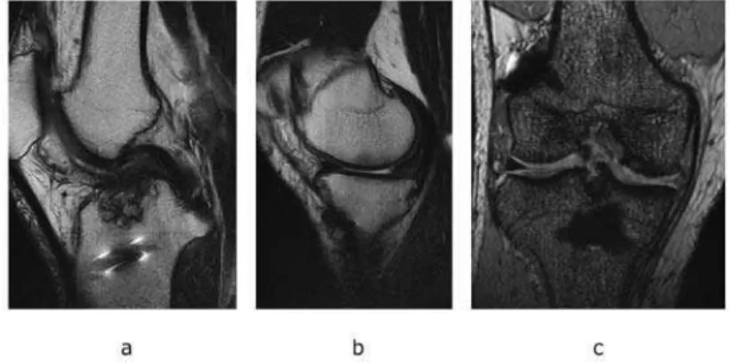
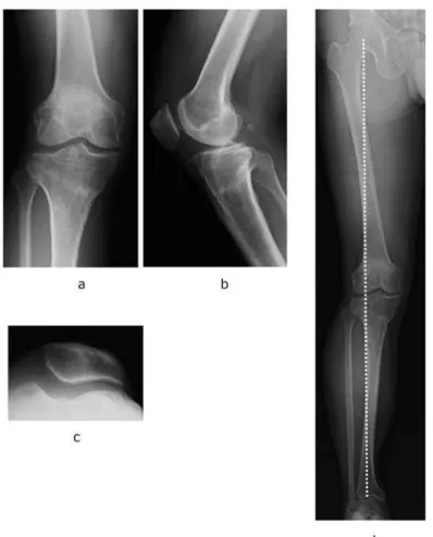
According to the radiological assessment, the P-A X-ray of Rosenbergʼs view showed that the medial joint space of the right knee was narrower than that of the left knee by 1.3 mm. There were osteophyte formations at the medial side of the tibia and the intercondylar space of the femur. The medial knee OA was Kellgren-Lawrence14 grade II (Figure 1). The long leg standing A-P X-ray showed that the femoro-tibial angle (FTA) was 186 degrees in the right knee and 179 degrees in the left knee. The hip knee angle (HKA, valgus is positive and varus is negative) was minus 10.4 degrees in the right knee and minus 5.8 degrees in the left knee. The per-centage of the mechanical axis (%MA) was 10% in the right knee and 28% in the left knee, indicating severe varus defor-mity in the right knee. The mechanical lateral distal femoral angle (mLDFA) was 86 degrees, the medial proximal tibial angle (MPTA) was 79 degrees, and the joint line conver-gence angle was 2.7 degrees, indicating that the reason for the varus knee was primarily the medial inclination of the tibial plateau. The lateral X-ray view showed that the posterior tibial slope (PTS), the complementary angle between the center shaft of the tibia and the tangential line of the medial tibial plateau, was 17 degrees (Table 2). The MRI of the right knee showed no primary reconstructed ACL graft intensity on T2-weighted imaging. Cartilage wear of the medial fem-oro-tibial (FT) joint and a horizontal tear of the medial meniscus were also seen (Figure 2).
This case was diagnosed as a tear of the primary recon-structed ACL graft with medial knee OA, and simultaneous HTO and revision ACLR were planned.
Pre operation Three years after operation
Right Left Right
Ballottement of patella (-) (-) (-)
extension [degrees] 0 0 0
flexion [degrees] 145 145 145
McMurray test (-) (-) (-)
Lachman test (+) (-) (-)
Pivot shift test (+) (-) (-)
Anterior translation of tibia
(KT-2000 manual max) [mm] 13 9 10
Pre operation Three years after operation
Right Left Right
Femoro-Tibial Angle [degrees] 186 179 168
Hip Knee Angle [degrees] -10.4 -5.8 6.2
% Mechanical Axis [%] 10 28 73
mechanical Lateral Distal Femoral Angle
[degrees] 86 85 87
Medial Proximal Tibial Angle [degrees] 79 80 96
Joint Line Convergence Angle [degrees] 2.7 1.6 2.8
Posterior Tibial Slope [degrees] 17 17 15
Table 2. X ray deformity analysis of lower legs
Figure 1 Preoperative X-ray
a. Rosenbergʼs view of the right knee: slight narrowing of medial joint space / osteophytes of the medial proximal tibia and intercondylar space
b. Rosenbergʼs view of the left knee
c. Lateral view of the right knee: primary ACL graft fixation device d. Lateral view of the left knee
e. Skyline view of the right knee f. Skyline view of the left knee
130 Akihiko Yonekura et al.: Simultaneous MOWHTO & revision ACLR
Preoperative planning
An ipsilateral BPTB graft for revision ACLR was planned because the ipsilateral hamstring tendons had already been used for primary ACLR. The rectangular bone tunnel tech-nique for revision ACLR developed by Shino et al15 was used. It creates a narrow tunnel that is just sufficient to achieve stabilization, which allows its flexible positioning and anatomical fiber recruitment. The Double Spike Plates (DSP; Smith-Nephew Endoscopy, Andover, MA) were used for femoral and tibial side graft fixation.
MOWHTO was planned for correction of varus alignment. The target postoperative %MA was 62%16. The correction angle and opening height were determined by Miniaciʼs methods17. It was planned to insert the wedge shaped β-TCP (OSferion 60; OLYMPUS Corporation, Tokyo, Japan) into the opening site, with fixation of the opening site by the To-moFix™ Medial High Tibial Standard plate (TomoFix plate; DePuy Synthes trauma, West Chester, PA).
Operation
On arthroscopy, the reconstructed ACL graft at primary ACLR was torn, thin, and slack (Figure 3a). The cartilage injury at the medial femoral condyle and medial tibial plateau was International Cartilage Repair Society (ICRS)18 grade Ⅱ (Figure 3b). The medial meniscus was partially resected due to the horizontal tear at the middle and posterior seg-ments. The lateral meniscus was intact. There was no carti-lage injury at the lateral FT joint or the patello-femoral joint.
The torn ACL graft was debrided, and the soft tissue around the previous femoral bone tunnels was dissected. The
previous tunnels were positioned within the anatomical ACL insertion. A skin incision was made at the lateral distal thigh, and the femoral bone surface was exposed. Two pieces of Endobutton-CL (Smith & Nephew plc, Andover, MA) that were used at primary ACLR were removed. A Kirschner wire (K-wire) was inserted into the previous femoral tunnel of the antero-medial (AM) bundle. Another K-wire was in-serted 5 mm distal from the K-wire of the AM tunnel. These K-wires were overdrilled by a 5-mm-diameter endoscopic drill. Two drill holes were dilated to create a rectangular bone tunnel by a 5 x 11-mm cannulated dilator (Smith & Nephew plc) using outside-in technique.
An 8-cm-long oblique skin incision was made at the prox-imal medial part of the lower leg and exposed pes anserinus. Two DSPs used at primary ACLR were removed. A K-wire was inserted into the previous tibial tunnel of the postero-lateral (PL) bundle, which should be avoided when placing a TomoFix plate and inserting screws.
HTO was then performed following the technique of MOWHTO 19 using a TomoFix plate. Biplane osteotomy was performed from the medial to the lateral side of the tibia (Figure 4a). The osteotomy site of the medial proximal tibia was spread 13 mm to achieve a postoperative %MA of 62%. A wedge-shaped β-TCP was inserted into the osteotomy site. The osteotomy site was fixed by a TomoFix plate with-out interfering with the inserted K-wire in the tibial drill hole (Figure 4b).
Finally, the revision ACLR was completed. Another K-wire was inserted 5 mm anterior from the temporary K-K-wire of the tibia. These K-wires were overdrilled by a 5-mm-di-ameter endoscopic drill. The two drill holes were dilated to create a rectangular bone tunnel by a 5 x 11-mm cannulated dilator using outside-in technique. The BPTB graft, a
rectan-Figure 2 Preoperative MRI of the right knee a. T2WI sagittal: reconstructed ACL graft tear
b. T2WI sagittal: cartilage wearing of the medial FT joint
gular bone plug, was inserted into the knee joint. First, the femoral bone plug was fixed by DSP. Then, the tibial bone plug was fixed by DSP under 30-N pretension for five min-utes using a tensioning boot (Meira Corporation, Aichi, Japan) (Figure 3c, Figure 4c, d).
Postoperative protocol
Range of motion exercise was started from one week after operation, partial weight-bearing gait exercise was started from two weeks after operation, and full weight-bearing gait exercise was permitted from five weeks after operation. The patient started sports activities such as jogging from four months after operation.
The DSPs, TomoFix plate, and screws were removed 18 months after operation. At second-look arthroscopy, there was good synovial covering of the reconstructed ACL graft.
It still remained at proper tension at proving and had no tears (Figure 3d). The cartilage surface of the medial FT joint had no progression from wear (Figure 3e).
The patient had no knee pain and no giving way when followed-up three years after operation. Range of motion was unlimited. The Lachman test and the pivot shift test were negative. Anterior translation of the tibia measured by the KT-2000 knee arthrometer improved from 13 mm to 10 mm (Table 1). The Japanese Orthopaedic Association score for osteoarthritic knees (JOA score, 100 points maximum)20 improved from 85 points to 100 points postoperatively. The β-TCP inserted into the osteotomy site was almost replaced by cancellous bone structure. There was no progression of knee OA (Figure 5). Postoperative FTA was 168 degrees, HKA was 6.2 degrees, and %MA was 73% (Table 2). The patient was satisfied with his knee condition because he could return to mountain climbing.
Figure 3 Arthroscopic findings of the right knee: a, b, c, at simultaneous HTO and revision ACLR; d, e, at hardware removal surgery
a. The ACL graft by hamstring tendons at primary ACLR (arrow) is torn. b. Cartilage wearing of the medial FT joint (*) / medial meniscus tear c. ACL graft by BPTB at revision ACLR (arrow)
d. Second look of c after 1 year 6 months (arrow)
e. Second look of b after 1 year 6 months (arrow) / no progression of cartilage wearing
Akihiko Yonekura Figure 3
c d
a
b e
*
*
*
132 Akihiko Yonekura et al.: Simultaneous MOWHTO & revision ACLR
Figure 4
a. X-ray image intensifier at operation; arrow: K-wire inserted temporarily into the tibial tunnel of primary ACLR, AR: alignment rod, BS: bone spreader b. TomoFix plate fixation avoiding interference with the K-wire
c. A-P view X-ray 1 week after operation d. Lateral view X-ray 1 week after operation
Figure 5 X-ray three years after operation
a. A-P view of the right knee b. Lateral view of the right knee
Discussion
This is the first report of simultaneous HTO and revision ACLR using a BPTB graft as far as we are aware. There have been a few reports of simultaneous HTO and revision ACLR using hamstring tendons12 or the quadriceps tendon21. On the other hand, there are quite a few reports of simultane-ous HTO and primary ACLR from the 1990s8–11. The combi-nation of closed wedge HTO and ACLR using BPTB8,9,11 or artificial ligaments 10 was performed in earlier periods. Aka-matsu et al reported 4 cases of simultaneous MOWHTO
fixed by a TomoFix plate and primary ACLR using a single-strand quadruped hamstring tendon13. They reported not only improvement of the clinical score for knee OA and knee instability, but also of the Tegner Activity Scale, which eval-uates sports activity. Their operation method is thought to be a gold standard for simultaneous HTO and primary ACLR, because it allows early weight-bearing gait and is less inva-sive. Similar results were reported after their report22. A BPTB graft was used in the present case because the ham-string tendons had already been extracted in the primary ACLR. The MOWHTO using a TomoFix plate allowed early postoperative rehabilitation and led to good results.
The lower limb alignment of medial knee OA is usually varus. The cadaveric study by van de Pol et al showed that the more varus the limb alignment, the larger was the tension of the ACL graft when axial compression force was added23. They concluded that HTO should be performed simultane-ously to prevent an ACL graft tear in a severe varus knee that has lateral thrust. The retrospective clinical study by Won et al showed that varus limb alignment was a risk factor of re-tear of a reconstructed ACL graft24. They showed that the limb alignment of the revision ACLR group (n=58) was more varus than that of the primary ACLR group (n=116). These reports support the rationale of simultaneous HTO and ACLR.
On the other hand, some reports questioned the necessity of ACLR. Mehl et al reported a sufficient result by HTO without ACLR, because there were no significant differences in the IKDC score25 and the KT-2000 value compared with simultaneous HTO and ACLR26. Meanwhile, Williams et al reported satisfactory results by both operative methods, but simultaneous HTO and ACLR had more cases with good or excellent results27. Lattermann et al analyzed the age, pain, and instability of operated patients28. They recommended simultaneous HTO and ACLR for patients from 20 to 35 years old with severe knee instability. They recommended HTO without ACLR for patients above 40 years old who had knee pain mainly with severe knee OA, such as exposure of
subchondral bone of the medial FT joint on arthroscopy. Al-though the present patient was 49 years old, he had high activity with his job and sports and did not have severe knee OA. Thus, he was considered a good candidate for simulta-neous HTO and revision ACLR.
The controversy over simultaneous versus separate opera-tions of HTO and ACLR has been discussed in the literature. Noyes et al reported 41 closed-wedge HTO cases of medial knee OA with ACL injury. They performed additional ACLR in 34 cases. The remaining 7 cases did not require ACLR because of their low sports activity29. Lattermann et al re-ported postoperative complications such as flexion contrac-ture, valgus overcorrection of lower leg alignment, and intra-articular fracture in 13% of the simultaneous HTO and ACLR cases. Thus, they recommended two-staged ACLR from 9 to 12 months after HTO for patients ranging in age from 25 to 40 years who initially had knee instability as well as knee pain if they still had knee instability28. Giffin et al reported in their cadaveric study that the increase of PTS significantly increases anterior translation of the tibia during axial loading30. This result indicates that decreasing PTS after HTO would reduce the symptoms of ACL insufficiency. We should evaluate postoperative complications, sports activity, and change of PTS in each patient. We should con-sider isolated HTO or isolated ACLR especially for cases of revision ACLR. In the present case, simultaneous surgery was performed because of the patientʼs high level of sports activity. Attention was also paid to the PTS change, as a de-crease of the PTS by two degrees might provide a protective effect for the reconstructed ACL graft.
134 Akihiko Yonekura et al.: Simultaneous MOWHTO & revision ACLR
References
1) Hefti FL, Kress A, Fasel J, Morscher EW. Healing of the transected anterior cruciate ligament in the rabbit. J Bone Joint Surg Am 73: 373-383, 1991
2) Andersson C, Odensten M, Good L, Gillquist J. Surgical or non-surgi-cal treatment of acute rupture of the anterior cruciate ligament. A ran-domized study with long-term follow-up. J Bone Joint Surg Am 71: 965-974, 1989
3) Blyth MJG, Gosal HS, Peake WM, Bartlett RJ. Anterior cruciate liga-ment reconstruction in patients over the age of 50 years: 2- to 8-year follow-up. Knee surgery, Sport Traumatol Arthrosc 11: 204-211, 2003 4) Stein DA, Brown H, Bartolozzi AR. Age and ACLR revisited.
Orthope-dics 29: 533-536, 2006
5) Koshino T, Yoshida T, Ara Y, Saito I, Saito T. Fifteen to twenty-eight yearsʼ follow-up results of high tibial valgus osteotomy for osteoarthrit-ic knee. Knee 11: 439-444, 2004
6) Akizuki S, Shibakawa A, Takizawa T, Yamazaki I, Horiuchi H. The long-term outcome of high tibial osteotomy: A TEN- TO 20-YEAR FOLLOW-UP. J Bone Jt Surg - Br Vol 90-B: 592-596, 2008
7) Takeuchi R, Umemoto Y, Aratake M, et al. A mid term comparison of open wedge high tibial osteotomy vs unicompartmental knee arthro-plasty for medial compartment osteoarthritis of the knee. J Orthop Surg Res 5: 65, 2010
8) OʼNeill DF, James SL. Valgus osteotomy with anterior cruciate liga-was 73%, FTA liga-was 168 degrees, and HKA liga-was 6.2 degrees. The first limitation of this study is that it is a case report. However, currently, the hamstring tendons are used for ACL grafts in 90% of primary ACLR cases34. Thus, the number of cases who need simultaneous HTO and revision ACLR by ipsilateral BPTB graft will increase. Verification of our operation method is expected. The second limitation is the short follow-up period. The arthritis from knee OA would adversely affect the ACL graft, and the valgus lower leg alignment would increase the tensile strength of the ACL graft. A long duration of follow-up will be needed in the present case.
The operation methods and clinical result of simultaneous MOWHTO and revision ACLR using ipsilateral BPTB were reported. The short-term clinical result 3 years after opera-tion was satisfactory. This procedure appears useful. How-ever, there are some pitfalls, such as interference between the tibial bone tunnel of ACLR and the locking screws of the TomoFix plate. There are also matters to be resolved, such as the indications for simultaneous surgery and the appropriate postoperative alignment of the lower leg.
Conclusion
Simultaneous MOWHTO and revision ACLR using an ipsilateral BPTB were performed, and the good clinical re-sult at 3 years indicates the usefulness of this procedure.
ment laxity. Clin Orthop Relat Res 278: 153-159, 1992
9) Neuschwander DC, Drez D, Paine RM, David C, Russell M. Simultane-ous high tibial osteotomy and ACLR for combined genu varum and symptomatic ACL tear. Orthopedics 16: 679-684, 1993
10) Boss A, Stutz G, Oursin C, Gächter A. Anterior cruciate ligament re-construction combined with valgus tibial osteotomy (combined proce-dure). Knee Surgery, Sport Traumatol Arthrosc 3: 187-191, 1995 11) Badhe NP, Forster IW. High tibial osteotomy in knee instability: the
rationale of treatment and early results. Knee Surgery, Sport Traumatol Arthrosc 10: 38-43, 2002
12) Zaffagnini S, Bonanzinga T, Grassi A, et al. Combined ACLR and clos-ing-wedge HTO for varus angulated ACL-deficient knees. Knee Sur-gery, Sport Traumatol Arthrosc 21: 934-941, 2013
13) Akamatsu Y, Mitsugi N, Taki N, Takeuchi R, Saito T. Simultaneous anterior cruciate ligament reconstruction and opening wedge high tibial osteotomy: Report of four cases. Knee 17: 114-118, 2010
14) Kellgren J, Jeffrey M, Ball J. The Epidemiology of Chronic Rheuma-tism. Volume II: Atlas of Standard Radiographs of Arthritis. Oxford: Blackwell Scientific Publications; 1963.
15) Shino K, Mae T, Nakamura N. Surgical technique: revision ACLR with a rectangular tunnel technique. Clin Orthop Relat Res 470: 843-852, 2012
16) Fujisawa Y, Masuhara K, Shiomi S. The effect of high tibial osteotomy on osteoarthritis of the knee. An arthroscopic study of 54 knee joints. Orthop Clin North Am 10: 585-608, 1979
17) Miniaci A, Ballmer FT, Ballmer PM, Jakob RP. Proximal tibial osteoto-my. A new fixation device. Clin Orthop Relat Res 246: 250-259, 1989 18) Brittberg M, Winalski C. Evaluation of cartilage injuries and cartilage
repair. J Bone Jt Surg 85: 58-69, 2003
19) Staubli AE, De Simoni C, Babst R, Lobenhoffer P. TomoFix: a new LCP-concept for open wedge osteotomy of the medial proximal tibia--early results in 92 cases. Injury 34 Suppl 2: B55-62, 2003
20) Okuda M, Omokawa S, Okahashi K, Akahane M, Tanaka Y. Validity and reliability of the Japanese Orthopaedic Association score for os-teoarthritic knees. J Orthop Sci 17: 750-756, 2012
21) Noyes FR, Barber-Westin SD. Anterior Cruciate Ligament Revision Reconstruction: Results Using a Quadriceps Tendon-Patellar Bone Au-tograft. Am J Sports Med 34: 553-564, 2006
22) Schuster P, Schulz M, Richter J. Combined Biplanar High Tibial Os-teotomy, Anterior Cruciate Ligament Reconstruction, and Abrasion/ Microfracture in Severe Medial Osteoarthritis of Unstable Varus Knees. J Arthrosc Relat Surg 32: 283-292, 2016
23) van de Pol GJ, Arnold MP, Verdonschot N, van Kampen A. Varus Align-ment Leads to Increased Forces in the Anterior Cruciate LigaAlign-ment. Am J Sports Med 37: 481-487, 2009
24) Won HH, Chang CB, Je MS, Chang MJ, Kim TK. Coronal Limb Align-ment and Indications for High Tibial Osteotomy in Patients Undergoing Revision ACLR. Clin Orthop Relat Res 471: 3504-3511, 2013 25) Irrgang JJ, Anderson AF, Boland AL, et al. Development and validation
of the international knee documentation committee subjective knee form. Am J Sports Med 29: 600-613, 2001
26) Mehl J, Paul J, Feucht MJ, et al. ACL deficiency and varus osteoarthri-tis: high tibial osteotomy alone or combined with ACLR? Arch Orthop Trauma Surg 137: 233-240, 2017
27) Williams RJ, Kelly BT, Wickiewicz TL, Altchek DW, Warren RF. The short-term outcome of surgical treatment for painful varus arthritis in association with chronic ACL deficiency. J Knee Surg 16: 9-16, 2003 28) Lattermann C, Jakob RP. High tibial osteotomy alone or combined with
ligament reconstruction in anterior cruciate ligament-deficient knees. Knee Surg Sports Traumatol Arthrosc 4: 32-38, 1996
29) Noyes FR, Barber-Westin SD, Hewett TE. High Tibial Osteotomy and Ligament Reconstruction for Varus Angulated Anterior Cruciate Liga-ment-Deficient Knees. Am J Sports Med 28: 282-296, 2000
31) Yasuda K, Majima T, Tsuchida T, Kaneda K. A ten- to 15-year follow-up observation of high tibial osteotomy in medial compartment osteoar-throsis. Clin Orthop Relat Res 282: 186-195, 1992
32) Kilger RHP, Stehle J, Fisk JA, Thomas M, Miura K, Woo SL-Y. Ana-tomical Double-Bundle Anterior Cruciate Ligament Reconstruction After Valgus High Tibial Osteotomy. Am J Sports Med 34: 961-967, 2006
33) Kawamura Demange M, Luis Camanho G, Ricardo Pécora J, Gomes Gobbi R, Eduardo Passarelli Tirico L, Freire da Mota Albuquerque R. Simultaneous anterior cruciate ligament reconstruction and computer-assisted open-wedge high tibial osteotomy: A report of eight cases. Knee 18: 387-391, 2011