Graft-Versus-Host Disease Amelioration by Human
Bone Marrow Mesenchymal Stromal/Stem
Cell-Derived Extracellular Vesicles Is Associated
with Peripheral Preservation of Naive
T Cell Populations
S
UMIEF
UJII,
a,bY
ASUOM
IURA,
a,cA
YAF
UJISHIRO,
a,dT
AKEROS
HINDO,
bY
UTAKAS
HIMAZU,
bH
IDEYOH
IRAI,
aH
IDETOSHIT
AHARA,
eA
KIFUMIT
AKAORI-K
ONDO,
bT
ATSUOI
CHINOHE,
c*
T
AIRAM
AEKAWAa*
Key Words.Mesenchymal stem cells•Graft-versus-host disease•Extracellular vesicles•
microRNA•Regulatory T cells•Naive T cells•Hematopoietic stem cell transplantation
A
BSTRACTA substantial proportion of patients with acute graft-versus-host disease (aGVHD) respond to cell therapy with culture-expanded human bone marrow mesenchymal stromal/stem cells (BM-MSCs). However, the mechanisms by which these cells can ameliorate aGVHD-associated complications remain to be clarified. We show here that BM-MSC-derived extracellular vesicles (EVs) recapitu-lated the therapeutic effects of BM-MSCs against aGVHD. Systemic infusion of human BM-MSC-derived EVs prolonged the survival of mice with aGVHD and reduced the pathologic damage in mul-tiple GVHD-targeted organs. In EV-treated GVHD mice, CD41and CD81T cells were suppressed. Importantly, the ratio of CD62L-CD441to CD62L1CD44- T cells was decreased, suggesting that BM-MSC-derived EVs suppressed the functional differentiation of T cells from a naive to an effector phenotype. BM-MSC-derived EVs also preserved CD41CD251Foxp31regulatory T cell popula-tions. In a culture of CD3/CD28-stimulated human peripheral blood mononuclear cells with BM-MSC-derived EVs, CD31T cell activation was suppressed. However, these cells were not suppressed in cultures with EVs derived from normal human dermal fibroblasts (NHDFs). NHDF-derived EVs did not ameliorate the clinical or pathological characteristics of aGVHD in mice, suggesting an immuno-regulatory function unique to derived EVs. Microarray analysis of microRNAs in BM-MSC-derived EVs versus NHDF-BM-MSC-derived EVs showed upregulation of miR-125a-3p and downregulation of cell proliferative processes, as identified by Gene Ontology enrichment analysis. Collectively, our findings provide the first evidence that amelioration of aGVHD by therapeutic infusion of BM-MSC-derived EVs is associated with the preservation of circulating naive T cells, possibly due to the unique microRNA profiles of BM-MSC-derived EVs.STEMCELLS2018;36:434–445
S
IGNIFICANCES
TATEMENTCell therapy with human bone marrow mesenchymal stromal/stem cells (BM-MSCs) is clinically effective for patients with intractable acute graft-versus-host disease (aGVHD) after allogeneic hematopoietic stem cell transplantation. This study revealed that BM-MSC-derived extracellular vesicles (EVs) recapitulated the therapeutic effects of BM-MSCs against aGVHD by inhibiting effector T cell induction and preserving peripheral naive T cells. Gene Ontology analysis of microRNAs differentially expressed in BM-MSC-derived EVs suggested that these microRNAs may downregulate cell proliferative processes. These findings suggest that BM-MSC-derived EVs mediate the anti-aGVHD effects of BM-MSCs, thereby raising the possibility of using EVs as a cell-free therapy for the treatment of aGVHD.
I
NTRODUCTIONAlthough molecularly-targeted therapies have improved the prognosis of patients with hemato-logical diseases, allogeneic hematopoietic stem cell transplantation (HSCT) remains the only
curative therapy for refractory disease. Acute graft-versus-host disease (GVHD) occurs when immune cells transplanted from a genetically non-identical donor recognize and are activated by alloantigens in HSCT recipients, resulting in aDepartment of Transfusion
Medicine and Cell Therapy, Kyoto University Hospital, Kyoto, Japan;bDepartment of Hematology and Oncology, Graduate School of Medicine, Kyoto University, Kyoto, Japan;cDepartment of Hematology and Oncology, Research Institute for Radiation Biology and Medicine, Hiroshima University, Hiroshima, Japan; d
Division of Gastroenterology and Hematology, Department of Medicine, Shiga University of Medical Science, Shiga, Japan;eDepartment of Cellular and Molecular Biology, Institute of Biomedical & Health Sciences, Hiroshima University, Hiroshima, Japan *Contributed equally.
Correspondence: Yasuo Miura, M.D., Ph.D., Department of Transfusion Medicine and Cell Therapy, Kyoto University Hospital, 54 Kawaharacho, Shogoin, Sakyo-ku, Kyoto 606-8507, Japan; Department of Hematology and Oncology, Research Institute for Radiation Biology and Medicine, Hiro-shima University, HiroHiro-shima 734-8553, Japan. Telephone: 81-75-751-3630; e-mail: ym58f5@ kuhp.kyoto-u.ac.jp
Received August 24, 2017; accepted for publication December 3, 2017; first published online inSTEMCELLS EXPRESSDecember 14, 2017.
http://dx.doi.org/ 10.1002/stem.2759 This is an open access article under the terms of the Creative Commons Attribution-NonCom-mercial-NoDerivs License, which permits use and distribution in any medium, provided the origi-nal work is properly cited, the use is non-commercial and no modifications or adaptations are made.
S
TEMC
ELLS2018;36:434–445 www.StemCells.com
VC 2017 The AuthorsSTEMCELLSpublished byorgan damage [1]. Various immunosuppressants have been clini-cally applied for the prevention of acute GVHD [2]. However, a substantial proportion of HSCT recipients develop this potentially life-threatening complication. The efficacy of standard primary therapy with corticosteroids is about 50%, and the complete response rate to secondary therapy with a variety of immunosup-pressants is about 30%, with a median overall survival of less than 1 year in steroid-refractory patients [3]. Therefore, develop-ment of novel treatdevelop-ment strategies is extremely important to improve the overall survival of recipients of HSCT.
Human mesenchymal stromal/stem cells (MSCs) are multi-potent cells that show a variety of biological characteristics, including immunomodulatory capacity [4–6]. Clinical studies have demonstrated the efficacy of systemic infusion of culture-expanded allogeneic human bone marrow (BM)-MSCs for the treatment of patients with steroid-refractory acute GVHD [7]. In some countries, allogeneic bone marrow mesen-chymal stromal/stem cell (BM-MSC) products are clinically used as an off-the-shelf product for steroid-resistant acute GVHD [8]. Previous lines of evidence have suggested that BM-MSCs inhibit the function of immune cells mainly through the local secretion of soluble immune modulators and partially through cell-to-cell contact-dependent mechanisms [9]. How-ever, because most infused BM-MSCs remain in the lungs, these observations do not clearly explain the mechanism by which MSCs can exert “remote” immunomodulatory effects in GVHD-affected organs.
Recently, extracellular vesicles (EVs) have gained interest as mediators of cell-to-cell communication based on their ability to transmit signals to sites of action regardless of phys-iological barriers [10, 11]. EVs are small membrane particles released from many types of cells. They contain various types of molecules, including microRNAs that regulate a diverse range of biological processes [12]. Several studies have dem-onstrated that BM-MSC-derived EVs contribute to the immu-nomodulatory and regenerative effects of MSCs [13]. Most of these reports show that MSC-derived EVs can recapitulate the therapeutic and regenerative effects of MSCs on damaged tis-sues/organs in models of myocardial ischemia [14], acute tubular injury [15], stroke [16], acute lung injury/ischemia [17, 18], and skin wounds [19]. More recently, a case report described the successful treatment of a patient with refrac-tory acute GVHD by intravenous infusions of EVs derived from BM-MSCs [20]. However, the biologic mechanisms by which BM-MSC-derived EVs exert these functions remain unknown.
In this study, we examined the effects of human BM-MSC-derived EVs on the clinical, pathological, and immunologic characteristics of acute GVHD model mice. A microarray analy-sis was also conducted to evaluate the biological processes mediated by microRNAs within human BM-MSC-derived EVs.
M
ATERIALS ANDM
ETHODSHuman BM-MSC Culture
Human MSCs were isolated from BM samples purchased from AllCells (Alameda, CA) using our previously published method [21, 22]. BM samples were obtained from three different healthy adult donors. A single-cell suspension of 13 106BM mononuclear cells was seeded into a 15 cm culture dish, and adherent cells were cultured in Advanced Minimal Essential
Medium (Invitrogen/Thermo Fisher Scientific, Waltham, MA) supplemented with 5% fetal bovine serum (FBS; Gibco/ Thermo Fisher Scientific), 100 mM ascorbic acid (Wako Pure
Chemical Industries, Osaka, Japan), 2 mML-glutamine, 100 U/ ml penicillin, and 100 mg/ml streptomycin (all from Gibco/
Thermo Fisher Scientific) (complete culture medium, CCM). Primary cultures were passaged to disperse the colony-forming cells (passage 1). Cells at passage 3 were used as BM-MSCs in this study. Prior to use in experiments, surface marker expression profiles of these cells were obtained to confirm that the cells fulfil the minimal definition criteria for human MSCs, as proposed by the International Society for Cellular Therapy [23]. In addition, the multilineage differentia-tion capacity and hematopoiesis-inducing capacity of these cells were confirmed as previously described [24, 25]. In some experiments, normal human dermal fibroblasts (NHDFs) pur-chased from Lonza (Basel, Switzerland) were cultured using the FGM-2 Bullket Kit (Lonza), which contains human fibro-blast growth factor-b, insulin, FBS, and gentamicin/amphoteri-cin-B.
EV Preparation
Human BM-MSCs (2 3 105 cells) were plated in T-75 culture flasks in CCM. Seven days later, the CCM was replaced with 10 ml serum-free medium (SFM). The conditioned SFM was collected after 24 hours and centrifuged at 2,000g for 30 minutes. The supernatant fraction was passed through a 0.2
mm filter, mixed with Total Exosome Isolation Reagent
(Invitro-gen/Thermo Fisher Scientific), incubated overnight at 48C, and centrifuged at 10,000g for 1 hour. The EV-containing pellet was resuspended in Dulbecco’s phosphate-buffered saline at 100ml per 1.03106BM-MSCs. The particle and protein
con-tents of the EVs were measured by tunable resistive pulse sensing (qNano, Izon Science Ltd, Oxford, U.K.) [26] or by Bradford protein assay (Thermo Fisher Scientific), respectively. The mean protein and particle contents of the EV prepara-tions were 4.48 mg/ml and 4.23 3 109 particles/ml, respectively.
Immunoblot Analysis and Transmission Electron
Microscopy
For immunoblot analysis, EVs solutions were lysed with 5X SDS sample buffer, separated on 4%–12% SDS-PAGE gels (Invi-trogen/Thermo Fisher Scientific), and transferred to PVDF membranes. Primary antibodies against CD63 (clone TS63, Abcam, London, U.K.) and CD81 (rabbit polyclonal, System Biosciences, Palo Alto, CA) were used to detect EVs. For trans-mission electron microscopy, EVs were placed on formvar-coated copper grids and allowed to absorb onto the grid overnight. After the excess solution was carefully removed using filter paper, the samples were stained with 1% uranyl acetate for 10 minutes, dried, and observed with a JEOL JEM-1230S transmission electron microscope operating at 80 KV (JEOL, Tokyo, Japan).
In Vitro T Cell Expansion Assay
Human peripheral blood mononuclear cells (PBMCs) were separated using Ficoll-Paque Plus (GE Healthcare Japan, Tokyo, Japan). PBMCs (1 3 106/well) were activated with anti-CD3 and anti-CD28 antibodies using Human T-Activator CD3/CD28 Dynabeads (Life Technologies/Thermo Fisher Scientific) in
24-Fujii, Miura, Fujishiro et al. 435
well plates (25 ml/well) according to the manufacturer’s
instructions (day 0). Five days later (day 5), the number of cells was counted by Trypan blue dye exclusion and their sur-face marker expression was assessed by flow cytometry. For co-culture assays, BM-MSCs (5 3104/well) were prepared in 24-well plates the day before adding the PBMCs (day -1). To examine the effects of EVs, 5 ml EVs prepared from
condi-tioned SFM derived from 5 3 104 BM-MSCs was added to the T cell expansion culture.
Murine Acute GVHD Model
Specific pathogen-free 7- to 9-week-old C57BL/6 mice (H-2Db), DBA/2 mice (H-2Dd), and B6D2F1 mice (C57BL/63DBA/2 F1; H-2Db3d) were obtained from CLEA Japan (Tokyo, Japan). For induction of GVHD, 1 3 107 spleen cells from parental C57BL/6 mice (B6) were intravenously injected via the tail vein into B6D2F1 (BDF1) mice that received 8 Gy total body irradiation prior to transplantation [27]. GVHD was indicated by a 10% loss of body weight in the recipient mice [28]. The GVHD mice were randomized into two groups 5 days after the injection of the spleen cells. The control group received a tail vein injection of 200 ll saline, and the EV-treated mice received an injection of 200ll saline containing EVs derived from 2 3 106 human BM-MSCs per kg of body weight; for example, for a mouse weighing 20 g, 4 ml EVs
derived from 4 3 104 human BM-MSCs were prepared and adjusted to a final volume of 200 ml with saline
(approxi-mately 1.6 3 107 particles containing 16 lg protein). The study was approved by the Committee for Animal Research of the Kyoto University Graduate School of Medicine (Medkyo #17284). All animal studies were performed in accordance with the relevant guidelines and regulations.
Histological and Clinical Assessment of GVHD
For histological assessment, mice were sacrificed 6 days after administration of EVs, and tissue samples from the large bowel, liver, small bowel, and skin were collected. The sever-ity of GVHD was scored by evaluating H&E-stained paraffin-embedded sections of these tissues/organs, as previously described [29]. Seven (for the large and small bowel) or 10 (for the liver) parameters were evaluated and scored as fol-lows: 0 (normal), 0.5 (rare, focal), 1 (mild, focal), 2 (mild, dif-fuse), 3 (moderate, difdif-fuse), or 4 (severe, diffuse). Scores from each parameter were summed for a total score of 0–28 for the large bowel and small bowel, and 0–40 for the liver. For evaluation of the skin, the severity of damage was evalu-ated as mild, moderate, or severe by previously described cri-teria, with some modifications [30]. Clinical assessment was performed 10 days after the injection of spleen cells and was based on five parameters, including weight loss, posture, activity, fur texture, and skin integrity, as previously described [28, 31]. A score of 0 (good) to 2 (poor) for each parameter was summed, for a total score of 0–10.
Flow Cytometric Analysis
For the analysis of surface marker expression of BM-MSCs, NHDFs, and murine peripheral blood (PB), a single-cell suspen-sion of these cells was stained with fluorescent-conjugated anti-bodies and analyzed on a FACS Canto II (BD Biosciences, Franklin Lakes, NJ). For the analysis of expanded cells in anti-CD3/CD28-stimulated human PBMC cultures, the Dynabeads were removed
with a magnet prior to staining. The primary antibodies used in this study are listed in Supporting Information Table S1. Dead cells were excluded by staining with propidium iodide. For the staining of the intracellular protein Foxp3, the Live/Dead Fixable Blue Dead Cell Stain Kit (Molecular Probes/Thermo Fisher Scien-tific) was used to exclude dead cells. Data were analyzed using FlowJo software (Tree Star, Ashland, OR). The CD3-negative pop-ulation, as determined by the staining of the isotype control Ab, included few CD41/CD81cells.
Microarray Analysis of microRNAs
Total RNA was extracted from human BM-MSC- or NHDF-derived EVs using the miRNeasy Mini Kit (Qiagen, Hilden, Ger-many). The integrity of the RNA was evaluated using an Agi-lent 2100 Bioanalyzer (AgiAgi-lent Technologies, Santa Clara, CA). The extracted total RNA was labeled with Cyanine5 using the 3D-Gene miRNA labeling kit (Toray Industries, Kamakura, Japan). The microRNA expression profiles were examined using the 3D-Gene miRNA microarray analysis system (Toray Industries). The mRNA targets of microRNAs were predicted using TargetScan (http://www.targetscan.org/). Gene Oncology (GO) enrichment and Kyoto Encyclopedia of Genes and Genomes (KEGG) pathway analyses were performed using GeneCodis tools with the predicted targets of each microRNA as the input (http://genecodis.cnb.csic.es/). The complete microarray data are available in the NCBI Gene Expression Omnibus. The microarray accession number is GSE99082.
Array Analysis of Soluble Factors
Soluble factors expressed by human BM-MSC- or NHDF-derived EVs were analyzed using the Proteome Profiler Human Cytokine Array (R&D Systems, Minneapolis, MN) according to the manu-facturer’s instructions. EVs were solubilized in Tween 20 (MP Bio-medicals, Illkirch-Graffenstaden, France) at a final concentration of 1% and sonicated for 60 seconds. Protein prepared from EVs (300mg) was mixed with a cocktail of biotinylated detection
anti-bodies and applied to membranes coated with immobilized cap-ture antibodies. Immunoreactive proteins were detected and visualized with streptavidin-horseradish peroxidase and chemilu-minescent detection reagents. Protein expression levels were measured using Quantity One software (Bio-Rad, Hercules, CA).
Statistical Analysis
Fisher’s exact test was used unless otherwise indicated. Bar graphs indicate the mean6SD. Statistical significance is expressed as follows: *,p<.05; **,p<.01. Survival was esti-mated using the Kaplan–Meier method, and survival estimates were compared by log-rank testing. Statistical analyses were performed with the EZR software program [32].
R
ESULTSCharacteristics of EVs and Their Originated Human
BM-MSCs
An overview of EV preparation from culture supernatants of human BM-MSCs is shown in Supporting Information Fig. S1A. EVs from three different lots of BM-MSCs expressed the EV-associated markers CD63 and CD81 by immunoblot analysis (Supporting Information Fig. S1B). Transmission electron microscopy of the prepared EVs identified particles with a
436 Human BM-MSC-Derived EVs Ameliorate GVHD
VC 2017 The AuthorsSTEMCELLSpublished by STEMCELLS
saucer-like morphology, as previously described [33, 34] (Sup-porting Information Fig. S1C). The size of the particles in the EV preparations ranged from 90 to 400 nm, with most par-ticles measuring approximately 110 nm (Supporting Informa-tion Fig. S1D).
When human PBMCs were stimulated with anti-CD3 and anti-CD28 antibodies in the presence of human BM-MSCs, the
number of expanded cells at 5 days was significantly lower than in cultures of PBMCs stimulated alone (Fig. 1A, 1B). Flow cytometric analysis showed that the expansion of total T cells as well as CD41T cells was substantially suppressed (Fig. 1C– 1E). This suppressive effect was also observed when activated PBMCs were co-cultured with different lots of BM-MSCs and when a different lot of activated PBMCs was co-cultured with Figure 1. Human BM-MSC-derived EVs suppress T cells and preserve naive Treg in vitro. Human peripheral blood mononuclear cells (PBMCs) were activated with anti-CD3/CD28-coated Human T-Activator Dynabeads. These cells were cultured alone (black bars in A, C, F; Controlin B, D, E, G), co-cultured with BM-MSCs (red bars in A, C, F;BM-MSCin B, D, E, G), or cultured with BM-MSC-derived EVs (purple bars in A, C, F;EVin B, D, E, G).(A):The number of viable cells was counted by Trypan blue dye exclusion.(B):Representative phase-contrast images of the cultures. Red arrows indicate PBMCs. Yellow arrows indicate Dynabeads. Bars, 50lm. Original magnifica-tion, 3200.(C):Flow cytometric analysis showing the percentage of CD31T cells and the percentages of CD41and CD81T cells out of total CD31T cells.(D, E): Representative contour plots are shown. The numbers represent the percentage of CD31T cells or the percentages of CD41and CD81T cells out of total CD31T cells. The CD3-negative cell population, as determined by staining with iso-type control antibodies, included few T cells (CD41or CD81).(F, G):Flow cytometric analysis of three fractions of Foxp3-expressing cells, Fraction 1 (Fr1; CD45RA1Foxp3lownaive Treg), Fraction 2 (Fr2; CD45RA-Foxp3highactivated Treg), and Fraction 3 (Fr3; CD45RA-Fox-p3lownon-Treg). (F) The frequency of each Foxp3-expressing cell fraction out of total CD41T cells and the ratio of CD81T cells to each Foxp3-expressing cell fraction are shown. (G) Representative contour plots are shown. The numbers indicate the percentage of each fraction out of all CD41T cells. Data in (A, C, F) represent the mean6SD. **,p<.01; *,p<.05. Abbreviations: BM-MSC, bone marrow mesenchymal stromal/stem cell; EVs, extracellular vesicles; SSC, side scatter.
Fujii, Miura, Fujishiro et al. 437
the BM-MSCs (Supporting Information Fig. S2A, 2B). The PBMCs and MSCs were mismatched by at least six out of eight human leukocyte antigen (HLA) alleles (Supporting Infor-mation Table S2). Therefore, BM-MSC lots used in this study could suppress the expansion of T cells in an HLA-independent manner, as previously demonstrated [6, 35].
Human BM-MSC-Derived EVs Suppress the Expansion
of Effector T Cells but Preserve Naive Regulatory T
Cells In Vitro
The effect of EVs on the expansion of functional T cell subsets was evaluated in cultures of anti-CD3/CD28-stimulated PBMCs. In the presence of human BM-MSC-derived EVs, the expansion of anti-CD3/CD28-stimulated human PBMCs was suppressed (Fig. 1A, 1B). The expansion of total T cells, includ-ing CD41 T cells, was suppressed in a manner similar to co-cultures with BM-MSCs (Fig. 1C–1E). The expansion of CD81 T cells was also suppressed in the presence of EVs, but not in co-cultures with BM-MSCs (Fig. 1C–1E). When the supernatant fraction generated in the process of EV preparation was used instead of EVs in the expansion cultures, these suppressive effects were not observed (Supporting Information Fig. S1A, 1E, 1F). The EV-mediated suppressive effects were observed when anti-CD3/CD28-stimulated PBMCs were cultured in the presence of different lots of BM-MSC-derived EVs (Supporting Information Fig. S2A), and when different lots of PBMCs were cultured in the presence of BM-MSC-derived EVs (Supporting Information Fig. S2B).
Previous studies suggested that regulatory T cells (Treg) may be associated with the immunomodulatory functions of BM-MSCs [6]. We therefore examined whether BM-MSC-derived EVs influence Treg. In the anti-CD3/CD28-stimulated PBMC expansion cultures, the frequency of CD45RA1Foxp3low naive Treg (Frac-tion 1) was higher, and the frequency of CD45RA-Foxp3high effec-tor Treg (Fraction 2) was lower, in the presence of EVs (Fig. 1F, 1G). In addition, the ratio of total CD81T cells to naive Treg was lower in the presence of EVs (Fig. 1F, 1G). Finally, the frequency of CD45RA-Foxp3lownon-Treg (Fraction 3) was lower in cultures with EVs compared to cultures of PBMCs alone, suggesting the suppression of non-Treg within the Foxp3-expressing T cell popu-lation by BM-MSC-derived EVs. Collectively, these data demon-strate that BM-MSC-derived EVs preserve naive Treg.
With regard to other hematopoietic cell populations, the frequency of CD191cells, CD3-CD561cells, CD3-CD161cells, CD11b1 cells, and CD3-CD4dim cells remained high in the expansion cultures of PBMCs containing EVs (Supporting Infor-mation Fig. S3). These results suggested that EVs specifically suppress the expansion of T cells, but not B cells, NK cells, or mature myeloid cell populations. In particular, B cells and mature myeloid cell populations seemed to be preserved in the expansion cultures with EVs (Supporting Information Fig. S3).
Human BM-MSC-Derived EVs Ameliorate the Clinical
and Pathological Characteristics of Acute GVHD in
Mice
To investigate the effects of BM-MSC-derived EVs on acute GVHD in vivo, a major histocompatibility complex (MHC)-hap-loidentical murine model was used in which 1 3 107 spleen cells from C57BL/6 mice (B6, H-2Db) were injected into B6D2F1 (BDF1, H-2Db3d) mice. These mice were randomly divided into two groups 5 days after the injection of spleen
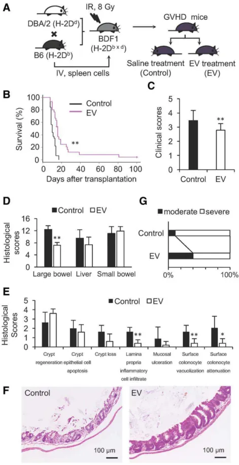
cells. One group of GVHD mice was treated with human BM-MSC-derived EVs, and the other group of mice received saline injections (Fig. 2A). GVHD mice that received systemic infu-sion of EVs showed prolonged survival, with a median survival of 16 days compared with 10 days in the control GVHD mice (Fig. 2B). The clinical scores of EV-treated mice were lower than those of the control mice (average of 2.8 vs. 3.5, Fig. 2C), indicating that EVs ameliorated the systemic symptoms of acute GVHD such as body weight loss. EV treatment also sig-nificantly mitigated GVHD-associated pathology in the large bowel, particularly the infiltration of the lamina propria by inflammatory cells and the vacuolization and attenuation of surface colonocytes (Fig. 2D–2F). The histological scores of the livers of the EV-treated GVHD mice were lower than those of the control mice, but this did not reach statistical significance, and the histological scores of the small bowels of the EV-treated mice were comparable to those of the control mice (Fig. 2D, Supporting Information Fig. S4B; Supporting Informa-tion Table S3). With regard to the skin, the percentage of mice with severe damage was lower in the EV-treated group than in the control group (60.0% vs. 88.8%) (Fig. 2G, Support-ing Information Fig. S4B; SupportSupport-ing Information Table S4). Collectively, systemic administration of human BM-MSC-derived EVs significantly prolonged the survival of mice with acute GVHD, which was accompanied by an amelioration of clinical GVHD symptoms and pathological damage in multiple GVHD-targeted organs, particularly the large bowel.
Human BM-MSC-Derived EVs Suppress the Functional
Differentiation of T Cells in Mice with GVHD
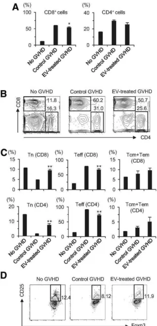
PB T cell subsets were analyzed in GVHD mice that received sys-temic administration of EVs. Flow cytometric analysis indicated that the expansion of CD81T cells was suppressed by treat-ment with human BM-MSC-derived EVs (Fig. 3A, 3B). In the con-trol GVHD mice, most of the CD81 and CD41 T cells were CD62L-CD441effector T cells (Fig. 3C, Supporting Information Fig. S5B, 5D). In GVHD mice treated with EVs, the frequency and number of effector T cells among CD41and CD81T cells were decreased, and conversely, the frequency and number of CD62L1CD44- naive T cells were increased (Fig. 3C, Supporting Information Fig. S5A–5D). These results suggest that systemic administration of human BM-MSC-derived EVs suppressed the functional differentiation of T cells from a naive phenotype to an effector phenotype. The frequency of CD41CD251Foxp31 Treg was also decreased in the GVHD control mice compared to mice without GVHD, whereas the EV-treated GVHD mice contained a substantial CD41CD251Foxp31population (Fig. 3D), suggesting that human BM-MSC-derived EVs preserve Treg. The populations of non-T cells, including CD191Gr-1- cells (B cells), Gr-1-CD11b1cells (monocytes), and Gr-11CD11b1 cells (neutrophils), were depleted in the control GVHD mice compared to mice without GVHD, but EV treatment ameliorated the depletion of these cell populations (Supporting Information Fig. S6).
NHDF-Derived EVs Do Not Suppress T Cell Expansion
In Vitro or Ameliorate Acute GVHD in Mice
We explored whether the anti-GVHD effects of BM-MSC-derived EVs could be induced by EVs BM-MSC-derived from other stromal cells. To address this, we used NHDFs because they are adherent stromal cells with a similar morphology to human BM-MSCs.
438 Human BM-MSC-Derived EVs Ameliorate GVHD
VC 2017 The AuthorsSTEMCELLSpublished by STEMCELLS
Flow cytometric analysis showed that NHDFs were negative for hematopoietic and endothelial markers, including CD45, CD34, CD11b, CD19, CD31, and HLA-DR, and were positive for mesen-chymal stem cell-associated markers, including CD105, CD73,
CD90, CD44, and CD166, which was a similar expression pattern as BM-MSCs (Supporting Information Fig. S7A, 7B). On the other hand, NHDFs were distinct from BM-MSCs in the expression of CD10, CD106, and CD146 (Supporting Information Fig. S8A). In addition, NHDFs did not show apparent multi-differentiation capability to osteogenic (Supporting Information Fig. S8B, 8C), chondrogenic (Supporting Information Fig. S8D), or adipogenic (Supporting Information Fig. S8E, 8F) cells in vitro. Moreover, unlike BM-MSCs, NHDFs did not form bone or induce hemato-poietic marrow when subcutaneously transplanted into immu-nocompromised mice (Supporting Information Fig. S8G). Thus, NHDFs were fundamentally different from BM-MSCs.
When anti-CD3/CD28-stimulated human PBMCs were cul-tured with NHDF-derived EVs, the expansion of total T cells, including CD41and CD81T cells, was comparable to that in cultures of PBMCs alone and in cultures with the supernatant fraction generated in the process of EV preparation from con-ditioned SFM of NHDFs (Supporting Information Fig. S9A, 9B). Therefore, NHDF-derived EVs did not have the suppressive effects on T cells seen with BM-MSC-derived EVs.
In accordance with these observations, when MHC-haploidentical GVHD mice received NHDF-derived EVs, the survival of the mice was not prolonged and was actually shortened compared to the control mice (Supporting Informa-tion Fig. S10A). The clinical scores of the GVHD mice treated with NHDF-derived EVs were comparable to those of the con-trol GVHD mice (Supporting Information Fig. S10B). In addi-tion, the histological scores for the large bowel, liver, and small bowel of GVHD mice treated with NHDF-derived EVs were comparable to those of the control GVHD mice (Sup-porting Information Fig. S10C). With regard to the skin, in the mice treated with NHDF-derived EVs, the percentage of mice with severely damaged skin was lower, and conversely, the percentage of mice with mildly or moderately damaged skin was higher compared to the control mice (Supporting Infor-mation Fig. S10D). Flow cytometric analysis of PB T cell sub-sets in GVHD mice indicated that the expansion of CD81 T cells was not suppressed by treatment with NHDF-derived EVs (Supporting Information Fig. S10E). In addition, the frequency of effector T cells, naive T cells, and Treg in GVHD mice treated with NHDF-EVs was comparable to that in the control GVHD mice (Supporting Information Fig. S10E, 10F). Collec-tively, these data indicate that systemic administration of NHDF-derived EVs had no significant effect on the functional differentiation of T cells from a naive phenotype to an effec-tor phenotype or on the preservation of Treg. These data sug-gest that the immunologic and anti-GVHD effects of human BM-MSC-derived EVs are unique, at least in comparison to NHDF-derived EVs.
Proliferation-Related Gene Signatures Are
Downregulated by microRNAs in Human
BM-MSC-Derived EVs
The expression of multiple soluble factors such as cytokines, chemokines, and growth factors in NHDF-derived and BM-MSC-derived EVs was compared (Supporting Information Fig. S11). Differences in the expression of several molecules, including macrophage migration inhibiting factor, interleukin (IL)-13, plasminogen activator inhibitor-1, and CXCL12, were observed.
Figure 3. Human bone marrow mesenchymal stromal/stem cell (BM-MSC)-derived EVs preserve peripheral naive T cells and Treg in mice with GVHD. Flow cytometric analysis of T cell subsets in PB from the GVHD mice 6 days after the administration of EVs. (A): The frequency of CD81and CD41T cells. (B): Representa-tive contour plots are shown. (C): The frequency of naive (Tn; CD62L1CD44-), effector (Teff; CD62L-CD441), and central mem-ory and effector memmem-ory (Tcm and Tem; CD62L1CD441) T cells out of total CD81and CD41T cells.(D):Flow cytometric analysis of Treg. The numbers indicate the percentage of Treg out of total CD41T cells in the PB. No GVHD, BDF1 mice that did not receive spleen cells from B6 mice; Control GVHD, BDF1 mice that received spleen cells from B6 mice (H-2Db) but did not receive human BM-MSC-derived EVs; EV-treated GVHD, BDF1 mice that received spleen cells from B6 mice and then received human BM-MSC-derived EVs. Data in (A, C) indicate the mean6SD. **,
p<.01; *, p<.05. Abbreviations: EVs, extracellular vesicles; GVHD, graft-versus-host disease.
440 Human BM-MSC-Derived EVs Ameliorate GVHD
VC 2017 The AuthorsSTEMCELLSpublished by STEMCELLS
Finally, microarray analysis of microRNAs in BM-MSC-derived EVs was performed (Supporting Information Fig. S12). This analysis identified 336 and 337 microRNAs that were upregulated or downregulated, respectively, in human BM-MSC-derived EVs compared to NHDF-derived EVs (Fig. 4A, Supporting Information S-File 1). Among them, miR-125a-3p was identified as the most highly upregulated microRNA in BM-MSC-derived EVs (Fig. 4B). TargetScan analysis predicted 11726 mRNAs as targets of the top 10 upregulated microRNAs (Supporting Information S-File 2). Among them, 3477 mRNAs
were commonly targeted by three or more microRNAs and were selected for further analysis. GO enrichment analysis showed that these commonly targeted genes were highly involved in the regulation of proliferation-related processes (Table 1). A similar analysis was performed on 12553 mRNAs that were predicted as targets of the top 10 downregulated microRNAs (Fig. 4B, Supporting Information S-File 3). Among them, 4356 mRNAs were commonly targeted by three or more microRNAs, and these mRNAs were found to be highly involved in inhibition of proliferation by GO enrichment Figure 4. Microarray analysis of microRNAs in BM-MSC-derived EVs versus NHDF-derived EVs.(A):Scatter plot of microRNAs expressed in BM-MSC-derived EVs compared with NHDF-derived EVs. Blue lines indicate the 2.0-fold threshold. Circles represent the top 10 upre-gulated microRNAs in BM-MSC-derived EVs. Squares represent the top 10 downreupre-gulated microRNAs in BM-MSC-derived EVs.(B):List of the top 10 microRNAs that were up- or downregulated in BM-MSC-derived EVs. The color of the numbers corresponds to the color of the circles or squares in the scatter plot in (A). (C):Kyoto Encyclopedia of Genes and Genomes pathway analysis of mRNAs that were commonly targeted by three or more microRNAs among the top 10 up- or downregulated microRNAs. Abbreviations: BM-MSC, bone marrow mesenchymal stromal/stem cell; EVs, extracellular vesicles; NHDF, normal human dermal fibroblasts.
Fujii, Miura, Fujishiro et al. 441
analysis (Table 1). Collectively, these analyses suggest that microRNAs within BM-MSC-derived EVs downregulate cell pro-liferation. Furthermore, KEGG pathway analysis identified gene groups involved in cell cycle regulation, T cell receptor signaling, and GVHD (Fig. 4C). These pathways were consid-ered to be downregulated because they include genes tar-geted by commonly upregulated microRNAs.
D
ISCUSSIONA series of clinical studies have examined the efficacy of sys-temic infusion of culture-expanded BM-MSCs for acute GVHD patients and shown overall response rates ranging from about 30%–80% [36, 37]. Attempts to improve the outcome of BM-MSC therapy have been based on the concept that this form of therapy is dependent on the number of infused cells that can successfully traffic to sites of damaged or diseased tissue [38]. However, systemic administration of an increased num-ber of cells did not augment the therapeutic effects of BM-MSCs in GVHD [39]. In addition, another approach, in which BM-MSCs were directly delivered into the gut via the mesen-teric artery, was not more effective than systemic injection [40]. These clinical results underscore the current molecular understanding that the therapeutic effects of BM-MSCs are, at least in acute GVHD, attributed mainly to secreted immune-modulatory factors [9, 41]. In the present study, we found, for the first time, that human BM-MSC-derived EVs ameliorated GVHD symptoms and pathology in GVHD-targeted organs, and prolonged the survival of GVHD mice in association with preservation of circulating naive T cell populations.
BM-MSCs suppress the expansion of human naive T cells as well as activated CD41 and CD81 T cells regardless of MHC compatibility [41, 42]. It has also been reported that BM-MSCs induce human Treg, which inhibit allogeneic lym-phocyte proliferation [43, 44]. In line with these reports, we found that BM-MSC-derived EVs suppressed the proliferation of CD41 and CD81 T cells in anti-CD3/CD28-stimulated expansion cultures of human PBMCs and in PB obtained from the GVHD mice. In addition, BM-MSC-derived EVs suppressed the functional differentiation of T cells from a naive to an
effector phenotype in the GVHD mice. Moreover, naive Treg were preserved in the presence of BM-MSC-derived EVs in cultures of anti-CD3/CD28-stimulated human PBMCs and in PB of the GVHD mice. Collectively, it is conceivable that the anti-GVHD effect of BM-MSCs is, at least partially, associated with an EV-mediated immunomodulatory effect on T cells.
However, a number of studies suggest that MSC-derived EVs mimic the therapeutic effects of their MSCs of their origin to a large extent, even though the underlying mechanisms may not necessarily be similar [13]. In our in vitro study, BM-MSCs suppressed the expansion of CD31 T cells to a lesser extent than EVs derived from the same number of the BM-MSCs from which they originated. BM-MSC-derived EVs may, therefore, have a stronger effect on T cell suppression than BM-MSCs. This is supported by the observation that the sup-pressive effect of EVs on T cell expansion was dose-dependent (data not shown). With respect to the Foxp3-expressing T cell population, the suppression of CD45RA-Foxp3low cells (non-Treg) by BM-MSC-derived EVs might reflect the suppression of activated CD41 T cells. Given that CD45RA1Foxp3low naive Treg become CD45RA-Foxp3high Treg upon anti-CD3/CD28 stimulation, it is possible that BM-MSC-derived EVs had the enhanced suppressive effect on the ability of Treg to shift from a naive to an activated status or that BM-MSC-derived EVs induced more naive Treg within the CD41 T cell popula-tion compared to BM-MSCs. Previous in vitro studies on the modulation of functional T cell populations by BM-MSCs have been contradictory [45–47]. As for in vivo studies, a recent report suggested that EVs contribute to the suppression of Th1 cells in a human-to-mouse xenogeneic chronic GVHD model [48]. Further studies are needed to determine the con-tribution of EVs to the BM-MSC-mediated T cell modulation.
Various types of cells release EVs. To address the unique effects of human BM-MSC-derived EVs, we used NHDF-derived EVs as a control because human BM-MSCs and fibro-blasts share many properties, including a plastic-adherent spindle-like morphology and a similar surface marker expression profile [49]. Nevertheless, NHDFs are functionally different from BM-MSCs. In our analysis, NHDFs lacked multi-differentiation capacity and hematopoiesis induction. In addition, NHDF-derived EVs did not suppress T cell expansion Table 1.List of enriched GO terms. GO analysis of commonly targeted genes by up- or downregulated microRNAs in bone marrow mes-enchymal stromal/stem cell-derived extracellular vesicles
GO accession GO term (number of genes included)
Corrected
p-value microRNA
0008284 Positive regulation of cell proliferation (93) 4.20 E –16 Upregulated 0008285 Negative regulation of cell proliferation (86) 2.28 E –09 Downregulated
0007049 Cell cycle (79) 2.49 E –06 Upregulated
0008283 Cell proliferation (64) 5.82 E –07 Upregulated
0051301 Cell division (47) 2.11 E –03 Upregulated
0000278 Mitotic cell cycle (43) 1.39 E –02 Upregulated
0000082 G1/S transition of mitotic cell cycle (29) 1.49 E –03 Upregulated
0007067 Mitosis (29) 1.42 E –02 Upregulated
0007050 Cell cycle arrest (28) 6.70 E –03 Downregulated
0030307 Positive regulation of cell growth (13) 6.91 E –03 Upregulated 0051781 Positive regulation of cell division (13) 2.43 E –03 Upregulated 0045786 Negative regulation of cell cycle (11) 1.48 E –02 Downregulated 0045787 Positive regulation of cell cycle (8) 8.83 E –03 Upregulated 0045930 Negative regulation of mitotic cell cycle (5) 4.67 E –02 Downregulated 0071158 Positive regulation of cell cycle arrest (5) 3.04 E –02 Downregulated
Abbreviation: GO, Gene Oncology.
442 Human BM-MSC-Derived EVs Ameliorate GVHD
VC 2017 The AuthorsSTEMCELLSpublished by STEMCELLS
in cultures with anti-CD3/CD28-stimulated PBMCs. Most importantly, NHDF-derived EVs did not exhibit the anti-GVHD effects of BM-MSC-derived EVs, including amelioration of clini-cal symptoms, resolution of the pathology in the gut and liver, or prolongation of survival. These results suggest that BM-MSC-derived EVs have a unique therapeutic effect in acute GVHD. Interestingly, systemic infusion of human BM-MSC-derived EVs was effective in resolving the pathology in the large bowel and skin of the GVHD mice. This is consistent with the clinical characteristics of BM-MSC therapy for acute GVHD [7, 8, 50, 51]. The mechanisms that underlie the differ-ences in the efficacy of BM-MSC-derived EVs among organs/ tissues have not been well-studied.
There are limitations to this study. First, the effects of BM-MSC-derived EVs on GVHD-targeted organs were modest. Only the suppression of GVHD pathology in the large intestine was sig-nificantly associated with clinical improvement in GVHD mice treated with BM-MSC-derived EVs. Although multiple organs can be affected in GVHD, pathologic damage to the large intestine can be lethal; therefore, favorable effects of BM-MSC-derived EVs on the large intestine might predominantly contribute to the improved clinical outcomes. Second, the survival of GVHD mice treated with NHDF-derived EVs was less than that of the control GVHD mice, despite the lack of effects on PB T cells. This suggests the possibility that the distribution of T cell subsets in the PB does not reflect the distribution in GVHD-targeted organs.
Comprehensive characterization of EVs would provide the information necessary to explore their therapeutic potential [52]. A previous study by Phinney et al. reported the microRNA expres-sion profiles of MSC-derived exosomes [53]. Because they com-pared microRNA expression levels between exosomes and their parental MSCs, the most highly up- or downregulated microRNAs were different from the most highly regulated microRNAs in our analysis. However, it is of interest that such microRNAs in that study were also upregulated in EVs in our analysis. MicroRNAs that are expressed differentially in EVs compared with the parental MSCs might contribute to MSC-associated functions.
Microarray analysis of microRNAs in BM-MSC-derived EVs versus NHDF-derived EVs revealed the differential expression of multiple microRNAs, suggesting the involvement of multiple biological processes. Previous studies have demonstrated that BM-MSCs inhibit the proliferation of activated T cells via cell cycle arrest [54, 55] and modulate the expression of various cytokines, including proinflammatory (e.g., IL-2, IFN-g, TNF-a) and anti-inflammatory cytokines (e.g., IL-4, IL-10), which play important roles in the pathogenesis of acute GVHD [6, 9, 56–59]. The GO enrichment analysis identified the down-regulation of multiple cell proliferation-related processes but not altered production of these cytokines. Furthermore, KEGG analysis suggested the downregulation of pathways associated with GVHD and T cell receptor signaling. These comprehensive analyses support our in vitro and in vivo results, where T cell expansion was suppressed by BM-MSC-derived EVs and acute GVHD was suppressed clinically and pathologically in GVHD mice.
Of the microRNAs that were differentially expressed in human BM-MSC-derived EVs and NHDF-derived EVs, miR-125a-3p was the most highly altered microRNA. Notably, miR-125a-miR-125a-3p suppressed cell proliferation in several cell lines, consistent with our in vitro observations [60–62]. It has also been shown that miR-125a is downregulated in PBMCs from patients with systemic
lupus erythematosus [63]. The downregulation of miR-125a con-tributed to the elevated expression of the inflammatory chemo-kine regulated upon activation, normal T cell expressed and secreted by targeting Kruppel-like factor 13 in activated T cells [64]. Therefore, the upregulation of miR-125a-3p in BM-MSC-derived EVs might contribute to the suppression of T cells. Collino et al. performed microarray analysis of microRNAs contained in EVs released from both BM-MSCs and liver-resident stem cells [65]. Although miR-125a was not specifically discussed by the authors, it was one of the most abundantly expressed microRNAs in microvesicles and their cells of origin. Their analysis indicated that abundantly expressed microRNAs in BM-MSCs contributed to multiple cell processes, including regulation of the immune system, differentiation-related functions, and processes related to the regulation of cell cycle progression, proliferation, and cell death. One microRNA can regulate many target genes, while con-versely one gene can be targeted by many microRNAs; thus mul-tiple relationships between microRNAs and target genes elaborately determine the fate of T cells. It will be important to use a conditional microRNA-125a-3p knockout mouse model to more conclusively demonstrate the association between upregu-lated microRNA-125a-3p and the anti-GVHD effect.
Array analysis of multiple soluble factors identified several molecules that were more highly expressed in BM-MSC-derived EVs than in NHDF-BM-MSC-derived EVs. Among them, CXCL12 could potentially be associated with the amelioration of GVHD, as was reported in a previous study [66]. EVs contain a variety of bioactive molecules, and further analysis of immune-modulatory factors is warranted to fully understand the anti-GVHD mechanism of BM-MSC-derived EVs.
Use of BM-MSC-derived EVs may have potential advan-tages over their cell of origin in some aspects. From a safety standpoint, concerns about the transformation of BM-MSCs to malignant cells in patients do not need to be considered with the use of EVs. From a therapeutic standpoint, BM-MSCs may become trapped in capillaries, particularly in the lungs, and become unable to reach damaged or diseased tissues/organs, which is necessary for them to exert their therapeutic effects. This is also not a concern with EVs. On the other hand, BM-MSCs are activated by proinflammatory cytokines present in the microenvironment of damaged or diseased tissues/organs [67]. This implies that the immunomodulatory effects of BM-MSCs might not be exerted in the absence of abnormal envi-ronmental conditions. Our study suggests that BM-MSC-derived EVs have therapeutic effects in acute GVHD. However, the EVs showed strong suppression of T cell activation in vitro, and extreme immunosuppression by BM-MSC-derived EVs may increase susceptibility to infection in a clinical setting. In addition, because EVs are small, spherical membrane frag-ments and may include exosomes, microvesicles, and apopto-tic bodies [33], there is a possibility that the BM-MSC-derived EV-concentrated fractions in this study contain the above par-ticles [68]. Therefore, additional research incorporating more homogenous EV preparations is warranted.
C
ONCLUSIONHuman BM-MSC-derived EVs exerted immunomodulatory effects on T cells in vitro and anti-GVHD effects in mice. Amelioration of acute GVHD by BM-MSC-derived EVs was associated with the
Fujii, Miura, Fujishiro et al. 443
preservation of circulating naive T cells. Microarray analysis iden-tified microRNAs associated with the immunomodulatory effects of BM-MSC-derived EVs. Further studies are needed to increase the potency of BM-MSC-derived EVs and to better understand the mechanisms of action of this novel potential therapeutic for acute GVHD.
A
CKNOWLEDGMENTSWe thank Yoko Nakagawa (Kyoto University) and Ikuko Fukuba (Hiroshima University) for their excellent technical assistance. We also thank Drs. Mariko Ikuo and Shigeyuki Teranishi (Hiro-shima University) for their EV analysis. This work was sup-ported in part by Grants-in-Aid from the Ministry of Education, Culture, Sports, Science, and Technology of Japan (#15K09453, #16K07171, and #17H04264 to Y.M., T.M., and T.I., respectively) and from the Japan Agency for Medical Research and Development (AMED) (17ek0510022s0601, to T.I.). This work was also supported in part by the Program of the Network-Type Joint Usage/Research Disaster Medical Sci-ence of Hiroshima University, Nagasaki University, and Fukush-ima Medical University (Y.M., S.F., and T.I.).
A
UTHORC
ONTRIBUTIONSS.F., A.F., T.S., and Y.S.: conception and design, collection of study material, collection and assembly of data, data analysis and inter-pretation, manuscript writing; Y.M.: conception and design, finan-cial support, collection of study material, collection and assembly of data, data analysis and interpretation, manuscript writing; H.H.: conception and design, collection of study material, assem-bly of data, data analysis and interpretation, manuscript writing; H.T.: collection and assembly of data, data analysis and interpreta-tion; A.T.-K., T.I., and T.M.: conception and design, financial sup-port, administrative supsup-port, provision of study material, data analysis and interpretation, manuscript writing; S.F., A.F., T.S., Y.S., Y.M., H.H., H.T., A.T.-K., T.I., and T.M.: final approval of manuscript.
D
ISCLOSURE OFP
OTENTIALC
ONFLICTS OFI
NTERESTH.H. received research funding from Kyowa Hakko Kirin and Novartis Pharma. H.T. is a founder and the chairman/chief exec-utive officer of MiRTeL Co. LTD., and owns stock in MiRTeL Co. LTD. T.M. received research funding from Bristol-Meyers Squibb. The other authors indicated no potential conflicts of interest.
R
EFERENCES1 Blazar BR, Murphy WJ, Abedi M. Advan-ces in graft-versus-host disease biology and therapy. Nat Rev Immunol 2012;12:443–458.
2 Deeg HJ. How I treat refractory acute GVHD. Blood 2007;109:4119–4126.
3 Martin PJ, Rizzo JD, Wingard JR et al. First- and second-line systemic treatment of acute graft-versus-host disease: Recommen-dations of the American Society of Blood and Marrow Transplantation. Biol Blood Marrow Transplant 2012;18:1150–1163.
4 Lazarus HM, Haynesworth SE, Gerson SL et al. Ex vivo expansion and subsequent infu-sion of human bone marrow-derived stromal progenitor cells (mesenchymal progenitor cells): Implications for therapeutic use. Bone Marrow Transplant 1995;16:557–564.
5 Pittenger MF, Mackay AM, Beck SC et al. Multilineage potential of adult human mesenchymal stem cells. Science 1999;284: 143–147.
6 Aggarwal S, Pittenger MF. Human mes-enchymal stem cells modulate allogeneic immune cell responses. Blood 2005;105: 1815–1822.
7 Le Blanc K, Frassoni F, Ball L et al. Mes-enchymal stem cells for treatment of steroid-resistant, severe, acute graft-versus-host dis-ease: A phase II study. Lancet 2008;371: 1579–1586.
8 Muroi K, Miyamura K, Okada M et al. Bone marrow-derived mesenchymal stem cells (JR-031) for steroid-refractory grade III or IV acute graft-versus-host disease: A phase II/III study. Int J Hematol 2016;103:243–250.
9 Le Blanc K, Mougiakakos D. Multipotent mesenchymal stromal cells and the innate immune system. Nat Rev Immunol 2012;12: 383–396.
10 Raposo G, Stoorvogel W. Extracellular vesicles: Exosomes, microvesicles, and friends. J Cell Biol 2013;200:373–383.
11 Pitt JM, Kroemer G, Zitvogel L. Extracellular vesicles: Masters of intercellular communication and potential clinical interventions. J Clin Invest 2016;126:1139–1143.
12 El Andaloussi S, M€ager I, Breakefield XO et al. Extracellular vesicles: Biology and emerging therapeutic opportunities. Nat Rev Drug Discov 2013;12:347–357.
13 Phinney DG, Pittenger MF. Concise Review: MSC-derived exosomes for cell-free therapy. STEMCELLS2017;35:851–858. 14 Lai RC, Arslan F, Lee MM et al. Exosome secreted by MSC reduces myocardial ische-mia/reperfusion injury. Stem Cell Res 2010;4: 214–222.
15 Bruno S, Grange C, Deregibus MC et al. Mesenchymal stem cell-derived microvesicles protect against acute tubular injury. J Am Soc Nephrol 2009;20:1053–1067.
16 Doeppner TR, Herz J, G€orgens A et al. Extracellular vesicles improve post-stroke neuroregeneration and prevent postischemic immunosuppression. STEM CELLS TRANSLATIONAL
MEDICINE2015;4:1131–1143.
17 Lee C, Mitsialis SA, Aslam M et al. Exo-somes mediate the cytoprotective action of mesenchymal stromal cells on hypoxia-induced pulmonary hypertension. Circulation 2012;126:2601–2611.
18 Zhu YG, Feng XM, Abbott J et al. Human mesenchymal stem cell microvesicles for treatment of Escherichia coli endotoxin-induced acute lung injury in mice. STEMCELLS
2014;32:116–125.
19 Zhang B, Wang M, Gong A et al. HucMSC-exosome mediated-Wnt4 signaling is required for cutaneous wound healing. STEM
CELLS2015;33:2158–2168.
20 Kordelas L, Rebmann V, Ludwig AK et al. MSC-derived exosomes: A novel tool to treat therapy-refractory graft-versus-host disease. Leukemia 2014;28:970–973.
21 Yoshioka S, Miura Y, Yao H et al. CCAAT/ enhancer-binding protein b expressed by
bone marrow mesenchymal stromal cells reg-ulates early B-cell lymphopoiesis. STEM CELLS
2014;32:730–740.
22 Sugino N, Miura Y, Yao H et al. Early osteoinductive human bone marrow mesen-chymal stromal/stem cells support an enhanced hematopoietic cell expansion with altered chemotaxis- and adhesion-related gene expression profiles. Biochem Biophys Res Commun 2016;469:823–829.
23 Dominici M, Le Blanc K, Mueller I et al. Minimal criteria for defining multipotent mesenchymal stromal cells. The International Society for Cellular Therapy position state-ment. Cytotherapy 2006;8:315–317.
24 Yao H, Miura Y, Yoshioka S et al. Parathy-roid hormone enhances hematopoietic expansion via upregulation of cadherin-11 in bone marrow mesenchymal stromal cells. STEMCELLS2014;32:2245–2255.
25 Iwasa M, Miura Y, Fujishiro A et al. Bor-tezomib interferes with adhesion of B cell precursor acute lymphoblastic leukemia cells through SPARC up-regulation in human bone marrow mesenchymal stromal/stem cells. Int J Hematol 2017;105:587–597.
26 Maas SL, De Vrij J, Broekman ML. Quantification and size-profiling of extracellu-lar vesicles using tunable resistive pulse sens-ing. J Vis Exp 2014;92:e51623.
27 Murai M, Yoneyama H, Ezaki T et al. Peyer’s patch is the essential site in initiating murine acute and lethal graft-versus-host reaction. Nat Immunol 2003;4:154–160.
28 Cooke KR, Kobzik L, Martin TR et al. An experimental model of idiopathic pneumonia syndrome after bone marrow transplanta-tion: I. The roles of minor H antigens and endotoxin. Blood 1996;88:3230–3239.
29 Hill GR, Cooke KR, Teshima T et al. Inter-leukin-11 promotes T cell polarization and prevents acute graft-versus-host disease after allogeneic bone marrow transplantation. J Clin Invest 1998;102:115–123.
444 Human BM-MSC-Derived EVs Ameliorate GVHD
VC 2017 The AuthorsSTEMCELLSpublished by STEMCELLS
30 Wu H, Allan AE, Harrist TJ. Noninfectious vesiculobullous and vesiculopustular diseases. In: Elder DE, ed. Lever’s Histopathology of the Skin. 11th ed. Philadelphia, PA: Wolters Kluwer, 2015:315–317.
31 Takashima T, Kadowaki M, Aoyama K et al. The Wnt agonist R-spondin1 regulates systemic graft-versus-host disease by protect-ing intestinal stem cells. J Exp Med 2011; 208:285–294.
32 Kanda Y. Investigation of the freely avail-able easy-to-use software ‘EZR’ for medical statistics. Bone Marrow Transplant 2013;48: 452–458.
33 L€otvall J, Hill AF, Hochberg F et al. Mini-mal experimental requirements for definition of extracellular vesicles and their functions: A position statement from the International Society for Extracellular Vesicles. J Extracell Vesicles 2014;3:26913.
34 Nakai W, Yoshida T, Diez D et al. A novel affinity-based method for the isolation of highly purified extracellular vesicles. Sci Rep 2016;6:33935.
35 Le Blanc K, Tammik L, Sundberg B et al. Mesenchymal stem cells inhibit and stimulate mixed lymphocyte cultures and mitogenic responses independently of the major histo-compatibility complex. Scand J Immunol 2003;57:11–20.
36 Kaipe H, Erkers T, Sadeghi B et al. Stro-mal cells–are they really useful for GVHD? Bone Marrow Transplant 2014;49:737–743.
37 Hashmi S, Ahmed M, Murad MH et al. Survival after mesenchymal stromal cell ther-apy in steroid-refractory acute graft-versus-host disease: Systematic review and meta-analysis. Lancet Haematol 2016;3:e45–e52.
38 Sugino N, Ichinohe T, Takaori-Kondo A et al. Pharmacological targeting of bone mar-row mesenchymal stromal/stem cells for the treatment of hematological disorders. Inflamm Regen 2017;37:7.
39 Kebriaei P, Isola L, Bahceci E et al. Adult human mesenchymal stem cells added to corticosteroid therapy for the treatment of acute graft-versus-host disease. Biol Blood Marrow Transplant 2009;15:804–811.
40 Arima N, Nakamura F, Fukunaga A et al. Single intra-arterial injection of mesenchymal stromal cells for treatment of steroid-refractory acute graft-versus-host disease: A pilot study. Cytotherapy 2010;12:265–268.
41 Di Nicola M, Carlo-Stella C, Magni M et al. Human bone marrow stromal cells sup-press T-lymphocyte proliferation induced by cellular or nonspecific mitogenic stimuli. Blood 2002;99:3838–3843.
42 Tse WT, Pendleton JD, Beyer WM et al. Suppression of allogeneic T-cell proliferation by human marrow stromal cells: Implications in transplantation. Transplantation 2003;75: 389–397.
43 Maccario R, Podesta M, Moretta A et al. Interaction of human mesenchymal stem cells with cells involved in alloantigen-specific immune response favors the differentiation of CD41 T-cell subsets expressing a regula-tory/suppressive phenotype. Haematologica 2005;90:516–525.
44 Melief SM, Schrama E, Brugman MH et al. Multipotent stromal cells induce human regulatory T cells through a novel pathway involving skewing of monocytes toward anti-inflammatory macrophages. STEM
CELLS2013;31:1980–1991.
45 Blazquez R, Sanchez-Margallo FM, de la Rosa O et al. Immunomodulatory potential of human adipose mesenchymal stem cells derived exosomes on in vitro stimulated T cells. Front Immunol 2014;5:556.
46 Conforti A, Scarsella M, Starc N et al. Microvescicles derived from mesenchymal stromal cells are not as effective as their cel-lular counterpart in the ability to modulate immune responses in vitro. Stem Cell Dev 2014;23:2591–2599.
47 Gouveia de Andrade AV, Bertolino G, Riewaldt J et al. Extracellular vesicles secreted by bone marrow- and adipose tissue-derived mesenchymal stromal cells fail to suppress lymphocyte proliferation. Stem Cells Dev 2015;24:1374–1376.
48 Amarnath S, Foley JE, Farthing DE et al. Bone marrow-derived mesenchymal stromal cells harness purinergenic signaling to toler-ize human Th1 cells in vivo. STEMCELLS2015; 33:1200–1212.
49 Kundrotas G. Surface markers distin-guishing mesenchymal stem cells from fibro-blasts. Acta Med. Litu. 2012;19:75–79.
50 Prasad VK, Lucas KG, Kleiner GI et al. Efficacy and safety of ex vivo cultured adult human mesenchymal stem cells (Prochymal) in pediatric patients with severe refractory acute graft-versus-host disease in a compas-sionate use study. Biol Blood Marrow Trans-plant 2011;17:534–541.
51 Kurtzberg J, Prockop S, Teira P et al. Allogeneic human mesenchymal stem cell therapy (remestemcel-L, Prochymal) as a res-cue agent for severe refractory acute graft-versus-host disease in pediatric patients. Biol Blood Marrow Transplant 2014;20:229–235.
52 Katsuda T, Kosaka N, Takeshita F et al. The therapeutic potential of mesenchymal stem cell-derived extracellular vesicles. Prote-omics 2013;13:1637–1653.
53 Phinney DG, Di Giuseppe M, Njah J et al. Mesenchymal stem cells use extracellu-lar vesicles to outsource mitophagy and shut-tle microRNAs. Nat Commun 2015;6:8472.
54 Glennie S, Soeiro I, Dyson PJ et al. Bone marrow mesenchymal stem cells induce divi-sion arrest anergy of activated T cells. Blood 2005;105:2821–2827.
55 Cuerquis J, Romieu-Mourez R, Francois M et al. Human mesenchymal stromal cells transiently increase cytokine production by activated T cells before suppressing T-cell proliferation: Effect of interferon-gamma and tumor necrosis factor-alpha stimulation. Cytotherapy 2014;16:191–202.
56 Ferrara JL, Cooke KR, Teshima T. The pathophysiology of acute graft-versus-host disease. Int J Hematol 2003;78:181–187.
57 Sotiropoulou PA, Perez SA, Gritzapis AD et al. Interactions between human mesen-chymal stem cells and natural killer cells. STEMCELLS2006;24:74–85.
58 Krampera M, Cosmi L, Angeli R et al. Role for interferon-gamma in the immuno-modulatory activity of human bone marrow mesenchymal stem cells. STEM CELLS 2006;24:
386–398.
59 Itamura H, Shindo T, Tawara I et al. The MEK inhibitor trametinib separates murine host disease from graft-versus-tumor effects. JCI Insight 2016;1:e86331.
60 Jiang L, Huang Q, Zhang S et al. Hsa-miR-125a-3p and hsa-miR-125a-5p are downregu-lated in non-small cell lung cancer and have inverse effects on invasion and migration of lung cancer cells. BMC Cancer 2010;10:318.
61 Hashiguchi Y, Nishida N, Mimori K et al. Down-regulation of miR-125a-3p in human gastric cancer and its clinicopathological sig-nificance. Int J Oncol 2012;40:1477–1482.
62 Ninio-Many L, Grossman H, Shomron N et al. microRNA-125a-3p reduces cell prolifer-ation and migrprolifer-ation by targeting Fyn. J Cell Sci 2013;126:2867–2876.
63 Wang H, Peng W, Ouyang X et al. Circu-lating microRNAs as candidate biomarkers in patients with systemic lupus erythematosus. Transl Res 2012;160:198–206.
64 Zhao X, Tang Y, Qu B et al. MicroRNA-125a contributes to elevated inflammatory chemokine RANTES levels via targeting KLF13 in systemic lupus erythematosus. Arthritis Rheum 2010;62:3425–3435.
65 Collino F, Deregibus MC, Bruno S et al. Microvesicles derived from adult human bone marrow and tissue specific mesenchy-mal stem cells shuttle selected pattern of miRNAs. PLoS ONE 2010;5:e11803.
66 Gauthier SD, Leboeuf D, Manuguerra-Gagne R et al. Stromal-derived factor-1aand interleukin-7 treatment improves homeo-static proliferation of na€ıve CD4(1) T cells after allogeneic stem cell transplantation. Biol Blood Marrow Transplant 2015;21:1721– 1731.
67 Ren G, Chen X, Dong F et al. Concise Review: Mesenchymal stem cells and transla-tional medicine: Emerging issues. STEM CELLS
TRANSLATIONALMEDICINE2012;1:51–58.
68 Zeringer E, Li M, Barta T et al. Methods for the extraction and RNA profiling of exo-somes. World J Methodol 2013;3:11–18
See www.StemCells.com for supporting information available online.
Fujii, Miura, Fujishiro et al. 445