Authors Anniek De Witte Elaine Lea-Chou Jim Collins
Agilent Technologies Santa Clara, CA
A G A G A A G G A A A A A A
G A A A G A
C C C C C C C
C C C C C C C C A A G
T T T T T T T T T T
T T G T T T A A T T T G T G
GENOMICS
I N F O R M A T I C S P R O T E O M I C S M E T A B O L O M I C SCopy Number Analysis of Archival FFPE
Tumor Samples by Oligo Array CGH
Abstract
Genomic instability is a classic hallmark of cancer and genetic disorders. Microarray-based comparative genomic hybridization (aCGH) has become the accepted technique for studying DNA sequence copy number alterations. While DNA extracted from freshly frozen tissues is optimal for aCGH analysis, in many cases only formalin-fixed, paraffin-embedded (FFPE) tissue material is available. Because DNA extracted from FFPE tissue is often degraded or damaged, these samples present a significant challenge to label and subsequently analyze. The labeled targets from these samples are often short in length with low specific activity and, as a result, can generate noisy aCGH data. To address this critical issue, we have developed an FFPE labeling protocol for Agilent’s high-performance CGH microarray platform that produces exceptional data quality from a variety of FFPE tissues. This protocol is based on Kreatech’s Universal Linkage System (ULS™) technology, a non-enzymatic direct labeling methodology, and can be used with degraded FFPE tissue-derived DNA. This new labeling technology also features a simple, single-tube protocol, reducing cost per sample and experimental time. When used on the Agilent inkjet-printed array platform, DNA from a variety of archived FFPE tumor samples yields precise, robust, and high-quality aCGH data.
Introduction
In recent years, array-based Comparative Genomic Hybridization (aCGH) has evolved to analyze chromosomal changes at progressively higher resolutions (Barrett et al., 2004). Improvements in array quality and data analysis have encouraged many to reanalyze historical samples. However, challenging samples such as degraded or FFPE tissues have remained inaccessible for aCGH technology. Addressing this issue is critical because FFPE samples represent the largest source of archival biological material available for large retrospective prognostic studies of human cancer. In fact, it is estimated that over 400 million FFPE samples exist to date.
FFPE samples are challenging to use due to cross-linking between nucleic acid strands, DNA adducts with histones or nucleic acid binding proteins, DNA strand breaks, and acid depurination of DNA. The extent of damage increases with the amount of time after exposure to the fixative and is also dependent on the particular fixing protocol employed. The situation is further complicated because there are currently no consistent standards for tissue fixation.
Because enzymatic labeling of DNA samples potentially introduces bias and may further decrease DNA fragment sizes, a one-step chemical labeling method, called the Universal Linkage System (ULS), was chosen to label FFPE-extracted DNA. Its two advantages are labeling independent of fragment length and direct labeling of DNA. This eliminates the need for amplification or other manipulations of the genomic DNA. Additionally, the ULS system is easier to use and results in much higher quality data as compared to enzymatic approaches. For this series of aCGH experiments, a wide variety of FFPE samples from various cancer types (e.g. infiltrating ductal breast carcinoma, large cell lung carcinoma, and colon adenocarcinoma) with differing storage times (up to 10 years) were used. We isolated genomic DNA using an optimized protocol based on the method described by van Beers et al., and the Qiagen DNeasy Blood and Tissue Kit, then determined the degree of DNA degradation by agarose gel electrophoresis. First, using a male/female (XY/XX) model system, DNA isolated from normal tissue FFPE blocks was hybridized against sex-mismatched pooled reference DNA. In this model system, XY/XX ratios for X-chromosome probes have a theoretical value of 0.5 (log2ratio of -1). The log2ratios obtained from the normal FFPE tissues were close to the expected values, verifying the high quality aCGH data obtained using the non-enzymatic labeling protocol. Next, we illustrated that this method is capable of detecting the same amplifications and deletions in matched samples (DNA extracted from FFPE blocks as well as fresh frozen tissue from the same tumor). Then, we demonstrated that the non-enzymatic labeling protocol could produce higher quality data for FFPE samples, compared to the traditional enzymatic labeling protocol. Lastly, the frequent amplification seen in the human epithelial receptor 2 oncogene (HER-2, also known as HER-2/neu or erbB-2) was clearly detected in DNA isolated from 1-, 4-, and
microarrays for routine analysis of archival pathology specimens. This capability allows multilayered and more comprehensive retrospective analysis of sample biobanks and clinical repositories.
Materials and methods DNA isolation and gel analysis
See the Agilent Oligonucleotide Array-Based CGH for Genomic DNA Analysis (for FFPE Samples) user guide (Version 1.0, P/N G4410-90020) available at www.agilent.com/chem/dnamanuals- protocolsfor a detailed description of the protocol used. In short, genomic DNA was isolated from 2-3 20 µm-thick paraffin-embedded tissue sections. After heat
deparaffinization, the tissue was incubated in 1 M sodium thiocyanate overnight at 37°C, followed by Proteinase K treatment for two days at 55°C (fresh aliquots of Proteinase K were added at 17 and 24 hours). After a total Proteinase K incubation of ~44 hours, genomic DNA was isolated using the DNeasy Blood and Tissue Kit (Qiagen P/N 69504). Final elution of DNA was into water. 20 ng of each DNA sample was analyzed on Clear E-Gel 1.2% agarose gels (Invitrogen P/N G5518-01) post stained with SYBR Gold (Invitrogen P/N S11494). Reference genomic DNA from normal male 46, XY and normal female 46, XX was obtained from Promega (P/Ns G1471 and G1521). The reference DNA was heat fragmented for 10 minutes at 95°C.
DNA labeling, hybridization, and washing
The Oligo aCGH Labeling Kit for FFPE Samples (Agilent P/N 5190-0419) was used to chemically label 500 ng of genomic DNA with either ULS-Cy5 or ULS-Cy3 dye for 30 minutes at 85°C. Cy5- and Cy3-labeled samples were purified using Agilent-KREApure™columns (a component of the Oligo aCGH Labeling Kit for FFPE Samples). Purified, labeled samples were then combined and mixed with human Cot-1 DNA (Invitrogen P/N 15279-011), Agilent 10X Blocking Agent, and Agilent 2X Hybridization Solution (both components of the Agilent Oligo aCGH Hybridization Kit P/N 5188-5220). Prior to array hybridization, hybridization mixtures were denatured at 95°C for 3 minutes and incubated at 37°C for 30 minutes. Agilent- CGHblock (a component of the Oligo aCGH Labeling Kit for FFPE Samples) was added and samples were hybridized to the Agilent Human Genome CGH 4 x 44K Microarray (P/N G4426B, AMADID 014950). Each slide contains four identical
sequences with an average spatial resolution of ~35 kb. Hybridization was carried out at 65°C for 40 hours before washing in Agilent Oligo aCGH Wash Buffer 1 (P/N 5188- 5221) at room temperature for 5 minutes, followed by washing in Agilent Oligo aCGH Wash Buffer 2 (P/N 5188-5222) at 37°C for 1 minute.
Microarray scanning and data analysis
Scanning and image analysis were done on an Agilent DNA Microarray Scanner (P/N G2565BA) according to the Agilent Oligonucleotide Array-Based CGH for Genomic DNA Analysis (for FFPE Samples) user guide (Version 1.0, P/N G4410-90020). Feature Extraction Software (Version 9.5) was used for data extraction from raw microarray image files. Agilent CGH Analytics Software (Version 3.4) was used to visualize, detect, and analyze chromosomal patterns within the microarray profiles.
Results
Non-enzymatic labeling of FFPE samples
A comparison of the new non-enzymatic labeling FFPE sample protocol to the traditional enzymatic labeling protocol is outlined in Figure 1A. The non-enzymatic ULS technology is based on the stable binding properties of a platinum complex to DNA (Figure 1B). The ULS labeling system avoids enzyme processivity problems due to crosslinking/complexing that can result in enzymatic biases. Compared to enzymatic labeling, it is independent of fragment length, four times faster, contains fewer steps, and is easier to automate. Multi-pack microarray formats are also very cost effective. Processing time and cost is further reduced in the new non-enzymatic labeling FFPE protocol by replacing the 2-hour restriction enzyme digestion by a 10-minute heat fragmentation of the reference channel DNA.
Figure 1A. Workflow diagram: Non-enzymatic labeling FFPE sample protocol versus traditional enzymatic labeling protocol. Comparison of the new non-enzymatic labeling FFPE sample protocol (on right) versus the traditional enzymatic labeling protocol (on left).
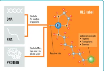
Figure 1B. ULS labeling mechanism. At its reactive site, ULS will react with genomic DNA and label it by formation of a coordinative bond on the N7 position of guanine. A detection moiety (green circle), such as Cy3 or Cy5, is linked via a spacer to the ULS molecule.
ENZYMATIC ULS
+ Agilent-CGHblock 2 hr Restriction Digestion
Random primers + 3’ Denaturation 2 hr Klenow Labeling
Microcon Clean-Up
10 min Heat Fragmentation
30 min ULS Labeling Agilent-KREApure Clean-Up De-Paraffinization
De-Crosslinking + Proteinase K DNA Purification (Qiagen)
40 hr Hybridization Wash
Scan
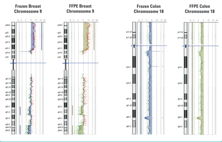
female/male genomic reference DNA (Figure 3). We used DNA isolated from two tumor samples to compare final CGH analysis profiles between fresh and FFPE samples. CGH Analytics software results of parallel DNA extractions from these matched FFPE and fresh frozen samples identified the same amplifications or deletions (Figure 4).
Higher quality data with new non-enzymatic labeling FFPE sample protocol
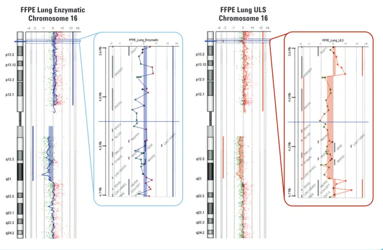
We used DNA isolated from the same FFPE tumor sample and labeled it with both the new non-enzymatic labeling FFPE sample protocol and the traditional enzymatic labeling protocol (Figure 5). The probe-to-probe log ratio noise was lower with the new non-enzymatic FFPE sample protocol and allowed the detection of smaller aberrations that could not be detected with the traditional enzymatic labeling protocol.
Prior to hybridization, the degree of DNA degradation was evaluated by agarose gel electrophoresis. As expected, analysis of DNA isolated from frozen and FFPE tissues from the matched tumor samples showed that genomic DNA from frozen tumors was of much higher molecular weight and less degraded than that from FFPE tissues (Figure 2).
FFPE results comparable to fresh tissue
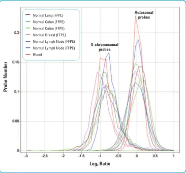
Using a male/female (XY/XX) model system, DNA isolated from six different normal tissue FFPE blocks—normal lung, normal colon (2 samples), normal breast (2 samples) and lymph node—was hybridized against sex-mismatched pooled reference DNA. The log2ratios obtained from FFPE tissues were close to expected values. Furthermore, FFPE values were also similar to those obtained with high-quality, commercially available DNA when hybridized against sex-mismatched
Figure 3. Hybridization of DNA isolated from normal FFPE tissues against sex-mismatched reference DNA.DNA isolated from normal tissue FFPE blocks was hybridized against sex-mismatched pooled reference DNA. X- chromosome probes have an XY/XX ratio theoretical value of 0.5 (log2ratio of -1). Autosomal probes have an XY/XX ratio theoretical value of 1.0 (log2ratio of 0). The log2ratios obtained from FFPE tissues were close to expected values. FFPE values were also similar to those obtained with high-quality, isolated from blood, commercially available DNA (red) when hybridized against sex-mismatched reference DNA.
Figure 2. DNA Characterization of matched frozen versus FFPE tumor samples. Genomic DNA was isolated from various tumors using the Qiagen DNeasy Blood and Tissue Kit and 20 ng analyzed by gel electrophoresis. Lane 1, marker; Lane 2, reference female genomic DNA; Lanes 3-5, tumor set A (infiltrating ductal breast carcinoma; frozen followed by duplicate FFPE); Lanes 6-8, tumor set B (large cell lung carcinoma; frozen followed by duplicate FFPE); Lanes 9-11, tumor set C (colon adenocarcinoma; frozen followed by duplicate FFPE).
1
Lanes
2 3 4 5 6 7 8 9 10 11
12,216 bp–
500 bp–
Log2 Ratio
Autosomal probes
X-chromosomal probes
Probe Number
Normal Lung (FFPE) Normal Colon (FFPE) Normal Colon (FFPE) Normal Breast (FFPE) Normal Lymph Node (FFPE) Normal Lymph Node (FFPE) Blood
Figure 4. Chromosomal aberration analysis comparing fresh and frozen FFPE samples from the same tumor.CGH Analytics v3.4 chromosome views of Chromosome 9 and Chromosome 18 analysis of fresh frozen (blue) and FFPE (green) tissue blocks from the identical tumor. Left panel: Infiltrating ductal breast carcinoma. Right panel: Colon adenocarcinoma. Log2ratio values for all oligonucleotide probes are plotted as a function of their chromosomal position. Each point represents a single probe, with a 1 MB moving average (thin wavy colored vertical lines). Aberration calls identified by the ADM-1 algorithm (thick straight vertical colored lines and colored shaded areas) are shown.
Frozen Breast Chromosome 9
FFPE Breast Chromosome 9
Frozen Colon Chromosome 18
FFPE Colon Chromosome 18 HER-2 gene amplification detection in 1-, 4-, and
10-year-old samples
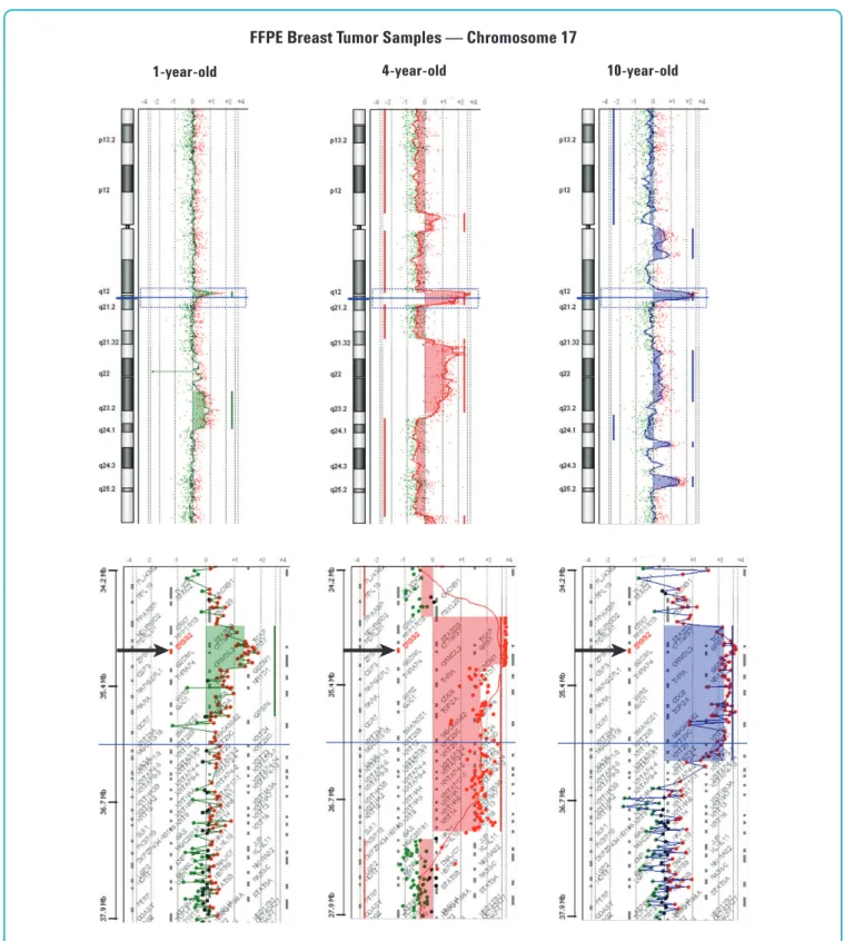
To test the archival range limit of how long stored FFPE tissues can remain useful for high-quality aCGH analysis, we isolated DNA using 1-, 4-, and 10-year-old archival breast tumor FFPE blocks and analyzed them for the well-documented
amplification of the HER-2 oncogene. HER-2 gene amplification and over-expression in breast cancer has been associated with more aggressive clinical behavior and HER-2 amplification status is currently used as criteria for Herceptin therapy. Analysis of these samples showed that the probe-to-probe log ratio noise was very similar for the 1- and 4-year-old FFPE samples. While the 10-year-old FFPE sample showed higher probe-to-probe log ratio noise, the 60-mer probes still enabled detection of the HER-2 amplification with a high level of confidence (Figure 6).
Discussion
Despite the fact that FFPE tissues represent the largest source of archival biological material available for large retrospective studies of human cancer, the use of archival FFPE tissues for aCGH studies has been limited because appropriate methods did not exist for getting good quality data out of degraded DNA. In this report we demonstrated that high-quality aCGH data can be generated on the Agilent platform from
significantly degraded FFPE tissue-derived DNA using the ULS non-enzymatic labeling methodology. When DNA isolated from normal tissue FFPE blocks was hybridized against sex-
mismatched pooled reference DNA, log2ratios obtained were close to the expected values. We further demonstrated that the same amplifications and deletions can be detected in DNA extracted from FFPE blocks as well as fresh frozen tissue from the same tumor. We also detected the frequent HER-2 gene
Figure 5. Non-enzymatic labeling FFPE sample protocol versus traditional enzymatic labeling protocol.Detection of DNA aberrations on Chromosome 16 in a large cell lung carcinoma FFPE sample. 500 ng of genomic DNA was labeled with both the traditional enzymatic labeling protocol (blue) and the new non- enzymatic labeling FFPE sample protocol (brown). Log2ratio values for all oligonucleotide probes are plotted as a function of their chromosomal position. Each point represents a single probe, with a 1 MB moving average (thin wavy colored vertical lines) on the chromosome panels and no moving average on the zoom-in
FFPE Lung Enzymatic Chromosome 16
FFPE Lung ULS Chromosome 16 amplification in DNA isolated from breast tumor FFPE samples
as old as 10 years. We conclude that the ULS non-enzymatic labeling protocol described here enables routine analysis of archival pathology specimens at a fraction of the processing time and cost, allowing large retrospective studies of genetic aberrations.
Figure 6. DNA aberrations in 1-, 4-, and 10-year-old FFPE breast tumor samples. Detection of Chromosome 17 aberrations in a 1- (green), 4- (red) and 10-year- old (blue) infiltrating ductal carcinoma samples showing the common human epithelial receptor 2 (HER-2) gene amplification. Log2ratio values for all
oligonucleotide probes are plotted as a function of their chromosomal position. Each point represents a single probe, with a 1 MB moving average (thin wavy colored vertical lines) on the chromosome panels and no moving average on the zoom-in (3.71 MB) panels. Aberration calls identified by the ADM-1 algorithm (thick straight vertical colored lines and colored shaded areas) are shown. Black arrows show location of HER-2 gene.
1-year-old 4-year-old 10-year-old
FFPE Breast Tumor Samples — Chromosome 17
About Agilent Technologies
Agilent Technologies is a leading supplier of life science research systems that enable scientists to understand complex biological processes, determine disease mechanisms, and speed drug discovery. Engineered for sensitivity, reproducibility, and workflow productivity, Agilent’s life science solutions include instrumentation, microfluidics, software, microarrays, consumables, and services for genomics, proteomics, and metabolomics applications.
Buy online:
www.agilent.com/chem/store
Find an Agilent customer center in your country: www.agilent.com/chem/contactus
U.S. and Canada 1-800-227-9770
[email protected] Asia Pacific
[email protected] Europe
This information contained in this publication is intended for Research Use Only and not to be followed as a diagnostic procedure. Information, descriptions, and specifications in this publication are subject to change without notice. Agilent Technologies shall not be liable for errors contained herein or for incidental or consequential damages in connection with the furnishing, performance or use of this material.
Universal Linkage System (ULS)™and KREApure™are both registered trademarks of KREATECH Biotechnology.
© Agilent Technologies, Inc. 2007 Printed in the U.S.A. August 10, 2007 5989-7120EN
Acknowledgements
The authors want to thank Ida Bukholm, Vessela N. Kristensen, and Anne-Lise Borresen-Dale at the Institute for Cancer Research, Oslo, Norway, for providing FFPE breast cancer tissue blocks.
References
1. Barrett M. et al., (2004) Comparative genomic hybridization using oligonucleotide microarray and total genomic DNA. PNAS101(51): 17765-70. 2. van Beers E. et al., (2006) A multiplex PCR predictor for aCGH success of
FFPE samples. Br J Cancer94(2):333-7.