functions.
A uthor(s )
S , Mohri; H, T akahashi; M, S akai; S , T akahashi; N, W aki; K ,
A izawa; H, S uganuma; T , A ra; Y , Matsumura; D , S hibata; T ,
Goto; T , K awada
C itation
PloS one (2018), 13(1)
Is s ue D ate
2018-01-12
UR L
http://hdl.handle.net/2433/230702
R ig ht
©
2018 Mohri et al. T his is an open access article distributed
under the terms of the C reative C ommons A ttribution L icense,
which permits unrestricted use, distribution, and reproduction
in any medium, provided the original author and source are
credited.
T ype
J ournal A rticle
Wide-range screening of anti-inflammatory
compounds in tomato using LC-MS and
elucidating the mechanism of their functions
Shinsuke Mohri1, Haruya Takahashi1,2, Maiko Sakai1, Shingo Takahashi3, Naoko Waki2,3, Koichi Aizawa3, Hiroyuki Suganuma3, Takeshi Ara2, Yasuki Matsumura4,
Daisuke Shibata2,5, Tsuyoshi Goto1,6, Teruo Kawada1,6*
1Laboratory of Molecular Function of Food, Graduate School of Agriculture, Kyoto University, Uji, Kyoto, Japan,2KAGOME Tomato Discoveries Laboratory, Graduate School of Agriculture, Kyoto University, Kyoto, Japan,3Innovation Division, KAGOME CO., LTD., Tochigi, Japan,4Laboratory of Quality Analysis and Assessment, Graduate School of Agriculture, Kyoto University, Uji, Kyoto, Japan,5Kazusa DNA Research Institutes, Kazusa-Kamatari, Kisarazu, Chiba, Japan,6Research Unit for Physiological Chemistry, Kyoto University, Kyoto, Japan
Abstract
Obesity-induced chronic inflammation is a key factor in type 2 diabetes. A vicious cycle involving pro-inflammatory mediators between adipocytes and macrophages is a common cause of chronic inflammation in the adipose tissue. Tomato is one of the most popular veg-etables and is associated with a reduced risk of diabetes. However, the molecular mecha-nism underlying the effect of tomato on diabetes is unclear. In this study, we focused on anti-inflammatory compounds in tomato. We found that the extract of tomato reduced plasma glucose and inflammatory markers in mice. We screened anti-inflammatory frac-tions in tomato using lipopolysaccharide-stimulated RAW264.7 macrophages, and active compounds were estimated by liquid chromatography-mass spectrometry over a wide range. Surprisingly, a large number of compounds including oxylipin and coumarin deriva-tives were estimated as anti-inflammatory compounds. Especially, 9-oxo-octadecadienoic acid and daphnetin suppressed pro-inflammatory cytokines in RAW264.7 macrophages inhibiting mitogen-activated protein kinase phosphorylation and inhibitor of kappa Bα pro-tein degradation. These findings suggest that tomato containing diverse anti-inflammatory compounds ameliorates chronic inflammation in obese adipose tissue.
Introduction
Obesity is a major risk factor for the development of numerous complications, including type 2 diabetes and cardiovascular diseases[1,2]. Lifestyle-related diseases result from abnormal glu-cose and lipid metabolism, which are primarily caused by obesity[2]. Obesity is an excessive accumulation of adipose tissue that has become a worldwide concern. In recent years, several studies have reported that obesity has been closely associated with low-grade chronic inflam-mation in the adipose tissue[3,4–6]. The inflaminflam-mation state of obesity increases infiltration of
a1111111111 a1111111111 a1111111111 a1111111111 a1111111111 OPEN ACCESS
Citation:Mohri S, Takahashi H, Sakai M, Takahashi S, Waki N, Aizawa K, et al. (2018) Wide-range screening of anti-inflammatory compounds in tomato using LC-MS and elucidating the mechanism of their functions. PLoS ONE 13(1): e0191203.https://doi.org/10.1371/journal. pone.0191203
Editor:Nobuyuki Takahashi, Tokyo University of Agriculture, JAPAN
Received:August 28, 2017
Accepted:December 30, 2017
Published:January 12, 2018
Copyright:©2018 Mohri et al. This is an open access article distributed under the terms of the Creative Commons Attribution License, which permits unrestricted use, distribution, and reproduction in any medium, provided the original author and source are credited.
Data Availability Statement:All relevant data are within the paper and its Supporting Information files.
Funding:This study was funded by KAGOME CO., LTD. The funders had no role in study design, data collection and analysis, decision to publish, or preparation of the manuscript.
macrophages in the adipose tissue that are recruited by the monocyte chemoattractant protein (MCP)-1 released from hypertrophied adipocytes[7–9]. These infiltrating macrophages cause the secretion of various inflammatory cytokines such as nitric oxide (NO) and tumor necrosis factor (TNF)-α, which can cause insulin resistance[10,11]. The interaction between adipocytes and infiltrating macrophages in the adipose tissue contributes to the vicious cycle that leads to chronic inflammation and glucose metabolism disorder under conditions of obesity[12,13]. Thus, in order to improve glucose metabolism, it is important to inhibit the production of these inflammatory cytokines and suppress the low-grade inflammation in obese adipose tissue.
Tomato is one of the most popular and commonly consumed fresh vegetables in the world. Previous studies have indicated that the dietary intake of tomato is linked to a reduced risk of chronic diseases such as cardiovascular diseases and type 2 diabetes[14–16]. Furthermore, tomato consumption reduces inflammation by decreasing inflammatory cytokines in over-weight and obese humans[17,18]. These interesting effects of tomato consumption have been elucidated, but the active compounds in tomato are not fully understood. In recent years, we demonstrated that tomato contained fatty acid derivatives (oxylipin) that activated lipid metabolism via peroxisome proliferator-activated receptor (PPAR)αactivation[19–21], which is important for fatty acid oxidation[22–24]. Although the effect of compounds in tomato on lipid metabolism has been elucidated, little is known about the effect on glucose metabolism. The aim of this study was to identify anti-inflammatory compounds in tomato and to show their mechanisms of action. In the present study, we demonstrated that tomato extract had the ability to reduce plasma glucose level in mice. We focused on anti-inflammatory compounds with the potential to reduce inflammatory cytokines associated with glucose metabolism disor-der in obesity. To unravel the active compounds in tomato, we screened anti-inflammatory fractions of tomato extract by measuring NO production in lipopolysaccharide (LPS)-stimu-lated RAW264.7 macrophages. In addition, we attempted to identify active compounds from the anti-inflammatory fractions using liquid chromatography-mass spectrometry (LC-MS). This wide-range screening revealed that tomato contained a large number of anti-inflamma-tory fractions and diverse anti-inflammaanti-inflamma-tory compounds, including oxylipin and coumarin. Representative compound of oxylipin and coumarin derivatives (9-oxo-ctadecadienoic acid (9-oxo-ODA) and daphnetin (7,8-dihydroxycoumarin)) inhibited inflammatory cytokines by suppressing mitogen-activated protein kinase (MAPK) phosphorylation and inhibitor kappa B (IκB)-αprotein degradation in RAW264.7 macrophages. Moreover, 9-oxo-ODA was detected
in vivoand tended to increase in the white adipose tissue (WAT) under tomato extract
treat-ment. These findings indicated that various anti-inflammatory compounds in tomato inhib-ited chronic inflammation between adipocytes and macrophages in obese adipose tissue, suggesting that tomato might be a valuable food to ameliorate glucose metabolism disorder under conditions of obesity.
Materials and methods
Plant materials and chemicals
In this study, we used tomatoes that were provided by KAGOME CO., LTD. (Nasushiobara, Tochigi, Japan) (identifier No.KTP001). All the other chemicals used were from Invitrogen Corp. (Carlsbad, CA, USA), Nacalai Tesque Inc. (Kyoto, Japan), or Wako (Osaka, Japan) and were guaranteed to be of reagent-, high-performance liquid chromatography (HPLC)-, or tis-sue culture-grade.
Animal experiments
Mice were kept in individual cages in a temperature-controlled room at 23±1˚C and main-tained under a constant 12 h light/dark cycle. Male C57BL/6 mice were purchased from CLEA Japan (Tokyo, Japan). 7-week-old mice were maintained for 7 days on a normal diet (ND) (Research Diet Inc., New Brunswick, NJ, USA) and then divided into two groups of similar average body weight. Each group was maintained on a high-fat diet (HFD) containing 60% kcal fat (Research Diet Inc.) or HFD containing 1% tomato extract (section2.3) for 10 weeks. This animal experiment was performed under free-feeding conditions. At the end of the treat-ment period, anesthetized mice were sacrificed by cervical dislocation, and blood and organ samples were collected. Plasma triglyceride (TG), glucose, and non-esterified fatty acid (NEFA) levels were measured using the TG E-test Wako kit (Wako), Glucose CII-test (Wako), and NEFA C-test (Wako), respectively. All animal experiments were approved by the Kyoto University Animal Care Committee (approval code: 28–76).
Extract preparation and fractionation of crude extract
The extraction and fractionation of tomatoes were performed as follows. The components in tomato were extracted from freeze-dried tomato powder using ethanol (EtOH) at room tem-perature for 24 h. The EtOH extract was partitioned with ethyl acetate (EtOAc) (tomato extract) and water mixture. The large-scale tomato extract for animal experiments was pre-pared in TOKIWA Phytochemical Co., Ltd. (Chiba, Japan). On the guidance of NO assay
(sec-tion2.7), the soluble portion of tomato extract was further partitioned withn-hexane (Hexane
extract) and 90% methanol (90% MeOH extract) mixture. Each soluble portion was fraction-ated by silica gel open column chromatography (eluted with Hexane-EtOAc and MeOH, Hex-ane: EtOAc = 100: 0 (I), 75: 25 (II), 50: 50 (III), 25: 75 (IV), 0: 100 (V), and 100% MeOH (VI)). After silica gel open column chromatography, the Hexane and 90% MeOH extract (H-III, M-II, III, and IV) was fractionated by reverse-phase HPLC on a 5C18-AR-II octa decyl silyl
(ODS) column (6.0×150 mm; Nacalai Tesque) using a mobile phase of water (solvent A) and acetonitrile (solvent B) with 0.1% v/v formic acid added to both solvents. The Hexane extract (H-V) was fractionated by reverse-phase HPLC on a 5C8-MS ODS column (6.0×150 mm;
Nacalai Tesque) using the same mobile phase. The program began with 1% solvent B in solvent A followed by a linear elution gradient from 1 to 100% solvent B in solvent A for 130 min. To monitor HPLC elution, a diode array detector was used in the range of 200–700 nm. Flow rate was set at 1.0 mL/min. Eluted fractions were collected at 1 mL/min. The above-mentioned sol-vents in extracts and eluted fractions were evaporated under vacuum at 37˚C using a rotary evaporator. The effects of evaporated samples re-dissolved in EtOH on the production of the pro-inflammatory mediators were examined.
mRNA expression levels
from GenBank, and the sequences are shown inTable 1. All mRNA expression data are pre-sented as ratios relative to the control in each experiment.
LC-MS analysis
The compounds of HPLC fractions and white adipose tissue were assessed using a LC-MS sys-tem as previously described[21,25]. Briefly, each HPLC fraction was dissolved in 1mL of extraction solvent (99.5% EtOH). Each white adipose tissue was homogenized in 1mL of extraction solvent with mixer, and then the solvent was centrifuged. After centrifugation (15,000 rpm, 10 min, 4˚C), the supernatant was collected for use as an extract. The extract was filtered through a 0.2-μm-pore polyvinylidene difluoride (PVDF) membrane (Whatman, Brentford, UK), and the filtrate was used for LC-MS. LC-MS was performed using a Waters Acquity UPLC system (Milford, MA, USA) coupled to a Xevo QTOF-MS equipped with an electrospray ionization source (ESI).
An aliquot of the extracted sample (3μL) was injected into an Acquity UPLC BEH-C18 reversed-phase column (2.1×100 mm column size; 1.7μm particle size). Mobile phases A (water and 0.1% formic acid) and B (acetonitrile and 0.1% formic acid) were used. The column temperature was set at 40˚C. The buffer gradient consisted of 30% to 50% B for 0–4 min, 50% to 85% B for 4–14 min, 99% B for 14–17 min, and 30% B for 3 min at a flow rate of 300μL/ min. The buffer gradient for daphnetin and esculetin analysis consisted of 1% B for 0–1 min, 1% to 50% B for 1–6 min, 50% to 99% B for 6–6.1 min, 99% B for 6.1–10 min, and 1% B for 5 min. Data were acquired with MassLynx software (Waters, Manchester, UK). External mass calibration was performed following the manufacturer protocol.
Cell culture
Cell culture was performed as previously described[26]. Briefly, the RAW264.7 macrophage was cultured in Dulbecco’s modified Eagle’s medium (DMEM) with 10% fetal bovine serum (FBS) and 100 U/mL penicillin and 100μg/mL streptomycin at 37˚C under a humidified 5% CO2atmosphere. To measure MCP-1, TNF-α, and NO levels, RAW264.7 cells were treated
with 100 ng/mL LPS and tomato extract, fraction, or authentic sample at various concentra-tions in serum-free medium for 24 h. 3T3-L1 preadipocytes were subcultured in DMEM with 10% FBS supplemented with 100 U/mL penicillin and 100μg/mL streptomycin at 37˚C under a humidified 5% CO2atmosphere. The differentiation of 3T3-L1 preadipocytes was induced
using adipogenic agents [0.5 mM 3-isobutyl-1-methylxanthine, 0.25μM dexamethasone, and 10μg/mL insulin] in DMEM containing 10% FBS for 2 days after the cells reached confluence (day 0). Then, the medium was replaced with DMEM containing 10% FBS and 5μg/mL insu-lin, which was replaced with fresh medium every 2 days. Twenty days after the differentiation induction, the cells that accumulated large lipid droplets were used as hypertrophied 3T3-L1 adipocytes.
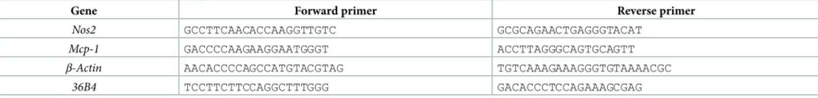
Table 1. Oligonucleotide primers used for mRNA analysis.
Gene Forward primer Reverse primer
Nos2 GCCTTCAACACCAAGGTTGTC GCGCAGAACTGAGGGTACAT
Mcp-1 GACCCCAAGAAGGAATGGGT ACCTTAGGGCAGTGCAGTT
β-Actin AACACCCCAGCCATGTACGTAG TGTCAAAGAAAGGGTGTAAAACGC
36B4 TCCTTCTTCCAGGCTTTGGG GACACCCTCCAGAAAGCGAG
In the co-culture system, adipocytes and macrophages were co-cultured in a contact system as previously described[12]. Briefly, RAW264.7 macrophages (1×10⁵cells/mL) were plated onto dishes with serum-starved and hypertrophied 3T3-L1 cells, and the co-culture was incu-bated in serum-free DMEM for 24 h. RAW264.7 and 3T3-L1 cells of equal numbers to those in the co-culture were cultured separately as control cultures. 9-oxo-ODA, daphnetin, or tomato extract was added to the co-culture at various concentrations as shown in each figure. After 24 h of treatment, culture supernatants were collected, and inflammatory mediators were measured as described below.
Measurement of inflammatory mediators
The concentrations of MCP-1 and TNF-αin the culture supernatants were determined by enzyme-linked immunosorbent assay (ELISA) conducted using a Ready-SET-Go mouse MCP-1 and TNF-αkit (eBioscience, San Diego, CA, USA) according to the manufacturer pro-tocol. The amount of NO in the cell-free culture supernatants was measured using Griess reagent[27]. Briefly, 100μL of supernatant were mixed with an equivalent volume of Griess reagent [1:1 (v/v) of 0.1%N-(1-naphthyl)-ethylenediamine in distilled water and 1% sulfanil-amide in 5% phosphoric acid] in a 96-well flat-bottom plate. After 10 min, absorbance at 550 nm was measured, and the amount of NO was calculated from the sodium nitrite (NaNO2)
standard curve.
Western blotting
Proteins from RAW264.7 macrophages were solubilized in lysis buffer containing 20 mM Tris HCl (pH 7.5), 15 mM NaCl, 1% Triton X100, and a protease and phosphatase inhibitor cock-tail (Nacalai Tesque). The protein concentration of the cell lysate was determined using deter-gent compatible (DC) protein assay (BioRad Laboratories, Hercules, CA, USA). Protein samples were subjected to sodium dodecyl sulfate-polyacrylamide gel electrophoresis (SDS-PAGE), and separated products were transferred to PVDF membrane (Millipore, Bed-ford, MA, USA). After blocking with 5% skim milk in TBS and 0.1% Tween20, the membrane was incubated with an anti-extracellular signal-regulated kinase (anti-ERK), anti-phosphory-lated ERK (anti-pERK), anti-c-Jun N-terminal kinase (anti-JNK), anti-p38, anti-βactin (Cell Signaling Technology, Beverly, MA, USA), or anti-IκB-α(Santa Cruz Biotechnology, Santa Cruz, CA, USA) antibody overnight, and then with a secondary antibody conjugated to horse-radish peroxidase (HRP) (Santa Cruz Biotechnology). The secondary antibody was visualized using chemiluminescent HRP substrate (Millipore). For band quantification, ImageJ (National Institutes of Health, Bethesda, MD, USA) was used.
Statistical analysis
The data are presented as means±standard error of the mean (SEM). Data were assessed by Student’st-test or one-way ANOVA and Dunnett’s multiple comparison tests. Differences were considered significant atp<0.05.
Results
Effects of tomato extract on metabolism in vivo
1B and 1C). In the white adipose tissue, we observed that the mRNA expression ofNos2 involved in NO production was markedly decreased by tomato extract treatment (Fig 1E). Fur-thermore, the expression ofMcp-1tended to decrease under tomato extract treatment (Fig 1F). These results suggested that anti-inflammatory effect of tomato extract improved glucose metabolism disorder in mice.
Screening of anti-inflammatory fraction by silica gel column
chromatography and HPLC in NO assay
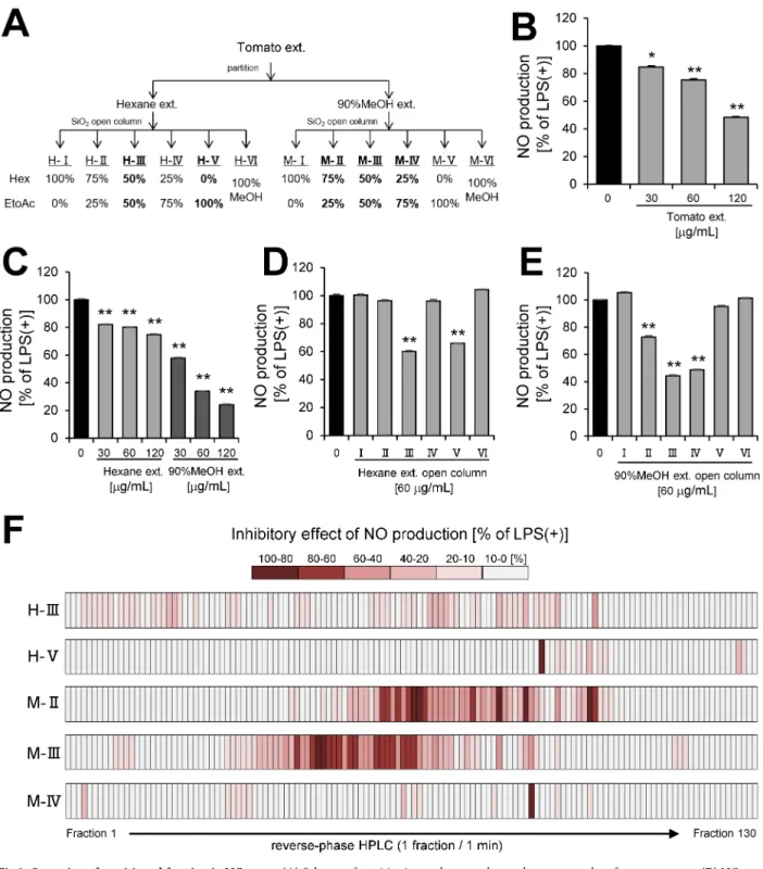
To determine tomato extract (Fig 2A) has anti-inflammatory effect, we investigated whether tomato extract inhibited NO production in LPS-stimulated RAW 264.7 macrophages. Tomato extract significantly inhibited NO production in a dose-dependent manner (Fig 2B). To iden-tify anti-inflammatory compounds in tomato extract, the extract was partitioned with n-hex-ane and 90% MeOH mixture (Fig 2A), and the inhibitory effects of these portions (Hexn-hex-ane
Fig 1. Effect of tomato extract on metabolism in mice. (A)Body weight gain, plasma(B)glucose,(C)TG, and(D)NEFA levels in C57BL/6 mice. Effect of tomato extract on(E)Nos2and(F)Mcp-1mRNA expression levels in white adipose tissue. Data are presented as the mean±SEM (n = 8–10/group),p<0.05 vs. HFD group. HFD; high fat diet, Tomato; tomato extract.
https://doi.org/10.1371/journal.pone.0191203.g001
Table 2. Effect of tomato extract on tissue weight in mice.
Tissue weight (g) HFD Tomato
WAT 3.76±0.41 3.55±0.36 BAT 0.14±0.01 0.13±0.01 Liver 1.26±0.05 1.26±0.07 Kidney 0.32±0.01 0.33±0.01
Fig 2. Screening of partitioned fraction in NO assay. (A)Scheme of partitioning and open column chromatography of tomato extract.(B)NO production by RAW264.7 cell stimulated with LPS (100 ng/mL) and incubated with tomato extract.(C)NO production by RAW264 cell stimulated with LPS and incubated with partition fractions (Hexane, 90% MeOH extract) of tomato extract. NO production by RAW264.7 cell stimulated with LPS and incubated with purified fractions of(D)Hexane or(E)90% MeOH extract by open column chromatography.(F)Heat map of inhibitory effect of NO production by RAW264.7 cell stimulated with LPS and incubated with HPLC fractions. Data are presented as means±SEM (n = 3/ group).p<0.05, p<0.01 vs. LPS alone.
extract and 90% MeOH extract) on NO production by LPS-stimulated RAW264.7 macro-phages were examined. The assay revealed that each portion inhibited NO production in a dose-dependent manner (Fig 2C). Therefore, we further fractioned each portion by silica gel open column chromatography (Fig 2A) and obtained five anti-inflammatory fractions (H-III, H-V, M-II, M-III, and M-IV) (Fig 2D and 2E). To purify the active compounds in the five anti-inflammatory fractions of open column chromatography, we separated the fractions by reverse-phase HPLC and obtained 650 HPLC fractions (Fig 2F). NO assay was performed on the 650 fractions acquired by HPLC. In consequence, a lot of anti-inflammatory fractions were presented in HPLC fractions, including 9 fractions suppressed 80–100% of NO production, 21 fractions suppressed 60–80% of NO production, 27 fractions suppressed 40–60% of NO pro-duction, 39 fractions suppressed 20–40% of NO propro-duction, and 89 fractions suppressed 10– 20% of NO production. Moreover, an overview of anti-inflammatory effect of tomato repre-sented by the heat map indicated that 90% MeOH extract contained more potent anti-inflam-matory fractions (Fig 2F).
Identification of anti-inflammatory compounds in HPLC fractions by
LC-MS
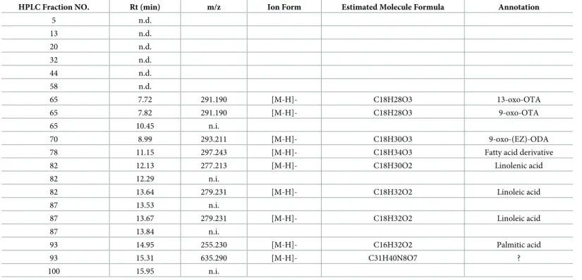
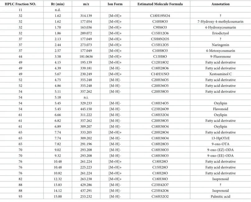
To identify the anti-inflammatory compounds in active HPLC fractions, we analyzed these fractions by LC-MS. The results showed that a wide variety of active compounds were esti-mated in HexaneIII (Table 3), HexaneV(Table 4), 90% MeOHII (Table 5), 90% MeOHIII (Table 6), and 90% MeOHIV (Table 7)-HPLC fractions. Interestingly, we noticed that a num-ber of lipid and coumarin analogs were present in these compounds (Tables3–7). Therefore,
Table 3. LC-MS analysis data of Hexane III—HPLC fraction.
HPLC Fraction NO. Rt (min) m/z Ion Form Estimated Molecule Formula Annotation
5 n.d.
13 n.d. 20 n.d. 32 n.d. 44 n.d. 58 n.d.
65 7.72 291.190 [M-H]- C18H28O3 13-oxo-OTA 65 7.82 291.190 [M-H]- C18H28O3 9-oxo-OTA 65 10.45 n.i.
70 8.99 293.211 [M-H]- C18H30O3 9-oxo-(EZ)-ODA 78 11.15 297.243 [M-H]- C18H34O3 Fatty acid derivative 82 12.13 277.213 [M-H]- C18H30O2 Linolenic acid 82 12.29 n.i.
82 13.64 279.231 [M-H]- C18H32O2 Linoleic acid 87 13.53 n.i.
87 13.67 279.231 [M-H]- C18H32O2 Linoleic acid 87 13.84 n.i.
93 14.95 255.230 [M-H]- C16H32O2 Palmitic acid 93 15.31 635.290 [M-H]- C31H40N8O7 ? 100 15.95 n.i.
n.d.; not ditected
n.i.; not identified parent ion ?; not annotated from database
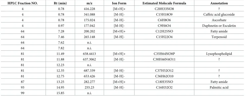
we attempted to identify oxylipin and coumarin analogs in 90% MeOHII, III, and IV extracts containing more potent anti-inflammatory fractions by LC-MS. Consequently, we identified 3 compounds of oxylipin (9-oxo-ODA (Fig 3A), 9-oxo-OTA (Fig 3B), and 13-HpOTrE
(Fig 3C)), and 5 compounds of coumarin derivatives (daphnetin (Fig 3D), esculetin (Fig 3D),
Table 4. LC-MS analysis data of Hexane V—HPLC fraction.
HPLC Fraction NO. Rt (min) m/z Ion Form Estimated Molecule Formula Annotation
81 11.52 n.i. 81 11.95 n.i. 81 12.33 n.i.
87 12.79 256.263 [M+H]+ C16H33NO Fatty amide 87 13.29 282.278 [M+H]+ C18H35NO Fatty amide 87 13.58 n.i.
93 14.97 255.230 [M-H]- C16H32O2 Palmitic acid 100 15.96 n.i.
114 16.08 n.i.
122 16.29 819.556 [M+H]+ C39H70N12O7 ?
n.d.; not ditected
n.i.; not identified parent ion ?; not annotated from database
https://doi.org/10.1371/journal.pone.0191203.t004
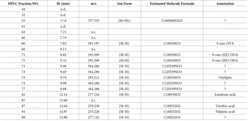
Table 5. LC-MS analysis data of 90%MeOH II—HPLC fraction.
HPLC Fraction NO. Rt (min) m/z Ion Form Estimated Molecule Formula Annotation
44 n.d. 52 n.d.
55 17.6 537.535 [M+H]+ C34H68N2O2 ? 61 n.d.
63 7.15 n.i. 66 7.74 n.i.
66 7.83 291.197 [M-H]- C18H28O3 9-oxo-OTA 66 8.13 n.i.
71 9.02 293.209 [M-H]- C18H30O3 9-oxo-(EZ)-ODA 71 9.32 293.209 [M-H]- C18H30O3 9-oxo-(EE)-ODA 73 9.46 364.286 [M-H]- C22H39NO3 ? 73 9.69 364.286 [M-H]- C22H39NO3 ? 73 9.74 293.212 [M-H]- C18H30O3 Oxylipin 73 9.98 364.286 [M-H]- C22H39NO3 ? 77 9.68 364.286 [M-H]- C22H39NO3 ? 82 12.16 277.216 [M-H]- C18H30O2 Linolenic acid 87 13.60 n.i.
87 13.64 279.230 [M-H]- C18H32O2 Linoleic acid 93 14.97 253.228 [M-H]- C16H32O2 Palmitic acid 99 15.88 277.142 [M-H]- C16H22O4 ?
n.d.; not ditected
n.i.; not identified parent ion ?; not annotated from database
4-hydroxycoumarin (Fig 3E), 4-methoxycoumarin (Fig 3F), and 7-hydroxy-4-methylcoumarin (Fig 3G)). At first, the ion chromatogram peak of estimated molecule with the formula C9H6O4(annotated as daphnetin and esculetin,Table 7) in 90% MeOHIV F4 extract did not
separate, and we could not identify the compound relative to this fraction. Therefore, we attempted to separate the peak in the 90% MeOH extract, and consequently both daphnetin and esculetin were included (Fig 3D). Next, the inhibitory effects of these compounds (Fig 3I) on NO production by LPS-stimulated RAW264.7 macrophages were examined. We showed that 9-oxo-ODA and daphnetin significantly inhibited NO production (Fig 3H). Thus, we
Table 6. LC-MS analysis data of 90%MeOH III—HPLC fraction.
HPLC Fraction NO. Rt (min) m/z Ion Form Estimated Molecule Formula Annotation
11 n.d.
32 1.62 314.139 [M+H]+ C18H19NO4 ?
32 1.62 177.054 [M+H]+ C10H8O3 7-Hydroxy-4-methylcoumarin 32 1.70 163.036 [M+H]+ C9H6O3 4-Hydroxycoumarin 32 1.86 289.072 [M+H]+ C15H12O6 Eriodictyol 37 2.13 177.049 [M+H]+ C5H8N2O5 ? 37 2.44 273.073 [M+H]+ C15H12O5 Naringenin 37 2.57 177.049 [M+H]+ C10H8O3 4-Metoxycoumarin 44 3.58 181.0636 [M+H]+ C13H8O 9-Fluorenone 49 4.15 195.139 [M+H]+ C12H18O2 Fatty acid derivative 49 4.39 339.181 [M-H]- C18H28O6 Fatty acid derivative 49 5.67 230.249 [M+H]+ C14H31NO Xestoaminol C 52 4.75 355.248 [M-H]- C20H36O5 Fatty acid derivative 52 4.86 355.248 [M-H]- C20H36O5 Fatty acid derivative 54 5.11 357.262 [M-H]- C20H38O5 Fatty acid derivative 54 5.18 n.i.
54 5.45 329.233 [M-H]- C18H34O5 Oxylipin 54 5.45 445.150 [M-H]- C23H26O9 Flavonoid 61 6.66 311.222 [M-H]- C18H32O4 Oxylipin 61 6.82 357.262 [M-H]- C20H38O5 Fatty acid derivative 61 6.89 309.207 [M-H]- C18H30O4 Oxylipin 65 7.74 333.205 [M+H]+ C20H28O4 Fatty acid derivative 65 7.74 309.202 [M-H]- C18H30O4 13-HpOTrE 65 7.82 291.196 [M-H]- C18H28O3 9-oxo-OTA 70 9.02 293.208 [M-H]- C18H30O3 9-oxo-(EZ)-ODA 70 9.32 293.208 [M-H]- C18H30O3 9-oxo-(EE)-ODA 76 10.48 261.224 [M+H]+ C18H28O Fatty acid derivative 76 10.48 225.223 [M+H]+ C15H28O Fatty acid derivative 76 10.82 261.224 [M+H]+ C18H28O Fatty acid derivative 82 12.32 263.238 [M+H]+ C18H30O Isoprenoid 88 13.83 429.286 [M-H]- C23H42O7 ? 88 14.12 437.291 [M-H]- C25H42O6 Isoprenoid 93 15.00 253.232 [M-H]- C16H32O2 Palmitic acid
n.d.; not ditected
n.i.; not identified parent ion ?; not annotated from database
used 9-oxo-ODA and daphnetin on the behalf of lipid and coumarin derivatives derived from tomato extract to elucidate the potential to suppress the production of pro-inflammatory mediators.
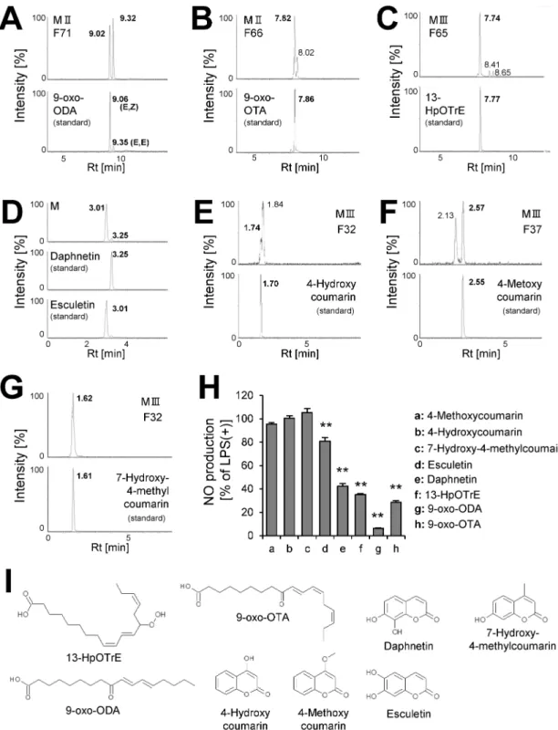
Effects of tomato extract, 9-oxo-ODA and daphnetin on pro-inflammatory
cytokine production in LPS-stimulated macrophages
We demonstrated that tomato extract had the ability to decrease not only NO production (Fig 4A) but also TNF-αand MCP-1 production (Fig 4B and 4C) in a dose-dependent manner. We investigated whether 9-oxo-ODA and daphnetin derived from tomato extract inhibited NO, MCP-1, and TNF-αproduction in activated macrophages stimulated with LPS. Our data showed that 9-oxo-ODA and daphnetin suppressed LPS-induced NO production in a dose-dependent manner (Fig 4D). In addition, we observed that the mRNA expression ofNos2was decreased by 9-oxo-ODA treatment (S1 Fig). Furthermore, 9-oxo-ODA inhibited LPS-induced TNF-αand MCP-1 production in a dose dependent manner (Fig 4E and 4F). Although daphnetin inhibited LPS-induced TNF-αand MCP-1 production, its effect was weaker than that of 9-oxo-ODA (Fig 4E and 4F). These results indicated that tomato extract, 9-oxo-ODA and daphnetin suppressed pro-inflammatory mediators in LPS-stimulated macrophages.
Effects of tomato extract, 9-oxo-ODA and daphnetin on inflammation by
co-culture of adipocytes and macrophages
The vicious cycle that augments inflammation in obese adipose tissue was mimicked by the co-culture of differentiated 3T3-L1 and RAW264.7 cells using a contact system. Indeed, the co-culture of these cells exhibited a significant increase in NO, TNF-α, and MCP-1 production
Table 7. LC-MS analysis data of 90%MeOH IV—HPLC fraction.
HPLC Fraction NO. Rt (min) m/z Ion Form Estimated Molecule Formula Annotation
4 0.78 416.228 [M+H]+ C20H33NO8 ? 4 0.78 341.088 [M-H]- C15H18O9 Caffeic acid glucoside 4 0.78 175.024 [M-H]- C6H8O6 Ascorbate 4 0.97 177.042 [M-H]- C9H6O4 Daphnetin or Esculetin 64 7.28 200.202 [M+H]+ C12H25NO Fatty amide 64 7.46 265.148 [M-H]- C15H22O4 Terpenoid 64 7.62 n.i.
64 7.82 n.i.
81 11.49 658.4413 [M+H]+ C35H64NO8P Lysophospholipid 81 11.88 637.3062 [M-H]- C30H46N4O11 ? 81 12.23 n.i.
81 12.35 687.339 [M-H]- C37H52O12 ? 81 12.75 653.426 [M-H]- C36H62O10 ? 87 13.25 282.277 [M+H]+ C18H35NO Fatty amide 93 14.95 255.23 [M-H]- C16H32O2 Palmitic acid 99 15.85 n.i.
n.d.; not ditected
n.i.; not identified parent ion ?; not annotated from database
Fig 3. Identification of anti-inflammatory compounds.The extracted ion chromatogram of(A)9-oxo-ODA (m/z = 293.209),
(B)9-oxo-OTA (m/z = 291.196),(C)13-HpOTrE (m/z = 309.20),(D)daphnetin and esculetin (m/z = 177.020),(E)
4-hydroxycoumarin (m/z = 163.036),(F)4-methoxycoumarin (m/z = 177.049), and(G)7-hydroxy-4-methylcoumarin (m/ z = 177.049).(H)Effect of identified compounds (50μM) on NO secretion in RAW264.7 cell stimulated with LPS. Data are presented as means±SEM (n = 3/group).p<0.05, p<0.01 vs. LPS alone.(I)Structure of identified compounds. M; 90% MeOH extract, H; hexane extract, F; HPLC fraction.
(Fig 5A–5F). We demonstrated that tomato extract decreased NO, TNF-α, and MCP-1 pro-duction (Fig 5A–5C). 9-oxo-ODA and daphnetin treatment in the co-culture notably inhibited NO production (Fig 5D). Although 9-oxo-ODA and daphnetin also suppressed NO produc-tion, these compounds had no effect on TNF-αand MCP-1 production (Fig 5E and 5F). These data indicated that 9-oxo-ODA and daphnetin derived from tomato extract mainly suppressed NO production on inflammation by co-culture of adipocytes and macrophages.
Mechanism of inhibition of pro-inflammatory mediators by tomato
extract, 9-oxo-ODA and daphnetin
To clarify the mechanism of inhibition of pro-inflammatory cytokines by tomato extract, 9-oxo-ODA, and daphnetin, MAPKs (JNK, ERK, and p38) phosphorylation was examined in LPS-stimulated RAW264.7 macrophages. The LPS treatment significantly facilitated the phos-phorylation of MAPKs, whereas tomato extract inhibited this phosphos-phorylation (Fig 6A). In addition, LPS-induced IκB-αdegradation, which leads to nuclear factor kappa B (NF-κB) acti-vation, was suppressed by tomato extract (Fig 6A). The quantification of western blot signals also showed that tomato extract inhibited the phosphorylation of MAPKs and IκB-α degrada-tion in LPS-stimulated RAW264.7 macrophages (S2A–S2F Fig). 9-oxo-ODA inhibited JNK
Fig 4. Effects of tomato extract, 9-oxo-ODA, and daphnetin on secretion of inflammatory mediators by LPS-stimulated RAW264.7 macrophages.RAW264.7 cells were stimulated with LPS (100 ng/mL) and incubated with tomato extract, 9-oxo-ODA, or daphnetin for 24 h. The levels of NO, TNF-α, and MCP-1 were measured. Effect of tomato extract on(A)NO,(B)TNF-α, and(C)MCP-1 secretion. Effect of 9-oxo-ODA and daphnetin on(D)NO,(E)TNF-α, and(F)MCP-1 secretion. Data are presented as means±SEM (n = 3/group).p<0.05,
p<0.01 vs. culture treated with LPS alone. BAY; positive control for anti-inflammatory effect.
and p38 phosphorylation, and IκB-αdegradation (Fig 6B), whereas daphnetin inhibited JNK, ERK, and p38 phosphorylation, and IκB-αdegradation (Fig 6B). These findings indicated that the anti-inflammatory effects of tomato extract, 9-oxo-ODA and daphnetin were through sup-pression of MAPKs phosphorylation and IκB-αdegradation.
Discussion
In this study, we demonstrated for the first time that tomato extract possessed many types of anti-inflammatory compounds (Fig 2F). We also showed that this extract was able to reduce plasma glucose and TG level in HFD-fed mice (Fig 1B and 1C). Previous studies have indicated that ingestion of tomatoes was related to suppression of various chronic diseases, including type 2 diabetes, cancer, and cardiovascular diseases[14–16]. These interesting effects of tomato are generally attributed to carotenoids, including lycopene[28,29]. It is well known that lyco-pene has anti-oxidative[30] and anti-inflammatory properties[31]. On the other hand, we showed that a large number of compounds in tomato, except carotenoids, have the ability to
Fig 5. Effects of tomato extract, 9-oxo-ODA, and daphnetin on inflammation induced by co-culture of 3T3-L1 adipocytes and RAW264.7 macrophages.Differentiated 3T3-L1 adipocytes were co-cultured with RAW264.7 macrophages for 24 h. The levels of NO, TNF-α, and MCP-1 in the co-culture medium were measured. Effect of tomato extract on(A)NO,(B)TNF-α, and(C)MCP-1 secretion. Effect of 9-oxo-ODA and daphnetin on(D)NO,(E)TNF-α, and(F)MCP-1 secretion. Data are presented as means±SEM (n = 3/group).
p<0.05, p<0.01 vs. non-treated co-culture. TNF-αin control (L1) and MCP-1 in control (RAW) are low limited of quantification.
BAY; positive control for anti-inflammatory effect.
Fig 6. Effects of tomato extract, 9-oxo-ODA, and daphnetin on JNK, ERK, and p38 phosphorylation, and IκB-α degradation in RAW264.7 macrophages stimulated with LPS.RAW264.7 cells were stimulated with LPS (100 ng/ mL) and incubated with tomato extract, 9-oxo-ODA, or daphnetin for 1 h. Total cell lysates were extracted from cultured RAW264.7 cells. Effect of(A)tomato extract,(B)9-oxo-ODA, and daphnetin on phosphorylated JNK, ERK, and p38, and IκB-αdegradation.(C)Schematic illustration of the mechanism of inhibition of pro-inflammatory mediators by tomato extract, 9-oxo-ODA, and daphnetin.
inhibit inflammation (Figs2and3). LC-MS and database analysis showed that almost all the compounds were specified as fatty acid and coumarin derivatives from HPLC fractions of the 90% MeOH extract (Tables5–7andFig 2F). In addition, we demonstrated that 9-oxo-ODA and daphnetin were identified as anti-inflammatory compounds from tomato extract (Fig 3). Furthermore, 9-oxo-ODA, daphnetin and lycopene had an ability to reduce inflammation on NO secretion (S3 Fig). Our results and previous findings raise the possibility that the effect of tomato on inflammation is explained by factors other than carotenoids.
9-oxo-ODA is an oxylipin and an linoleic acid (LIA) derivative. There is a possibility that enzymatic reaction participates in the production of 9-oxo-ODA[32]. It has been reported that free fatty acids are the substrates for lipoxygenases (LOXs)[33] and that 9-LOX activity oxi-dizes LIA at the C9 position to produce 9-hydroperoxy octadecatrienoic acid[34,35], which are possible precursors of 9-oxo-ODA. In a previous study, we have reported that 9-oxo-ODA activated PPARα[19]. It has been reported that PPARαpromotedβ-oxidation via enhance-ment of its target gene expression[36–38]. This resulted in reduced fat storage[39,40]. There-fore, PPARαis important for the regulation of lipid metabolism. In this study, we showed that plasma TG level decreased by tomato extract treatment. Probably, 9-oxo-ODA was involved in this process. On the other hand, we demonstrated for the first time that 9-oxo-ODA was not only a PPARαactivator but also showed anti-inflammatory properties. In addition, previous studies have reported that PPARγthe subtype of PPARα, is likely to be concerned with anti-inflammatory effect in LPS-stimulated macrophages[41,42]. Therefore, we investigated whether the anti-inflammatory activities of 9-oxo-ODA participated in PPARαand PPARγby using antagonist with reference to the previous studies[19,42]. The anti-inflammatory activi-ties of 9-oxo-ODA on NO production was unchanged by PPARαantagonist GW6471 and PPARγantagonist GW9662 (S4A and S4B Fig). Although our findings raise the possibility that 9-oxo-ODA is not related to PPARαand PPARγon the inhibition of NO secretion, further examination is necessary to elucidate the anti-inflammatory activities of 9-oxo-ODA via PPARαand PPARγ. Our previous and present studies showed the functional diversity of 9-oxo-ODA and suggested that 9-oxo-ODA contributed to the effect of tomato on health maintenance. In the tomato extract and white adipose tissue sample, we analyzed 9-oxo-ODA, which had the strongest effect on NO secretion in LPS-stimulated RAW264.7 macrophages (Fig 3H) using LC-MS. 9-oxo-ODA was detected in the tomato extract (Rt = 8.93 min,S1A Fig). The amount of 9-oxo-ODA in the tomato extract was approximately 160 ng/mg (data not shown). Furthermore, 9-oxo-ODA was also detected in the white adipose tissue and tended to increase in presence of the tomato extract treatment (HFD group: approximately 35 ng/mg WAT; Tomato extract group: approximately 60 ng/mg WAT;S1B Fig). We estimated that 9-oxo-ODA present at low level in HFD mice was an endogenous metabolite. The detection of 9-oxo-ODA in the white adipose tissue suggested that this compound acted directly as an anti-inflammatory factor.
for the identification of active compounds. We surmised that these fractions were likely to con-tain other anti-inflammatory compounds, which were difficult to detect under ESI condition.
Previous studies have reported that chronic inflammation in the adipose tissue was impor-tant for obesity[46] and that NF-κB[47] and MAPKs[48] were key factors of inflammation. NF-κB is present in the cytoplasm in an inactive form owing to the binding to α[49]. IκB-αdegradation induces NF-κB translocation to the nucleus, resulting in the secretion of pro-inflammatory factors[49]. Therefore, IκB-αdegradation is involved in regulating obesity-related inflammation[50,51]. MAPKs include three major groups: ERK, JNK, and p38 kinase [48]. MAPKs induce activation of AP-1 transcription factor, which stimulates the expression of inflammatory cytokine genes alone or in combination with NF-κB[48]. These findings sug-gest that NF-κB and MAPKs are important for activation of obesity-related inflammation. In this study, we demonstrated that tomato extract, 9-oxo-ODA and daphnetin had the ability to inhibit IκB-αdegradation and phosphorylation of MAPKs (Fig 6B and 6C).
InFig 5, although tomato extract inhibited NO, TNF-α, and MCP-1 secretion, 9-oxo-ODA and daphnetin had no effect on TNF-αand MCP-1 secretion. These findings suggested that other compounds in tomato were likely to contribute to decrease TNF-αand MCP-1 secretion. On the other hand, tomato extract, 9-oxo-ODA, and daphnetin inhibited NO secretion in the co-culture (Fig 5). In a previous study, it has been reported that the suppression of NO secre-tion contributes to improve glucose metabolism disorder[52]. We demonstrated that the tomato extract decreased plasma glucose level and the expression ofNos2involved in NO pro-duction (Fig 1). Both our results and previous findings raise the possibility that tomato extract, including 9-oxo-ODA and daphnetin, partly decreases plasma glucose level via suppression of NO secretion.
Whereas we identified 9-oxo-ODA and daphnetin as anti-inflammatory compounds from tomato extract, we also suggested that many other anti-inflammatory compounds remain to be identified. Not only the effect of a few compounds alone but also the additive or synergistic effect of many compounds should be taken into consideration when discussing more appro-priate food function. In fact, many metabolites are contained in tomato fruit[53]. Therefore, in this study we attempted to estimate many anti-inflammatory compounds by wide-range screening using LC-MS. In consequence, we showed that a large number of anti-inflammatory compounds may exist in tomato. Moreover, specified as fatty acid and coumarin derivatives were identified as anti-inflammatory compounds. Even though this study provides novel insights into the estimation of food function, further studies are necessary to elucidate the rela-tionship between these compounds and the effect of tomato on inflammation. In conclusion, wide-range screening using LC-MS and NO assay revealed that tomato possessed many anti-inflammatory compounds. In particular, 9-oxo-ODA and daphnetin inhibited the secretion of inflammatory cytokines via the suppression of NF-κB and MAPKs pathway. Furthermore, our study suggested that the effect of tomato on suppression of plasma glucose and expression of Nos2in the white adipose tissue was partly caused by 9-oxo-ODA.
Supporting information
S1 Fig. Effect of 9-oxo-ODA and daphnetin onNos2mRNA expression levels by LPS-stim-ulated RAW264.7 macrophage.RAW264.7 cells were stimulated with LPS (100 ng/mL) and incubated with 9-oxo-ODA or daphnetin (50μM) for 24 h. The levels ofNos2 mRNA expres-sion were measured. Data are presented as means±SEM (n = 4–6/group).p<0.05 vs. culture
S2 Fig. The quantification of western blot signals on MAPKs phosphorylation and IκB-α degradation in LPS-stimulated RAW264.7 macrophages treated with tomato extract. RAW264.7 cells were stimulated with LPS (100 ng/mL) and incubated with tomato extract for 1h. Total cell lysates were extracted from cultured RAW264.7 cells. The quantification of west-ern blot signals on(A)JNK1,(B)JNK2/3,(C)ERK1,(D)ERK2,(E)p38 phosphorylation, and (F)IκB-αdegradation. Data are presented as means±SEM (n = 3–4/group).p<0.05,
p<0.01 vs. culture treated with LPS alone.
(PPTX)
S3 Fig. Effect of 9-oxo-ODA, daphnetin and lycopene on secretion of NO by LPS-stimu-lated RAW264.7 macrophage.RAW264.7 cells were stimulated with LPS (100 ng/mL) and incubated with 9-oxo-ODA, daphnetin and lycopene (30μM) for 24 h. The levels of NO secre-tion were measured. Data are presented as means±SEM (n = 3/group).p<0.05,p<0.01
vs. culture treated with LPS alone. (PPTX)
S4 Fig. Effect of 9-oxo-ODA on secretion of NO by LPS-stimulated RAW264.7 macro-phage treated with or without PPARα, PPARγantagonist.LPS-stimulated (100 ng/mL) RAW264.7 cells were incubated with 9-oxo-ODA (30μM) and treated with or without(A) GW6471(10μM),(B)GW9662 (10μM) for 24h. GW6471 is a PPARαantagonist. GW9662 is a PPARγantagonist. Data are presented as means±SEM (n = 3/group). n.s.; Not significant vs. culture treated with LPS and 9-oxo-ODA.
(PPTX)
S5 Fig. LC-MS analysis of 9-oxo-ODA in tomato extract and white adipose tissue. (A)The extracted ion chromatogram (m/z = 293.209) in tomato extract sample.(B)The amount of 9-oxo-ODA in white adipose tissue. Data are presented as means±SEM (n = 8–10/group). (PPTX)
Acknowledgments
The authors thank Ms. S. Shinoto and Ms. R. Yoshii for secretarial support. The authors also thank Mr. S. Tamura and Dr. N. Yajima (KAGOME CO., LTD.) for supporting the research project.
Author Contributions
Conceptualization:Shinsuke Mohri, Yasuki Matsumura, Daisuke Shibata, Tsuyoshi Goto, Teruo Kawada.
Data curation:Shinsuke Mohri, Haruya Takahashi, Maiko Sakai.
Funding acquisition:Haruya Takahashi, Yasuki Matsumura, Daisuke Shibata, Tsuyoshi Goto, Teruo Kawada.
Investigation:Shinsuke Mohri, Haruya Takahashi, Takeshi Ara, Tsuyoshi Goto, Teruo Kawada.
Methodology:Shinsuke Mohri, Haruya Takahashi, Maiko Sakai.
Project administration:Teruo Kawada.
Resources:Shingo Takahashi, Naoko Waki, Koichi Aizawa, Hiroyuki Suganuma.
Writing – original draft:Shinsuke Mohri, Haruya Takahashi.
Writing – review & editing:Yasuki Matsumura, Daisuke Shibata, Tsuyoshi Goto, Teruo Kawada.
References
1. Reilly M P, Rader D J, The Metabolic Syndrome: more than the sum of its parts?Circulation, 2003, 108 (13), 1546–1551.https://doi.org/10.1161/01.CIR.0000088846.10655.E0PMID:14517150
2. Miranda P J, DeFronzo R A, Califf R M, Guyto J R, Metabolic syndrome: definition, pathophysiology, and mechanism.American Heart J., 2005, 149 (1), 33–45.
3. Gregor M F, Hotamisligil G S, Inflammatory mechanisms in obesity.Annu.Rev.Immunol, 2011, 29, 415–445.https://doi.org/10.1146/annurev-immunol-031210-101322PMID:21219177
4. Heibornn L K, Chambell L V, Adipose tissue macrophages, low grade inflammation and insulin resis-tance in human obesity.Current Pharmaceutical Design, 2008, 14 (12), 1225–1230. PMID:18473870 5. Schenk S, Saberi M, Olefsky J M, Insulin sensitivity: modulation by nutrients and inflammation.Journal of Clinical Investigation, 2008, 118 (9), 2992–3002.https://doi.org/10.1172/JCI34260PMID:18769626 6. Shoelson S E, Lee J, Goldfine A B, Inflammation and insulin resistance.J.Clin.Invest. 2006, 116 (8),
1793–1801.
7. Weisberg S P, McCann D, Desai M, Rosenbaum M, Leibel R L, Ferrante A W, Obesity is associated with mavrophage accumulation in adipose tissue.J.Clin.Invest. 2003, 112 (12), 1796–1808.https:// doi.org/10.1172/JCI19246PMID:14679176
8. Xu H, Barnes G T, Yang Q, Tan G, Yang D, Chou C J, et al. Chronic inflammation in fat plays a crucial role in the development of obesity-related insulin resistance.J.Clin.Invest. 2003, 112 (12), 1821– 1830.https://doi.org/10.1172/JCI19451PMID:14679177
9. Kanda H, Tateya S, Tamori Y, Kotani K, Hiasa K, Kitazawa R, et al. MCP-1 contributes to macrophage infiltration into adipose tissue, insulin resistance, and hepatic steatosis in obesityJ.Clin.Invest. 2006, 116 (6), 1494–1505.https://doi.org/10.1172/JCI26498PMID:16691291
10. Hotamisligil G S, Shargill N S, Spiegelman B M, Adipose expression of tumor necrosis factor-α: direct role in obesity-linked insulin resistance.Science, 1993, 259 (5091), 87–91. PMID:7678183 11. Engeli S, Boschmann M, Adams F, Franke G, Gorzelniak K, Janke J, et al. Dissociation between
adi-pose nitric oxide synthase expression and tissue metabolism.J.Clin.Endocrinol.Metab. 2007, 92 (7), 2706–2711.https://doi.org/10.1210/jc.2007-0234PMID:17456572
12. Suganami T, Nishida J, Ogawa Y, A paracrine loop between adipocytes and macrophages aggravates inflammatory changes: role of free fatty acids and tumor necrosis factor-αArterioscler Thromb.Vasc. Biol. 2005, 25 (10), 2062–2068.https://doi.org/10.1161/01.ATV.0000183883.72263.13PMID:
16123319
13. Odegaard J I, Chawla A, Pleiotropic actions of insulin resistance and inflammation in metabolic homeo-stasisScience, 2013, 339 (6116), 172–177.https://doi.org/10.1126/science.1230721PMID:23307735 14. Lazarus S A, Bowen K, Garg M L, Tomato juice and platelet aggregation in type 2 diabetes.JAMA
2004, 292 (7), 805–806.https://doi.org/10.1001/jama.292.7.805PMID:15315994
15. Blum A, Monir M, Wirsansky I, Ben-Arzi S, The benefical effects of tomatoes.European Journal of Inter-nal Medicine, 2005, 16 (6), 402–404.https://doi.org/10.1016/j.ejim.2005.02.017PMID:16198897 16. Kirstie C A, Jessica K C, Susan Z, Elizabeth H J, John W E, The Tomato As a Functional Food.
Ameri-can Society for Nutritional Sciences, 2005, 135 (5), 1226–1230.
17. Ghavipour M, Saedisomeolia A, Dialali M, Sotoudeh G, Eshraghyan M R, Moghadam A M, et al. Tomato juice consumption reduces systemic inflammation in overweight and obese females.Br.J. Nutr. 2013, 109 (11), 2031–2035.https://doi.org/10.1017/S0007114512004278PMID:23069270 18. Li Y F, Chang Y Y, Huang H C, Wu Y C, Yang M D, Chao P M, Tomato juice supplementation in young
women reduces inflammatory adipokine levels independently of body fat reduction.Nutrition, 2015, 31 (5), 691–696.https://doi.org/10.1016/j.nut.2014.11.008PMID:25837214
19. Kim Y I, Hirai S, Takahashi H, Goto T, Ohyane C, Tsugane T, et al. 9-oxo-10(E),12(E)-Octadecadienoic acid derived from tomato is a potent PPARαagonist to decrease triglyceride accumulation in mouse pri-mary hepatocytes.Mol.Nutr.Food Res. 2011, 55 (4), 585–593.https://doi.org/10.1002/mnfr.
201000264PMID:21462326
obese diabetic mice.PLoS One2012, 7 (2), e31317.https://doi.org/10.1371/journal.pone.0031317
PMID:22347463
21. Takahashi H, Kamakari K, Goto T, Hara H, Mohri S, Suzuki H, et al. 9-oxo-10(E),12(Z),15(Z)- octadeca-trienoic acid activates peroxisome proliferator-activated receptor-αin hepatocytes.Lipids. 2015, 50 (11), 1083–1091.https://doi.org/10.1007/s11745-015-4071-3PMID:26387026
22. Escher P, Wahli W, Peroxisome proliferator-activated receptors: insight into multiple cellular functions. Mutat.Res. 2000, 448 (2), 121–138. PMID:10725467
23. Desvergne B, Wahli W, Peroxisome proliferator-activated receptors: nuclear control of metabolism. Endocr.Rev. 1999, 20 (5), 649–688.https://doi.org/10.1210/edrv.20.5.0380PMID:10529898 24. Chinetti G, Fruchart J C, Staeles B, Peroxisome proliferator-activated receptors (PPARs): nuclear
receptors at the crossroads between lipid metabolism and inflammation.Inflamm.Res. 2000, 49 (10), 497–505.https://doi.org/10.1007/s000110050622PMID:11089900
25. Takahashi H, Goto T, Yamazaki Y, Kamakari K, Hirata M, Suzuki H, et al. Metabolomics reveal 1-palmi-toyl lysophosphatidylcholine production by peroxisome proliferator-activated receptorα.J.Lipid Res. 2015, 56 (2), 254–265.https://doi.org/10.1194/jlr.M052464PMID:25510248
26. Li Y, Goto T, Ikutani R, Lin S, Takahashi N, Takahashi H, et al. Xanthoangelol and 4-hydroxyderrcin suppress obesity-induced inflammatory responses.Obesity (Silver Spring). 2016, 24 (11), 2351–2360.
27. Granger D L, Taintor R R, Boockvar K S, Hibbs J B, Measurement of nitrate and nitrite in biological sam-ples using nitrate reductase and Griess reaction.Methods Enzymol. 1996, 268, 142–151. PMID:
8782580
28. Gouranton E, Thabuis C, Riollet C, Malezet-Desmoulins C, E l Yazidi C, Amiot M J, et al Lycopene inhib-its proinflammatory cytokine and chemokine expression in adipose tissue.J.Nutr.Biochem. 2011, 22 (7), 642–648.https://doi.org/10.1016/j.jnutbio.2010.04.016PMID:20952175
29. Luvizotto Rde A, Nascimento A F, Imaizumi E, Pierine D T, Conde S J, Correa C R, et al. Lycopene sup-plementation modulates plasma concentrations and epididymal adipose tissue mRNA of leptin, resistin and IL-6 in diet-induced obese rats.Br.J.Nutr. 2013, 110 (10), 1803–1809.https://doi.org/10.1017/ S0007114513001256PMID:23632237
30. Palozza P, Parrone N, Catalano A, Simone R, Tomato lycopene and inflammatory cascade: basic inter-actions and clinical implications.Curr.Med.Chem. 2010, 17 (23), 2547–2563. PMID:20491642 31. Fenni S, Hammou H, Astier J, Bonnet L, Karkeni E, Couturier C, et al. Lycopene and tomato powder
supplementation similarly inhibit high-fat diet induced obesity, inflammatory response, and associated metabolic disorders.Mol.Nutr.Food Res. 2017, Epub ahead of print.
32. Takahashi H, Kamakari K, Suzuki H, Mohri S, Goto T, Takahashi N, et al. Localization of 9- and 13-oxo-octadecadienoic acids in tomato fruit.Biosci.Biotechnol.Biochem. 2014, 78 (10), 1761–1764.https:// doi.org/10.1080/09168451.2014.930330PMID:25060034
33. Pulvera Z, M, Kitamura K, Hajika M, Shimada K, Matsui K, Oxylipin metabolism in soybean seeds con-taining different sets of lipoxygenase isozymes after homogenization.Biosci.Biotechnol.Biochem. 2006, 70 (11), 2598–2603. PMID:17090947
34. Kuo J M, Hwang A, Yeh D B, Pan M H, Tsai M L, Pan B S, Lipoxygenase from banana leaf: purification and characterization of an enzyme that catalyzes linoleic acid oxygenation at the 9-position.J.Agric. Food Chem. 2006, 54 (8), 3151–3156.https://doi.org/10.1021/jf060022qPMID:16608245
35. Mariutto M, Fauconnier M L, Ongena M, Laloux M, Wathelet J P, du Jardin P, et al. Reprogramming of fatty acid and oxylipin synthesis in rhizobacteria-induced systemic resistance in tomato.Plant Mol.Biol. 2014, 84 (4–5), 455–467.https://doi.org/10.1007/s11103-013-0144-yPMID:24146221
36. Tugwood J D, Issemann I, Anderson R G, Bundell K R, McPheat W L, Green S, The mouse peroxisome proliferator activated receptor recognizes a response element in the 5’ flanking sequence of the rat acyl CoA oxidase gene.EMBO J. 1992, 11 (2), 433–439. PMID:1537328
37. Roepstorff C, Halberg N, Hillig T, Saha A K, Ruderman N B, Wojtaszewski J F, et al. Malonyl-CoA and carnitine in regulation of fat oxidation in human skeletal muscle during exercise.Am.J.Physiol. Endocri-nol.Metab. 2005, 288 (1), E133–142.https://doi.org/10.1152/ajpendo.00379.2004PMID:15383373 38. Miller C W, Ntambi J M, Peroxisome proliferators induce mouse liver stearoyl-CoA desaturase 1 gene
expression.Proc.Natl.Acad.Sci.USA. 1996, 93 (18), 9443–9448. PMID:8790349
39. Bogna G G, Peroxisome proliferator-activated receptors and their ligands: nutritional and clinical impli-cations.Nutr.J. 2014, 13, 17.https://doi.org/10.1186/1475-2891-13-17PMID:24524207
40. Badman M K, Pissios P, Kennedy A R, Koukos G, Flier J S, Maratos-Flier E, Hepatic fibroblast growth factor 21 is regulated by PPARalpha and is a key mediator of hepatic lipid metabolism in ketotic states. Cell Metab. 2007, 5 (6), 426–437.https://doi.org/10.1016/j.cmet.2007.05.002PMID:17550778 41. Kang M S, Hirai S, Goto T, Kuroyanagi K, Lee J Y, Uemura T, et al. Dehydroabietic acid, a
Biophys Res Commun. 2008, 369 (2), 333–338.https://doi.org/10.1016/j.bbrc.2008.02.002PMID:
18267111
42. Lin C H, Lee S Y, Zhang C C, Du Y F, Hung H C, Wu H T, et al. Fenretinide inhibits macrophage inflam-matory mediators and controls hypertension in spontaneously hypertensive rats via the peroxisome pro-liferator-activated receptor gamma pathway.Drug Des Devel Ther. 2016, 10, 3591–3597.https://doi. org/10.2147/DDDT.S114879PMID:27843299
43. Yang E B, Zhao Y N, Zhang K, Mack P, Daphnetin, one of coumarin derivatives, is a protein kinase inhibitor.Biochem Biophys Res Commun. 1999, 260 (3), 682–685.https://doi.org/10.1006/bbrc.1999. 0958PMID:10403826
44. Yu W, Wang H, Ying H, Yu Y, Chen D, Ge W, et al. Daphnetin attenuates microglial activation and proin-flammatory factor production via multiple signaling pathways. Int Immunopharmacol. 2014, 21 (1), 1–9.
https://doi.org/10.1016/j.intimp.2014.04.005PMID:24747094
45. Kim Y, Park Y, Namkoong S, Lee J, Esculetin inhibits the inflammatory response by inducing heme oxy-genase-1 in cocultured macrophages and adipocytes.Food Funct. 2014, 5 (9), 2371–2377.https://doi. org/10.1039/c4fo00351aPMID:25088305
46. Wensveen F M, ValentićS, Sˇestan M, Turk Wensveen T, PolićB, The "Big Bang" in obese fat: Events initiating obesity-induced adipose tissue inflammation.Eur.J.Immunol. 2015, 45 (9), 2446–2456.
https://doi.org/10.1002/eji.201545502PMID:26220361
47. Benzler J, Ganjam G K, Pretz D, Oelkrug R, Koch C E, Legler K, et al. Central inhibition of IKKα/NF-κB signaling attenuates high-fat diet-induced obesity and glucose intolerance.Diabetes. 2015, 64 (6), 2015–2027.https://doi.org/10.2337/db14-0093PMID:25626735
48. Pearson G, Robinson F, Beers Gibson T, Xu B E, Karandikar M, Berman K, et al. Mitogen-activated pro-tein (MAP) kinase pathways: regulation and physiological functions.Endocr.Rev. 2001, 22 (2), 153– 183.https://doi.org/10.1210/edrv.22.2.0428PMID:11294822
49. Karin M, Yamamoto Y, Wang Q M, The IKK NF-kappa B system: a treasure trove for drug development. Nat.Rev.Drug Discov. 2004, 3 (1), 17–26.https://doi.org/10.1038/nrd1279PMID:14708018
50. Suganami T, Tanimoto-Koyama K, Nishida J, Itoh M, Yuan X, Mizuarai S, et al. Role of the Toll-like receptor 4/NF-kappaB pathway in saturated fatty acid-induced inflammatory changes in the interaction between adipocytes and macrophages.Arterioscler.Thromb.Vasc.Biol. 2007, 27 (1), 84–91.https:// doi.org/10.1161/01.ATV.0000251608.09329.9aPMID:17082484
51. Ravi R, Mookerjee B, van Hensbergen Y, Bedi G C, Giordano A, El-Deiry W S, et al. p53-mediated repression of nuclear factor-kappaB RelA via the transcriptional integrator p300.Cancer Res. 1998, 58 (20), 4531–4536. PMID:9788595
52. Tsuchiya K, Sakai H, Suzuki N, Iwashima F, Yoshimoto T, Shichiri M, et al. Chronic blockade of nitric oxide synthesis reduces adiposity and improves insulin resistance in high fat-induced obese mice. Endocrinology. 2007, 148 (10), 4548–4556.https://doi.org/10.1210/en.2006-1371PMID:17584959 53. Iijima Y, Nakamura Y, Ogata Y, Tanaka K, Sakurai N, Suda K, et al. Metabolite annotations based on