Revision of the Palearctic Species of the Myrmecophilous Genus Pella
(Coleoptera, Staphylinidae, Aleocharinae)
Munetoshi Maruyama
Department of Zoology, National Science Museum, 3–23–1 Hyakunin-chô, Shinjuku-ku, Tokyo, 169–0073 Japan
(http://www.myrmecophile.net/)
Abstract A biosystematic study of the Palearctic species of the myrmecophilous genus Pella (Coleoptera, Staphylinidae, Aleocharinae, Lomechusini, Myrmedoniina) is presented. The adult mor- phology of Pella is investigated into details in comparison with several other taxa of the Lomechusini. Pella is taxonomically revised, and is arranged as a distinct genus. Myrmelia, Pellochromonia and Lepla, formerly treated as subgenera of Zyras, are synonymized with Pella. Forty-one species includ- ing 15 new species (Pella horii, P. kidaorum, P. cooterorum, P. jelineki, P. schillhammeri, P. kinomurai, P. primorskyiana, P. hlavaci, P. iberica, P. kishimotoi, P. coreana, P. plutenkoi, P. interme- dia, P. masakoae, P. zhoui) are recognised in the genus, arranged into 16 species-groups and re- described/described with bionomical and host ant information. The following junior synonymies are proposed: Zyras (Pella) micropterus (⫽P. jureceki), Z. (P.) almaatensis (⫽P. bohaci), and Z. (P.) esau (⫽P. cinctipennis). Seven species are excluded from Pella, newly assigned to the other genera/sub- genera and redescribed. They are: Zyras (Zyras) coloratus, Z. (Z.) iridescens, Z. (Z.) quasar, Z. (Glos- sacantha) ceylonicus, Z. (G.) reelsi, Myrmoecia urartu, and Dvorakatheta wrasei (n. gen.). Life cy- cles and behaviour of the Japanese species of Pella are shortly reported based on the field and labora- tory observations.
INTRODUCTION
The myrmecophilous beetle genus Pella Stephens, 1835, belonging to the subtribe Myrme- doniina of the tribe Lomechusini (family Staphylinidae, subfamily Aleocharinae) is represented by about 30 named species described from the Palearctic region. They are about 3.5 to 7.0 mm in long and associated with ants of the genera Lasius Fabricius, 1804; Formica Linnaeus, 1758; Tapinoma Foerester, 1850; Liometopum Mayr, 1861; and Crematogaster Lund, 1831. Most of them are inquilines of Lasius (Dendrolasius) ants. Several species have been known as scav- engers in hosts’ nests or predators on host workers (Yasumatsu, 1937; Kistner, 1972; Hölldobler et al., 1981).
Pella beetles have been studied by several researchers from different points of view, especial- ly because of their myrmecophily. However, our current knowledge of Pella mainly consists of scattered descriptive works. On the other hand, myrmecophilous insects have recently been at- tractting attention of researchers as material for studies on host-inquiline relationships and co- evolution, and works using modern methods have been published (Axen, 2000; Elmes et al., 1999; Akino, 2002; Fraser et al., 2002; Schönrogge et al., 2002; Pierce et al., 2002, etc.). Be- cause the Pella beetles are among the commonest myrmecophiles in their distributional areas,
they are very useful for the study of biology of myrmecophily. A comprehensive systematic study of the beetles, therefore, would contribute to forward the study of myrmecophily, and the present study has been carried out in this perspective, dealing with morphology, taxonomy and bionom- ics of the Pella beetles.
MATERIALS AND METHODS
Material from Institutions and Private Collections
The majority of species of Pella were originally described without details useful for distin- guishing them, especially among closely related species. Therefore, examination of type series is necessary for reliable identification. Fortunately, the type specimens of most Pella species were accessible through loans or visits to museums or personal collections.
Most of the other specimens examined were collected by some colleagues and myself. In ad- dition, an amount of unidentified material from areas to which I was unable to have access for this study was supplied from some institutional or personal collections.
Depositories of the type specimens examined and the other specimens studied are as follows.
Institutions:
BMNH — The Natural History Museum, London (M. Brendell).
CNC — Canadian National Collection of Insects, Agriculture and Agri-food Canada, Ot- tawa, Ontario (A. Davies).
CNU — Department of Biology, Chungnam National University, Daejeon (K.-J. Ahn). DEI — Deutsches Entomologisches Institut, Müncheberg (L. Zerche).
EEEU — Laboratory of Environmental Entomology, Ehime University, Matsuyama (N. Ohbayashi, M. Sakai).
HUM — The Hokkaido University Museum, Sapporo (M. Ôhara).
IBSS — Institute of Biology and Soil Science, Vladivostok (V. Kuznetsov).
IRSNB — Institut Royal des Sciences Naturelles de Belgique, Bruxelles (D. Drugmand). IZAS — Institute of Zoology, Academia Sinica, Beijing (H.-Z. Zhou).
MHNG — Muséum d’Histore Naturelle, Genève (G. Cuccodoro). MHNL — Muséum d’Histore Naturelle, Lyon (M. Virgile). MNHN — Muséum national d’Histore Naturelle, Paris (T. Deuve). NHMW — Naturhistorisches Museum Wien (H. Schillhammer). NHRS — Naturhistoriska riksmuseet, Stockholm (B. Viklund). NMEG — Naturkundemuseum Erfurt, Erfurt (M. Hartmann). NMP — Národní Muzeum v Praze (J. Jelínek).
MNV — Museo Civico di Storia Naturale di Verona (A. Zanetti). NSMT — National Science Museum, Tokyo-to (S. Nomura). SCM — Sagamihara City Museum, Sagamihara (H. Moriya).
SEHU — Systematic Entomology, Hokkaido University, Sapporo (K. Yoshizawa). TUA — Tokyo University of Agriculture, Atsugi, Kanagawa (S. Okajima).
ZMHB — Museum für Naturkunde der Humboldt-Universität, Berlin (M. Uhlig, J. Frisch).
Private collections:
cAss — Volker Assing (Hannover, Germany). cDvor — Miroslav Dvorˇák (Praha, Czech Republic). cHlav — Peter Hlavácˇ (Kosˇce, Slovakia).
cMar — Munetoshi Maruyama (Tokyo-to, Japan). cRoug — Guilliame de Rougemont (Londinières, France). cSch — Michael Schülke (Berlin, Germany).
Material whose depository is not indicated belongs to my collection (cMar), SEHU, HUM and NSMT, or non-type material of common species that are represented by more than 50 speci- mens in the present study. Paratypes of some new species to be described in this paper and identi- fied specimens of common Japanese species will be distributed to the collections above.
Specimen Data
Specimen data is in many cases not followed the original spellings written on label but cor- rected for more precise and detailed data. But in the case of type material, and in the case of old specimens, e.g., which was collected before the beginning of 1900’s and was historically impor- tant, the original spellings on the label were transcribed.
All the types of Pella described by Gravenhorst (1802, 1806) and Märkel (1842, 1845) de- posited in ZMHB are labelled not any detailed data but a catalogue number. However, more de- tailed data are separately described in “Catalog. General. Musei Zoologici Berolinen” housed in the museum, and based on which actual data are transcribed in square brackets in Type material.
All the type specimens studied in the present paper are labelled, “HOLOTYPE [or, SYN- TYPE, LECTOTYPE, PARALECTOTYPE, PARATYPE] Myrmedonia. . . [name of species, au- thor and year in the original description] det. Maruyama, 200.. [year of examination]”.
Collecting
I investigated more than 120 nests of Lasius ants in Japan (Hokkaidô, Honshû, Shikoku), Korea, the Russian Far East and Slovakia, and collected about 7,000 specimens of Pella.
Pella beetles are usually abundant in the hosts’ nests, above all in the nests of the main host ants Lasius (Dendrolasius) spp. They also walk around the nests and columns (see, Chapter 3). Therefore, the most effective collecting method is to sift dead leaves around the nest and columns using a sieve and a white tray. Beetles dropped on the white tray should immediately be aspirat- ed. Another method, which was sometimes adopted, is pitfall traps that were set around the nest by the use of plastic cups. Beetles collected were transferred to a tube with a piece of cotton paper and a small quantity of ethyl acetate and were left in the tube for half a day. The killed in- sects were mounted each on a piece of paper card. Some beetles were kept alive for rearing.
Dissecting and Drawing
Dried or ethanol-preserved specimens were used for morphological observations. Dissection was made for observation of mouthparts and genital parts (abdominal terminalia), and dried specimens were softened before dissection by warming (60°C, 30–60 min.) each in a small ce- ramic bowl (2.5 cm in diameter) with a small amount of water.
Then the specimens were dissected in water under a high magnification stereomicroscope
Palearctic Species of Pella 3
(Olympus SZ-40, ⫻6.7–80). Eighth to 10th abdominal segments were removed and dissected for most specimens.
Mouthparts of some specimens were dissected: a sharp needle was thrust into the gular plate posterior to the mentum to destroy the muscular attachment of the mouthparts and to remove the mentum, labium, labrum, and maxilla from the head.
These body parts were soaked in warm 5–8% solution of potassium chloride (60°C, 20–60 min), cleaned in 30% ethanol (5 min) and dehydrated in 99% ethanol (5 min). When nec- essary, mouthparts were stained in a warm mixture of acetic fuchsine (5%) and lactic acid (60°C, 60–120 min) before cleaning. The dehydrated material was mounted in Euparal (Chroma- Gesellschaft) on a slide glass card, which was pinned under the insect from which the parts were removed (Maruyama, 2004).
Spermatheca was drawn in water before mounting in Euparal because it was usually transshaped by the osmotic pressure of the medium.
A transmission microscope (Olympus BH2, ⫻20–600, with a drawing device, or ocular mesh) was used for observations of microstructures and line drawings.
Drawing of forebody and antenna of each species was made under the stereomicroscope above-mentioned by the use of ocular mesh set in eyepiece. For drawings of antenna, 1st and 2nd segments were drawn in dorsal view, and 3rd to 11th segments were drawn in lateral view.
Symbiotic Hosts
Symbiotic hosts were mostly identified by myself and some by V. Assing, F. Ito and K. Kino- mura. The scientific names of ants generally follow Bolton (1995), but those of Dendrolasius ants follows Radchenko (2005) and Maruyama (2005). Lasius (Dendrolasius) fuji was formerly recognised as the eastern Palearctic population of Las. (D.) fuliginosus, and was erroneously as- signed to “Las. (D.) nipponensis” by Espadaler et al. (2001).
The species name of symbiotic hosts are abbreviated in “Type material” and “Other material” as follows: FP, Formica pratensis Retzius, 1783; LCAF, Lasius (Cautolasius) flavus (Fabricius, 1782); LCHU, Las. (Chthonolasius) umbratus (Nylander, 1846); LCH, Las. (Ch.) sp.; LDC, Las. (Dendrolasius) capitatus (Kuznetsov-Ugamsky, 1927); LDFJ, Las. (D.) fuji Radchenko, 2005; LDFL, Las. (D.) fuliginosus (Latreille, 1798); LDN, Las. (D.) nipponensis Forel, 1912; LDO, Las. (D.) orientalis Karawajew, 1912; LDS, Las. (D.) spathepus Wheeler, 1910a; LL, Las. (Lasius) sp.; LLB, Las. (Lasius) brunneus (Latreille, 1798); LLJ, Las. (Las.) japonicus Santschi, 1941; LM, Li- ometopum microcephalum (Panzer, 1798); TE, Tapinoma erraticum (Latreille, 1798).
Terminology and Abbreviations
Terminology of morphology used in this study follows Blackwelder (1936), Sawada (1970a, 1972, 1974), Yosii & Sawada (1976), Naomi (1987a, 1987b, 1988a, 1988b, 1988c, 1988d, 1989a, 1989b, 1989c, 1989d, 1990, 1997) and Hanley (2002). Numbers of setae, macrosetae, pores, and campaniform sensilla are confined to one body side, except for the pseudopores of the premen- tum and the pores of the medial pore field of epipharynx.
All measurements in the text are given in millimetres, and the following abbreviations are used for measurements: AL, antennal length; BL, body length, approximate whole length; EL, eye length, major axis; ELL, elytral length; ELW, elytral width; FBL, fore body length, from apex of clypeus to posterior margin of elytra; HL, head length, from apex of clypeus to the posterior
margin of head capsule, except for preocciput; HTL, length of hind tibia; HW, head width; PL, pronotal length; PW, pronotal width.
Collecting methods are abbreviated as follows: fit, flight interception trap; pt, pitfall trap; tt, truck trap.
Rearing
Rearing was made, above all, for observing immature stages in ant-free environment. Some beetles were reared with ant colonies (without queen) for observing their behaviour in the pres- ence of ants. The reared beetles belong to Pella humeralis, P. funesta, P. comes, P. socia, P. kidao- rum, P. japonica, P. kinomurai, P. lugens, P. masakoae, P. spreta, P. laticollis, and P. indiscreta.
Five to ten adult beetles were kept in a plastic box (L18 cm⫻W9 cm⫻H4.5 cm), which was bedded with mixture of peat moss and soil for gardening and provided with some small pieces of tree bark on the soil as shelters for beetles. The peat moss was used for preventing fungus growth and keeping moisture of the soil. The box was placed in room air temperature (18–25°C). Both adults and larvae were fed with dead worker ants of Lasius (Dendrolasius) fuji or L. (D.) spathep- us, which had been preserved in a freezer.
CHAPTER 1. MORPHOLOGY
This chapter describes the general external morphology of the adult Pella beetle in compari- son with other lomechusine species.
The tribe Lomechusini is composed of two subtribes the Lomechusina and Myrmedoniina, and more than 200 genera and subgenera from throughout the world (Maruyama, in prep.). In the Myrmedoniina the following species have been examined: Zyras (s. lat.) spp., Pedinopleurus spp., Diplopleurus spp., Tetrabothrus spp., Orphnebius spp., Drusilla spp., Amaurodera spp. and Apteranillus spp.; in the Lomechusina: Lomechusa spp., Lomechusoides spp. and Xenodusa spp. These species are referred to only by their generic names when character states are almost same within each of the genera. Other species lacking tribal status are all members of the Lomechusi- ni.
In the subfamily Aleocharinae, only a few works which study detailed morphology of whole body are known. The present study is expected to be one of the frameworks of morphological study of the Aleocharinae.
General Structure Body shape
The body shape (Fig. 1) in Pella species is slender or somewhat robust, more or less de- pressed above, and medium to large sized among aleocharine taxa. The body shape is highly gen- eralised in spite of its myrmecophily, i.e., non-limuloid, non-myrmecoid and lacking trichome or large secretary gland, and thus it is not very different from that of free-living aleocharines living in soil or under bark. However, somewhat wide pronotum in Pella species is common to many myrmecophilous aleocharines and indicative of their myrmecophily, though it is homoplastic states sometimes observed in non-myrmecophilous aleocharines, e.g., Aleochara spp. and Oxypo- da spp.
Palearctic Species of Pella 5
Colour
The ground colour in Pella species is usually pale brown to black, but exceptionally the pronotum of P. ruficollis is reddish orange. In colour or colour pattern, the members of the funes- ta group and P. ruficollis are very similar to their host ants, Lasius (Dendrolasius) spp. and Li- ometopum microcephalum, respectively. Most ants have been known to secret repellent sub- stances or to be capable of stinging against enemies, and most predacious animals avoid ants (Blum & Herman, 1978). Thus, it has been suggested that these colour patterns are their Batesian mimicry to the host ants, and it was currently proven by experiments using a predator of ants, Hyla japonica Günther, 1858 (Japanese tree-frog) (Taniguchi et al., 2005).
Body hairs
The body of Pella species is covered mainly with two kinds of hairs, the seta and the macroseta, which can be clearly distinguished. The seta is white or yellow, narrower than the macroseta and usually recumbent. The macroseta is black, stout and suberect. However, in the coreana group, the macrosetae are poorly differentiated from the setae in length and colour.
Fig. 1. Pella limbata (Paykull). Habitus.
Some kinds of secondary hairs are observed on the surface of hindwing, the scutoscutellar suture (Fig. 8 B) and the apex of female 8th sternite (Fig. 11 H) in some species, and they are re- ferred to as pubescence or sensory setae.
Head Head capsule
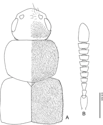
The head capsule (Figs. 2A, 2B, 3A) in Pella species is almost circular in dorsal and ventral views, wider than long, and convex dorsally around middle or posterior part. These states are commonly observed in most lomechusines and other aleocharines. In some species, head capsule is depressed above, especially in male.
Occipital suture. The occipital suture (Figs. 2 A, 2 B, 3 A) is well developed in Pella and
Palearctic Species of Pella 7
Fig. 2. Pella limbata (Paykull). — A, Head, dorsal view; B, head, lateral view.
extends from the ventro-lateral part of the head to near the posterior articulation of the hypos- toma. This character state is observed in most aleocharine tribes.
Postoccipital suture. The postoccipital suture (Figs. 2 A, 3 B) is in a generalised state in Pella species as in most aleocharines, and “neck” is not observed. In some genera of the Myrme- doniina, e.g., Drusilla, Tetrabothrus and Orphnebius, postoccipital suture is strongly constricted to form a “neck”.
Fig. 3. Pella limbata (Paykull). — A, Head, ventral view; B, antenna, dorsal view; C, 1st to 3rd segments of an- tenna, dorsal view; D, 10th and 11th segments of antenna, dorsal view.
Eyes
The compound eye (Figs. 2 A, 2 B) in Pella species is dorsolateral, tending to be situated on the anterior part of the head, and 0.29–0.48 times as long as the head width. Small setae appear among the facets. These states are common to most aleocharines.
The size of the eye is variable with species and is taxonomically informative.
The eye size probably depends on diurnal, nocturnal or day-and-night activity. While the species of Pella having smaller eyes (⬍0.4 times head width), e.g., the species of the humeralis group and the funesta group, are active day and night, the species having larger eyes (⬎0.40 times head width), e.g., the species of the lugens group and the spreta group, appeared to be noc- turnal in my field observations. The large-eyed species of the Lomechusini, e.g., Zyras spp., Pedinopleurus spp., Tetrabothrus spp. and Drusilla spp. are often attracted to light traps, and this suggests that they are also nocturnal.
Antennae
The antennae (Figs. 3 B–3 D) in Pella are usually shorter than the combined length of head, pronotum and elytra, except in P. comes (longer) and P. rambouseki (almost the same in length) and more or less depressed dorsoventrally. Fourth to 10th segments (Fig. 3 B) are usually wider than long and subquadrate in lateral view. Their surfaces are covered with setae and macrosetae, the setae becoming sparser and smaller toward the apex (Figs. 3 C, 3 D). These states are com- mon to most aleocharines.
Relative lengths of antennal segments, especially those of 1st to 3rd segments and the 11th are useful to diagnose species. Usually the 1st segment is the longest but sometimes the 11th is longer than it.
Mouthparts Labrum
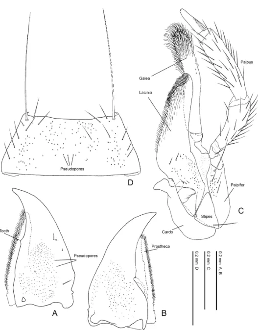
The labrum (Fig. 4 A) is relatively generalised in structure in Pella species. It is submembra- nous around the antero-medial part (sometimes also around the lateral margins). The surface is densely covered with pseudopores except in the lateral area and with 10–12 setae around the api- cal half.
Epipharynx
The epipharynx (Fig. 4 B) in Pella is generalised in structure. The number of lateral setae is normally 5, and these are situated on the mesolateral part of the epipharynx. The medial sensory field have 80–90 pores, and the lateral field is covered with several pores irregularly among scale- like sculptures.
Mandibles
The mandibles in Pella species (Figs. 5 A, 5 B) are simple in shape and lack mola or any pu- bescent patch. However, they are slightly asymmetric, and the right mandible has a small tooth around the middle of the inner margin.
Maxilla
The maxilla in Pella species (Fig. 5 C) is generalised in shape as in most aleocharines, except for the elongated galea.
Palearctic Species of Pella 9
The elongated galea is unique to the Lomechusini and has been considered to be an autapo- morphy of the tribe, though the maxilla is much shortened in the Lomechusina as in other ale- ocharines, and this is considered to be a secondary shortening probably due to their myrme- cophilous behaviour (Maruyama, in prep.).
Mentum
The general shape of the mentum in Pella species (Fig. 5 D) is trapeziform, and the anterior and posterior margins are completely truncate. The surface of the disc is densely covered with
Fig. 4. Pella limbata (Paykull). — A, Labrum, dorsal view; B, epipharynx (labrum, adoral view).
pseudopores except for the lateral area. Labium
The structure of labium is highly diversified in the Myrmedoniina, and this is phylogenetical- ly most informative character. Comparative morphology among lomechusine genera is provided in the following lines.
Prementum, antero-medial margin. The antero-medial margin is shallowly emarginate in Pella species (Fig. 6 A). In the Myrmedoniina, the depth and shape of the emargination is various
Palearctic Species of Pella 11
Fig. 5. Pella limbata (Paykull). — A, Right mandible, dorsal view; B, ditto, ventral view; C, maxilla, ventral view; D, mentum, ventral view.
Fig. 6. Pella limbata (Paykull). — A, Labium, ventral view; B, mentum and labium, lateral view; C, ligula, ven- tral view; D, antero-lateral part of prementum, ventral view; E, hypopharynx (labium, adoral view).
according to species.
Prementum, pseudopores. Location: the pseudopores are located around the antero-medial area of prementum in Pella species (Fig. 6 A). Most lomechusine species are in the same state as Pella.
Number: the number of pseudopores occurring around the antero-medial area is about 30–50 in Pella species (Fig. 6A). Most lomechusines are similar to Pella species, but the members of Myrmoecia and some species of Orphnebius completely lack pseudopores.
Size: the pseudopores in Pella are large and only slightly smaller than real pores. The pseudopores of the lomechusines examined are smaller than a half of real pore in diameter.
Prementum, real pores. Two real pores are located on the mesolateral area of the premen- tum in Pella species (Fig. 6 A). This state is common to most lomechusines and other ale- ocharines. In contrast, Diplopleurus sp., Tetrabothrus spp. and members of the Lomechusina have extra real pores, 3, 4 and 4 or 5 in total, respectively.
Prementum, setal pore. One setal pore occurs in the mesolateral area of the prementum in most aleocharines. That in Pella species (Fig. 6 A) is located slightly anteriorly, and this state is common to most lomechusines. The setal pore is located around the middle of the prementum in Zyras (Trachydonia) sp., Zyras (Rhynchodonia) sp. and Pedinopleurus sp.
Prementum, medial seta. A pair of medial setae are present at the antero-medial apex of the prementum in all lomechusines.
Prementum, internal ridge. Length: a pair of internal ridges are located at the backside of the prementum in the Aleocharinae. Those in Pella species (Fig. 6 D) are very small and only recognised around the backside of each medial seta, and a similar condition is observed also in Myrmoecia species. The length of the internal ridge is various in the Lomechusini. The ridge in most lomechusines is about 1/2 to 5/6 as long as the prementum, and in Diplopleurus sp. very long, extending to the postero-medial projection of the apodeme.
Shape: the internal ridge in most lomechusines is almost straight, but that in Diplopleurus spp., Tetrabothrus spp. and Zyras (Zyras) spp. is bifid anteriorly.
Apodeme. Lateral lobes (suspensoria): the general shape of the lateral lobe can be divided into the following two basic types in the Lomechusini: 1) not completely fused with the premen- tal disc and visible in ventral view; 2) completely fused with the premental disc and invisible in ventral view. Pella (Fig. 6 A) belongs to the type 1). The type 2) is limited to all members of the Lomechusina and part of the Myrmedoniina.
Base of prementum: the base of the prementum is roundly emarginate postero-medially and its lateral part is projected in Pella species (Fig. 6 A: arrow). The round emargination is observed in most lomechusines and some other tribes. The round margin is serrate, dentate or projected in some species of Drusilla, Zyras (Zyrastilbus) sp. and Myrmoecia spp. In contrast, the projection of Diplopleurus spp., Tetrabothrus spp. and Zyras (Zyras) spp. bears the internal ridges. This state is commonly observed in most aleocharines.
Palpus. General shape: the labial palpus in Pella species (Fig. 6 A) is 3-segmented, and the 2nd segment is almost as long as the 1st. The palpus in all species of the Lomechusini is 3-seg- mented, but usually the 2nd segment is shorter than the 1st. In the species of the Lomechusina, the 1st segment is much longer than the combined 2nd and 3rd, while it is shorter than the latter in the other species of the Lomechusini.
Membranous zone: the second segment of the labial palpus in Pella species bears a membra- nous zone (Fig. 6 A) on the ventrolateral area. This state is commonly observed in the Myrme- doniina and probably unique for the subtribe.
Palearctic Species of Pella 13
Setae: the seta a (Sawada, 1972) is situated near the apex of the 1st segment in Pella species. This state is also observed in Myrmoecia spp., Orphnebius spp. and Tetrabothrus spp.
Ligula. General shape: the ligula in Pella species (Figs. 6 A, 6 C) is bilobed, and the apex of each lobe is rounded. It is various in general shape in the Lomechusini. The ligulae of most species are bilobed and similar to that of Pella species, but those of Diplopleurus sp., Zyras (Zyrastilbus) adesi and some species of Drusilla are elongated and much longer than wide, and that of Zyras (Trachydonia) sp. is mount-shaped and only apically notched. The ligula in the species of the Lomechusina is very short, much wider than long and more or less flattened at the apex.
Fig. 7. Pella limbata (Paykull). — A, Prothorax, ventral view; B, meso- and metathoraces, ventral view; C, meso- and metasterna, internal view; D, anepisternum and epimeron of metathorax, internal, dorso-lateral view; E, ditto, internal view.
Palearctic Species of Pella 15
Fig. 8. Pella limbata (Paykull). — A, Scutellum, dorsal view; B, metanotum and 1st abdominal tergite, dorsal view; C, right elytron, dorsal view; D, ditto, ventral view; E, left hind wing, dorsal view; F, flabellum.
Apical setae, presence and number: the members of Pella has no seta at the apex of ligula (Figs. 6 A, 6 C), except for the presence of 3 campaniform sensilla at the apex of each lobe. The absence of setae is also observed in Myrmoecia spp. but the members of this genus even lack sockets. Most lomechusines, as well as most aleocharines, have 4 apical setae on each lobe. Diplopleurus spp. and Tetrabothrus spp. have 3 setae on each lobe.
Surface: the surface of the ligula is smooth in Pella species. In contrast, that of Zyras (Tra- chydonia) sp. is covered with minute granular sensilla.
Hypopharynx. A row of setae appears along the inner margin of the hypopharynx in Pella species (Fig. 6 E). This state is commonly observed in most lomechusines.
Thorax Pronotum
In Pella species, the pronotum is subquadrate, subtrapeziform or elliptical in dorsal view, and more or less flattened. The disc is weakly punctured or covered with microreticulations, and moderately to densely and uniformly covered with setae. Macrosetae are present along the poste- rior to lateral margins, and the number is useful in diagnosing species.
The general shape is various by species and very important in recognising Pella species at the first glance.
The structure of the pronotum is various in the Lomechusini. In the Myrmedoniina, the gen- eral structure is the same as in Pella species but the punctulation and the density of setae are very various by species. In most species of Zyras, pronotal disc is coarsely punctured and sparsely covered with erect and stout setae. In the canaliculata group of Drusilla, the disc is granulate- punctured and densely covered with stout setae. In contrast, the structure of pronotum is highly modified in the Lomechusina, in which the pronotum is distinctly depressed or caved mesally and very finely granulate-punctured.
Elytra
The elytra are subparallel-sided. The inner to posterior margin is more or less margined. The surface is similar to that of the pronotum but with setae becoming sparser apicad. Macrosetae are present along the shoulder to the lateral margin and their number is used for diagnosing species. Hind wing
The hind wing is entire in Pella species. The veins are weakly sclerotized and very obscure, but generalised as in most aleocharines. In contrast, some species of Drusilla, Tetrabothrus, Amaurodera and Zyras, and all species of Apteranillus are brachypterous and flightless.
Meso- and metathoraces
In Pella, the mesocoxal cavity is widely separated, and the metasternum is longer than mesosternum. These states are typical of the Lomechusini and well characterise the tribe. In most species of the Myrmedoniina, apex of the mesosternal process is rounded, but in some species of the Myrmedoniina and all species of the Lomechusina it is widely truncated.
Legs
The legs are generalised in Pella (Figs. 9 A–9 C) as in many other free-living species of the Aleocharinae, and the apices of the tibiae bear a ctenidium as in the other Myrmedoniina species.
Each femur bears a groove from near base to apex alongside the posterior margin to receive tibia when legs are squeezed together.
Palearctic Species of Pella 17
Fig. 9. Pella limbata (Paykull). — A, Foreleg; B, midleg; C, hindleg. A–C, left side, ventral view.
Abdomen Abdominal segments
General shape. In Pella, the abdomen (Figs. 10 A, 10 B) is highly generalised in shape, and its outline is subparallel-sided in dorsal view. However, P. excepta and P. maura have small pro- jections on the 3rd and 6th abdominal tergites. Many species of the Myrmedoniina bear projec- tions on male abdominal tergites or paratergites.
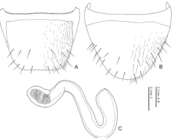
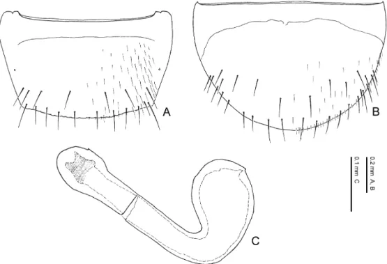
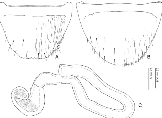
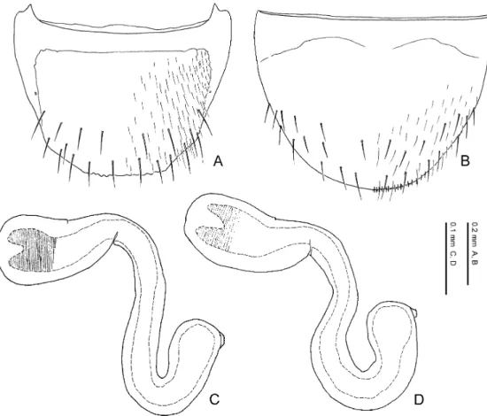
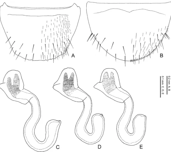
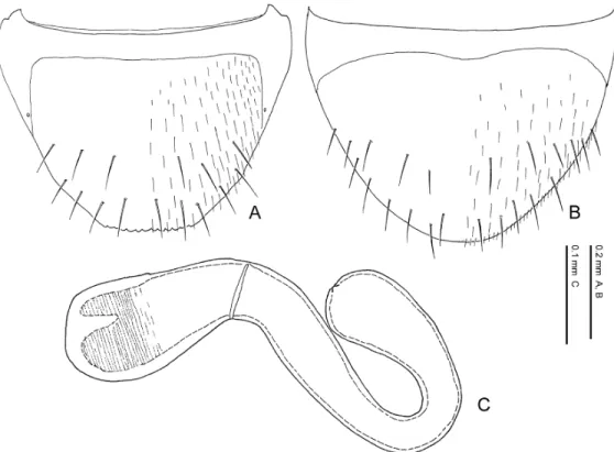
Eighth tergite. The 8th tergite is narrowed posteriad, truncated or emarginate at the apex which is crenate or dentate (Figs. 10 A, 11 A, 11 C, 11 E, 11 F). The general shape and number of macrosetae covering the surface are useful in diagnosing the species. Basal projection of the apodeme of the male (Fig. 11 E) is usually longer than that of the female (Fig. 11 F).
Eighth sternite. The 8th sternite is narrowed posteriad, rounded or truncated at the apex in Pella species (Figs. 10 B, 11 B, 11 D, 11 G, 11 H). The general shape and number of macrosetae covering the surface are useful for diagnosing the species. The 8th sternite is larger in the male than in the female. The apical setae in the female (Fig. 11 H) are longer and thicker than in the male (Fig. 11 G), and in some species the female apical setae are spindle-shaped and flattened. Minute pubescence appears along the apical margin (Fig. 11 H) in the females of most species.
Ninth tergite. The 9th tergite (Figs. 12 A, 16 A) covers the aedeagus or vagina from the dor- sal to the ventral side in Pella species, and this state is common to most aleocharines. The lateral lobes of the male 9th tergite (Fig. 12 A) is long and asymmetric in Pella species as well as in most aleocharines.
Ninth sternite. The male 9th sternite (Figs. 12 B, 12 C) is oblong oval in Pella species, and its apodeme is submembranous. The apex of the male 9th (Fig. 12 C) sternite is truncated or rounded, bearing a pair of macrosetae and 20–30 setae. The left side (ventral view) of the 9th sternite is connected with the 9th tergite in the male, and the aedeagus emerges out from the right side of the 9th sternite (Fig. 12 D). The female 9th sternite (Figs. 16 B, 16 C) is fused with the 9th tergite except around the apex (Fig. 16 C), which bears about 20 setae. These states are common to most aleocharines. The surface is covered with many setae and some macrosetae.
Tenth tergite. The 10th tergite in Pella species (Figs. 12 A, 16 A) is almost pentagonal and truncated or rounded at the apex. The surface is densely covered with setae, which become spars- er apicad.
These states are common to most aleocharines. The setose state is various by species in the Lomechusini.
Male genitalia
Median lobe of aedeagus. The aedeagal median lobe in Pella species (Figs. 13 A–13 E, 14 A–14 E) is mainly composed of the basal capsule and the apical lobe. The compressor plate (Fig. 13 C) is partly or completely covered by the basal capsule. The dorsal bridge (“athetine bridge”, sensu Seevers, 1978) is present. These structures are commonly observed in the Lomechusini. The distal crests (Figs. 13 C–13 E) are well developed and is fused with their sides in Pella species. The general shape is one of the most important characters in diagnosing species. The inner sac is a tubular structure and evaginate as shown in Figs. 14 D, 14 E. In my obser- vations of some couples of Pella beetles killed during copulation, the apical part of the apical lobe is inserted to the vagina from the ventral split line of the 9th sternite, and then the inner sac of aedeagus is evaginated in the vagina. A pair of the longitudinal bands is folded internally at the apical opening of the median lobe, and they cover the dorsal face of the inner sac in the pos-
Palearctic Species of Pella 19
Fig. 10. Pella limbata (Paykull). — A, Second to 8th tergites and paratergites, male, dorsal view; B, 3rd to 8th sternites, male, ventral view. A, B, abdominal segments, location of aedeagus indicated.
Fig. 11. Pella limbata (Paykull). — A, Eighth tergite, male, dorsal view; B, 8th sternite, male, ventral view; C, 8th tergite, female, dorsal view; D, 8th sternite, female, ventral view; E, antero-lateral part of 8th tergite, male, internal view; F, ditto, female; G, apical part of 8th sternite, male, dorsal view; H, ditto, female. A–H, abdomi- nal segments.
ture of evagination. The surface of the inner sac is covered with minute pubescence and projec- tions.
The copulatory piece (Figs. 13 F, 13 G) is very various according to species and important in discriminating allied species. It is not exposed when the inner sac is evaginated (Figs. 14 D, 14 E), and its dorsal part is covered with pubescence or spines.
Paramere. The paramere of aedeagus in Pella species (Figs. 15 A–15 D) is generalised in structure as in most aleocharines, in which it is attached to the median lobe as shown in Figs. 15 C, 15 D and composed of the condylite, paramerite and velum. The paramerite bears an apical
Palearctic Species of Pella 21
Fig. 12. Pella limbata (Paykull). — A, Ninth and 10th tergites, dorsal view; B, 9th sternite, ventral view; C, ditto, apical part; D, 7th to 10th segments in dorso-lateral view, aedeagus emerged out from between lateral sides of 9th tergite and sternite. A–D, male abdominal segments. Abbreviations: T, tergite; S, sternite.
lobe, with 4 setae around the apex. The apical lobe in Pella species is relatively long, exceeding the apex of the velum.
Female genitalia
Vagina. The vagina of Pella species (Fig. 16 A) is generalised in structure as in most ale- ocharines. The vaginal plate is weakly sclerotized, and its size is various probably in accordance with the size of the inner sac of aedeagus. The spermathecal duct is attached at the dorsal surface of the end of the vagina.
Fig. 13. Pella limbata (Paykull). — A, Median lobe of aedeagus, dorsal view; B, ditto, ventral view; C, ditto, lat- eral view; D, ditto, distal crest, dorsal view; E, ditto, lateral view; F, copulatory piece, lateral view; G, ditto, dorsal view.
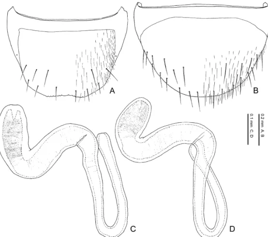
Spermatheca. The spermatheca in Pella species (Figs. 16 A, 16 D–16 F) is simplified and mainly composed of two parts: basal part and apical part. The basal part is attached to the sper- mathecal duct at its base, and sometimes its inner wall is wrinkled. The apical part is attached to the spermathecal gland and more or less notched at its connection with the latter. The inner wall is sparsely to densely wrinkled.
The general shape of the spermatheca is various by species and very important in diagnosing species.
Although the spermatheca in most lomechusines are generalised as in Pella, that in the species of Zyras (Zyras), Diplopleurus and Tetrabothrus are coiled many times.
Palearctic Species of Pella 23
Fig. 14. Pella limbata (Paykull). — A, Apical lobe of median lobe of aedeagus, dorsal view; B, ditto, lateral view; C, ditto, around apex, ventral view; D, evaginated inner sac of median lobe, ventral view; E, ditto, lateral view.
Fig. 15. Pella limbata (Paykull). — A, Paramere of aedeagus, lateral view; B, ditto, far side; C, position of para- mere on median lobe, ventral view; D, ditto, lateral view.
Palearctic Species of Pella 25
Fig. 16. Pella limbata (Paykull). — A, Ninth and 10th tergites, dorsal view, and spermatheca; B, 9th tergite and sternite, ventral view; C, 9th sternite, apical part, ventral view; D–F, spermathecae. A–C, female abdominal segments. Abbreviations: T, tergite; S, sternite.
CHAPTER 2. TAXONOMY
The identities of several Pella species are ambiguous or largely confused. For example, P. laeviceps (Eppelsheim), P. barbara (Fairmaire), P. excepta (Mulsant & Rey) and P. maura (Fauv- el), all described from the western Palearctic region, have been neither recorded nor redescribed since the original descriptions published more than a century ago. Judging from the original de- scriptions, the affiliation of several species described from southern Asia and Caucasia are appar- ently doubtful. Faunistic studies of Pella have well been done only in a few regions, e.g., Central Europe (Ganglbauer, 1895; Reitter, 1909; Lohse, 1974, 1989, etc.) and the Russian Far East (Dvorˇák, 1981). The present study also deals with material from poorly investigated areas in the Palearctic region, namely South Europe, North Africa, Turkey, Caucasia and East Asia. I have discovered 15 undescribed species from these areas.
In this chapter I will revise the Palearctic species of Pella taxonomically. Recently some species of the genus have been found in the Nearctic region, and they are separately revised by Klimaszewski et al. (2005). Detailed descriptions of these undescribed species and redescriptions of all the named species including above-mentioned ambiguous species are given. Because reli- able identification to Pella species is important to their ecological and behavioural studies of myrmecophily, this study would contribute much to biological study of Pella.
Brief History of Taxonomic Study of Pella
In this paper, I synonymize Myrmelia Mulsant & Rey, 1873b; Pellochromonia Reitter, 1909; and Lepla Tottenham, 1939, with Pella, which are formerly arranged as the subgenera of the genus Zyras Stephens, 1835.
The genus Pella was erected by Stephens (1835) to accommodate four British species of the genus Aleochara without designation of the type species. Later, Westwood (1838) designated Staphylinus limbatus Paykull, 1789, as the type species of Pella, and added two species to the genus, which had been arranged as members of Aleochara by Stephens (1835).
Erichson (1837) established the genus Myrmedonia to accommodate several Aleochara and Bolitochara species without designation of the type species. Duponchel (1841) designated Staphylinus canaliculatus (⫽Drusilla canaliculata) as the type species of Myrmedonia. However, Thomson (1859) was unconscious of Duponchel’s (1841) work and designated Aleochara humer- alis (⫽P. humeralis) as the type species of Myrmedonia. After that, under the influence of Thom- son (1859), many species of Pella were described under Myrmedonia in the 1860’s to the 1920’s. Myrmedonia is actually a junior synonym of Drusilla Leach, 1819.
Mulsant and Rey (1873b) established the subgenus Myrmelia for Myrmedonia excepta Mul- sant & Rey, 1861.
Reitter (1909) regarded Pella as a subgenus of the genus Myrmedonia and established a new subgenus Pellochromonia for Myrmedonia ruficollis Grimm, 1845.
Fenyes (1920) regarded Pella as a subgenus of the genus Zyras Stephens, 1835, together with Myrmedonia (sensu Mulsant & Rey, 1873a), Pellochromonia and Myrmelia and referred several species to each of them, which were described merely under Myrmedonia. Bernhauer and Scheerpeltz (1926) and Scheerpeltz (1934) in their catalogue almost followed Fenyes’ (1920) list. Tottenham (1939) established Lepla for the species, that had been included in Myrmedonia (sensu Mulsant & Rey, 1873a), and designated Myrmedonia lugens Gravenhorst, 1806 as the type species.
Kistner (1972) again raised Pella to the generic rank. However, current works (Lohse, 1974, 1989; Likovsky´, 1993; Smetana, 2004, etc.) did not follow Kistner’s view and regarded Pella as a subgenus of Zyras. Lohse (1972, 1989) is one of the most important identification guides of the Aleocharinae, and many researchers, not only in Central Europe but also in other countries of the Palearctic region, follow his arrangement.
Dvorˇák (1981) revised the Pella species of East Asia, but he misidentified some species and overlooked one species.
Gürlich (1981) recognised Pella as a distinct genus on the basis of the examinations of the chaetotaxy of abdominal macrosetae. However, he examined only the type species of the genus, and the chaetotaxy of abdominal macrosetae is highly homoplastic among lomechusine genera.
Descriptions
Tribe Lomechusini Fleming, 1821
Lomechusidae Fleming, 1821: 49 [type genus: Lomechusa Gravenhorst, 1806].
Myrmedoniini Ganglbauer, 1895: 196 [type genus: Myrmedonia Erichson, 1837]. — Seevers, 1978: 151.
Diagnosis. The Lomechusini has not been well defined, but it can be distinguished from the other tribes of Aleocharinae by a combination of the following character states: 1) galea elon- gate; 2) metasternal process longer than mesosternal process; 3) tarsal formula: 4–5–5; 4) ventral bridge of aedeagal median lobe present; 5) compressor plate of aedeagal median lobe covered by a plate, which extends from the bottom of the basal capsule.
Subtribe Myrmedoniina Thomson, 1867
Myrmedoniides Thomson, 1867: 209 [type genus: Myrmedonia Erichson, 1837]. Myrmedoniia Sharp, 1883: 170 [type genus: Myrmedonia Erichson, 1837].
Myrmedoniates Mulsant & Rey, 1873a: 34 [type genus: Myrmedonia Erichson, 1837]. Zyrini Bradley, 1930: 83 [type genus: Zyras Stephens, 1835].
Zyrasini Jeannel & Jarrige, 1949: 304 [type genus: Zyras Stephens, 1835]. Bolitocharinae Hatch, 1957: 146 [type genus: “Bolitochara” Westwood, 1838]. Myrmedoniinea Seevers, 1978: 18 [type genus: Myrmedonia Erichson, 1837].
Diagnosis. The Myrmedoniina has not yet been well defined and is estimated as a para- phyletic group (Maruyama, in prep.). However, it is distinguishable from the other subtribe of the Lomechusini, the Lomechusina, by the following character states: 1) galea elongate; 2) ligula ev- idently bifid; 3) trichome absent on 3rd to 5th abdominal segments.
Genus Pella Stephens, 1835
Pella Stephens, 1835: 434 (description, genus) [type species: Staphylinus limbatus Paykull, 1789, fixed by Westwood (1838)]. — Ganglbauer, 1895: 116 (subgenus of “Myrmedonia”, list). — Reitter, 1909: 42 (subgenus of
“Myrmedonia”, description). — Fenyes, 1920: 296 (subgenus of Zyras, description). — Bernhauer & Scheerpeltz, 1926: 694 (subgenus of Zyras, list). — Kistner, 1972: 150 (distinct genus, description). — Lohse, 1974: 224 (sub- genus of Zyras, description). — Gürlich, 1981: 211–212 (distinct genus, chaetotaxy).
“Myrmedonia”: Thomson, 1859: 255 (description). — Mulsant & Rey, 1873a: 50 (description). — Ganglbauer, 1895: 116 (description). — Reitter, 1909: 43 (key). — Fenyes, 1920: 297 (subgenus of Zyras, description). — Bernhauer & Scheerpeltz, 1926: 694 (subgenus of Zyras, list).
Myrmelia Mulsant & Rey, 1873b: 152 (subgenus of “Myrmedonia”, description) [type species: Myrmedonia excepta Mulsant & Rey, 1861, by monotypy]. — Ganglbauer, 1895: 116 (subgenus of “Myrmedonia”, list). — Fenyes, 1920:
Palearctic Species of Pella 27
299 (subgenus of Zyras, description). — Bernhauer & Scheerpeltz, 1926: 694 (subgenus of Zyras, list). N. syn. Pellochromonia Reitter, 1909: 43 (subgenus of “Myrmedonia”, description) [type species: Myrmedonia ruficollis
Grimm,1845, by monotypy]. — Fenyes, 1920: 298 (subgenus of Zyras, description). — Bernhauer & Scheerpeltz, 1926: 694 (subgenus of Zyras, list). — Lohse, 1974: 226 (subgenus of Zyras, description). N. syn.
Lepla Tottenham, 1939: 226 (new name for “Myrmedonia” sensu Mulsant & Rey, 1874) [type species: Aleochara lugens Gravenhorst, 1802, fixed by Tottenham, 1939]. — Gürlich, 1981: 211 (distinct genus, chaetotaxy). N. syn.
Distribution. Holoarctic region (except for tundra, desert or non-forest areas).
Diagnosis. The following character states are considered to be basal autapomorphies of the genus Pella (Maruyama, in prep.): 1) ligula without setae but with sensilla; 2) 2nd segment of labial palpus almost the same as the 1st in width. In addition, Pella can be distinguished from the other genera of the tribe Lomechusini by a combination of the following character states: 1) body 3.4–7.0 mm in length; 2) head almost circular in dorsal view; 3) head without “neck”; 4) head with occipital suture; 5) eyes 0.35–0.56 times as long as head width; 6) antenna generalised, but slightly depressed; 7) pronotum and elytra densely or moderately covered with setae; 8) pronotal and elytral surfaces smooth or with fine reticulations, without granulation or rugose punctulation; 9) hind wings entire; 10) paratergite generalised, without projection; 11) 10th tergite densely cov- ered with setae except for mesal area; 12) aedeagus simplified, pear-shaped or oval in ventral view.
Description. Body (Fig. 1). Slender or somewhat robust, subparallel-sided. Body length: 3.4–7.0 mm; fore body: 1.6–3.9 mm. Ground colour pale brown to black, or reddish orange.
Head. Head capsule (Figs. 2 A, 2 B, 3 A) almost circular. Occipital suture (Figs. 2 A, 2 B, 3 A) present, dorsally crossing hind part of head, continuing onto ventral side, and terminating on each side near posterior articulation of hypostoma; surface covered with setae. Eyes (Figs. 2 A, 2 B) oval in lateral view, more or less prominent, 0.35–0.56 times as long as head width; small setae present among facets. Antennae (Figs. 3 B–3 D) generalised in shape, almost as long as head and pronotum combined, more or less flattened dorsoventrally; setae on surface becoming denser and smaller apicad; 4th to 10th segments slightly dilated apicad or subparallel-sided, well margined postero-laterally; each segment with erect black setae, those on 3rd to 10th segments forming a row around apical rim; 11th segment oval to elongate oval.
Mouthparts. Labrum (Figs. 4 A, 4 B) much wider than long (W/L, 1.8–2.1), truncate or slightly emarginate antero-medially, submembranous around antero-medial area; surface covered with 100–130 pseudopores except on posterior and lateral areas; apodeme roundly projected pos- tero-medially, with lateral arm gently curved. Epipharynx (Fig. 4 B); medial sensory field with 80–90 pores and 50–80 grain-like sensory pores laterally; each mesolateral area irregularly with 10–15 pores among round projections; 5 pairs of lateral setae stout, about five times as long as wide. Mandibles somewhat asymmetric; right mandible (Figs. 5 A, 5 B) with a small tooth (Fig. 5 A) at middle of inner margin; 2 or 3 small setae present laterally; mesal areas of dorsal and ven- tral surfaces covered with numerous pseudopores; prostheca obtuse at apex, its inner margin densely pubescent. Maxilla (Fig. 5 C): cardo generalised, almost elliptical in ventral view, with 40–50 pseudopores. Stipes small, triangular, with 2 setae at base, without pseudopores. Palpifer triangular in lateral view, with a large seta, several small setae and 15–20 pseudopores near apex. Lacinia with 50–60 pores and 5 or 6 setae medially; apical inner margin with a row of setae forming a comb; surface of around comb densely pubescent; subgalea small, much smaller than last segment of maxillary palpus; galea long, about six times as long as wide, slightly curved at middle, with some pores near apex, densely pubescent apically. Maxillary palpus without pseudopores; 1st segment very small, with 1 seta; 2nd segment gently curved, and much dilated
apically; 3rd segment longest, slightly longer than 2nd, almost straight; 4th segment small, coni- cal. Mentum (Fig. 5 D) trapeziform; posterior and anterior margins truncate; around antero-later- al corner with a long seta; surface densely covered with pseudopores, with several setae laterally. Labium (Figs. 3 A, 6 A–6 E): prementum (Fig. 6 A) with 2 real pores and 1 setal pore mesolater- ally, and 40–50 medial pseudopores, which are relatively large and dense around the medial setae; internal ridge (Fig. 6 D) very small, its length less than 1/8 that of prementum; apodeme (Fig. 6 A) with postero-medial margin roundly emarginate, and its lateral part acutely projected (Fig. 6 A: arrow); lateral lobe of apodeme (Fig. 6 A) gently curved, pointed apically. Ligula (Figs. 6 A, 6 C) bilobed and each lobe rounded apically; apical setae absent, but 3 sockets present. Labi- al palpus (Figs. 6 A, 6 B) with 1st segment shortened and as long as 2nd; 2nd segment notched from base to apex, forming a membranous zone. Hypopharynx (Fig. 6 E) without apical seta; me- dial sensory field with small ridges around apex and scale-like sculptures near base.
Thorax. Pronotum (Figs. 1, 7 A) subtrapeziform, subquadrate or elliptical, much wider than long (PW/PL, 1.21–1.60); disc well margined, somewhat convex but more or less flattened above; surface moderately to densely covered with setae uniformly. Cervical sclerites (Fig. 7 A) gener- alised, rectangular in lateral and ventral view. Basisternum ⫹ preepisternum (Fig. 7 A) gener- alised, but lacking transverse suture. Scutellum (Fig. 8 A) generalised, its apex somewhat round- ed. Anepisternum and epimeron (Figs. 7 D, 7 E) of metasternum completely fused; anepisternum narrowed posteriad; epimeron subtriangular, widened posteriad. Metanotum (Fig. 8 B) weakly sclerotized; scutoscutellar suture with a pubescent patch along inner margin; transscutal suture obscure; scutoscutella generalised, triangular; scutelopostnotal suture almost straight. Mesoster- num (Fig. 7 B) with process short, slightly projected posteriad, rounded at apex. Metaepimeron (Fig. 7 B) small, semicircular. Metasternum (Fig. 7 B) large, three times longer than metaster- num; process well developed, narrowed apicad, its apex somewhat rounded. Metendosternite (Fig. 7 C) generalised; basal stalk convex dorsad, angled; furcal arm gently curved ventrad. Elytra (Figs. 8 C, 8 D) without epiplural suture, more or less margined from shoulder through inner mar- gin to posterior margin; surface almost uniformly covered with setae, which are slightly denser apicad. Hind wing (Figs. 8 E, 8 F) with its venation highly obscure; Sc⫹R, R1, Cu⫹Pcu and A1⫹A2 present, very weakly sclerotized; surface uniformly and moderately pubescent, with minute pubescence along posterior margin; flabellum (Fig. 8 F) with about 20 processes.
Legs. General shape as in Figs. 9 A–9 C. Coxae: fore coxa well developed, slightly shorter than fore femur, ventrally with a suture; mid coxa generalised, with a short suture ventrally; hind coxa subtriangular, rounded laterally. Trochanters: fore and mid trochanters small, subconical; hind trochanter rounded at apex. Femora stout, flattened, gently curved near apex. Tibiae dilated apicad from base to around apical 1/3; each apical margin with a ctenidium and 2 stout setae. Tarsi generalised; empodium with a pair of setae.
Abdomen. First segment (Fig. 8 B): tergite weakly sclerotized, with anterior margin deeply emarginate. Second segment (Fig. 10 A): tergite with posterior margin weakly emarginate, its postero-lateral corner rounded. Third to 6th segments (Figs. 10 A, 10 B): tergites with posterior margins almost straight; dorsolateral plates rectangular except that of 6th segment evidently nar- rowed posteriad; sternite with surface moderately to densely covered with setae. Seventh segment (Figs. 10 A, 10 B): tergite with a pair of gland openings at base; lateral plates fused, narrowed posteriad, pointed at apex. Eighth abdominal segment (Figs. 10 A, 10 B, 11A–11 H): tergite with basal suture curved laterally and continuing apicad or weakened around lateral sides; sternite with basal suture reaching lateral sides or weakened around lateral area. Ninth abdominal seg- ment (Figs. 12 A, 16 A): tergite with surface densely covered with setae. Tenth abdominal seg-
Palearctic Species of Pella 29
ment (Figs. 12 A, 16 A) with surface densely covered with setae except in mesal area; setae be- coming sparser and longer apicad.
Male characters. Head slightly depressed above; setae on dorsal surface denser and longer than in female. Eighth tergite (Figs. 11 A, 11 E) with basal projection of apodeme (Fig. 11 E) more or less larger than in female; posterior margin (Fig. 11 A) dentate or crenate. Eighth sternite (Figs. 11 B, 11 G) larger than in female; macrosetae more numerous than in female; apical senso- ry setae (Fig. 11 G) smaller than in female; apical margin lacking minute pubescence. Lateral projections of apodeme of 9th tergite (Fig. 12 A) asymmetrical; right projection longer than the left. Ninth sternite (Figs. 12 B, 12 C) oblong oval; apical margin more or less truncate; surface covered with setae, with a pair of macrosetae near posterior margin; left lateral margin attaching to tergite (Fig. 12 D). Aedeagus (Figs. 13 A–13 G, 14 A–14 E, 15 A–15 D): median lobe (Figs. 12 A–12 E, 14 A–14 E, 15 C, 15 D) with basal capsule bulbous; distal crests (Figs. 13 D, 13 E) fused on their sides, more or less projected; copulatory piece (Figs. 13 F, 13 G) small to medium- sized, pubescent or dentate dorsally, not exposed when inner sac evaginated; paramere (Figs. 15 A–15 D) with condylite almost straight; hinge zone distinct; apical lobe of paramerite short, sometimes exceeding apex of velum; velum densely ridged.
Female characters. Head slightly depressed above, less depressed than in male; setae on dorsal surface sparser and shorter than in male. Eighth tergite (Figs. 11 C, 11 F) with lateral pro- jection of apodeme (Fig. 11 F) smaller than in male; posterior margin (Fig. 11 C) crenate but weaker than in male and its crenate margin narrower than in male. Eighth sternite (Figs. 11 D, 11 H) smaller than in male; macrosetae less numerous than in male; apical sensory setae (Fig. 11 H) larger than in male, sometimes spindle-shaped and flattened; apical margin (Fig. 11 H) sometimes margined with minute pubescence. Ninth sternite (Figs. 16 B, 16 C) with posterior margin truncate; surface around posterior margin covered with setae. Vagina (Fig. 16 A) with vaginal plate weakly sclerotized. Spermatheca (Figs. 16 A, 16 D–16 F) large, as long as or longer than 10th tergite, clearly divided into basal and apical parts; spermathecal duct (Fig. 16 A) short, 3–5 times as long as spermatheca; spermathecal gland (Fig. 16 A) attaching to about middle of apical part.
Comments. Pella has been regarded as a subgenus of Zyras by authors but is recognised as a good genus in this study. Pella is a monophyletic group and far related to Zyras (Zyras) as a re- sult of phylogenetic analysis of the Lomechusini (Maruyama, in prep.).
I herewith synonymize Myrmelia, Pellochromonia and Lepla with Pella, which are also pre- viously regarded as subgenera of Zyras. This view is based on the adult external structure of all the known species of these genera/subgenera, there having been found no important difference of generic or subgeneric value.
As a result of type material examinations, it has been found that several species formerly re- garded as members of Pella (or Lepla) cannot be affiliated to the genus. They are: Zyras (Pella?) coloratus Cameron, 1939; Z. (P.) ceylonicus Cameron, 1939; Z. (P.) urartu Iablokoff-Khnzoryan, 1962; Z. (L.) iridescens (Sawada, 1970b); Z. (P.) wrasei Dvorˇák, 1988; Z. (P.) quasar Dvorˇák, 1996; and Z. (P.) reelsi Pace, 1998a. These species are transferred to other genera (or tribe) and redescribed at the foot of this chapter.
Check List of the Genus Pella The limbata group
Pella limbata (Paykull, 1789)
Staphylinus divisus Marsham, 1802 Aleochara laevis Gravenhorst, 1802 P. horii Maruyama, n. sp.
The similis group
P. similis (Märkel, 1845), n. comb. The humeralis group
P. humeralis (Gravenhorst, 1802), n. comb. P. laeviceps (Eppelsheim, 1880), n. comb. The funesta group
P. funesta (Gravenhorst, 1806), n. comb. Aleochara crassicornis Stephens, 1832 Myrmedonia atrata Heer, 1839 P. comes (Sharp, 1874)
P. socia (Sharp, 1874)
P. rambouseki (Bernhauer, 1929), n. comb. P. jureceki (Dvorˇák, 1981), n. comb.
Zyras (Pella) micropterus Pace, 1998a, n. syn. P. kidaorum Maruyama, n. sp.
P. cooterorum Maruyama, n. sp. The barbara group
P. barbara (Fairmaire, 1863), n. comb. P. leonhardi (Bernhauer, 1912), n. comb. P. jelineki Maruyama, n. sp.
The erratica group
P. erratica (Hagens, 1863), n. comb. Myrmedonia mustela Rottenberg, 1870 Myrmedonia Ehlersi Eppelsheim, 1884b The schillhammeri group
P. schillhammeri Maruyama, n. sp. The kinomurai group
P. kinomurai Maruyama, n. sp. P. primorskyiana Maruyama, n. sp. The hlavaci group
P. hlavaci Maruyama, n. sp. The cognata group
P. cognata (Märkel, 1842), n. comb. P. iberica Maruyama, n. sp. P. japonica (Sharp, 1888) P. kishimotoi Maruyama, n. sp. The ruficollis group
P. ruficollis (Grimm, 1845), n. comb. Myrmedonia fernandi Fairmaire, 1855 The coreana group
P. coreana Maruyama, n. sp. P. plutenkoi Maruyama, n. sp. The excepta group
P. excepta (Mulsant & Rey, 1861), n. comb.
Palearctic Species of Pella 31
P. maura (Fauvel, 1898), n. comb. Myrmedonia Ragusae Ragusa, 1921 P. kuluensis (Cameron, 1939), n. comb. P. bohaci (Dvorˇák, 1984), n. comb.
Zyras (Pella) almaatensis Pace, 2002, n. syn. P. cinctipennis (Eppelsheim, 1884), n. comb.
Zyras (Pella) esau Dvorˇák, 1984, n. syn. The lugens group
P. lugens (Gravenhorst, 1802), n. comb. P. beijingorum (Pace, 1998), n. comb. P. intermedia Maruyama, n. sp. P. masakoae Maruyama, n. sp. The spreta group
P. spreta (Sharp, 1888), n. comb. P. zhoui Maruyama, n. sp. The laticollis group
P. laticollis (Märkel, 1845), n. comb. P. hampei (Kraatz, 1862), n. comb. P. indiscreta (Sharp, 1888), n. comb. Incertae sedis
P. pumila (Fiori, 1914), n. comb.
Key to the Species Groups of the Genus Pella
1. Eleventh antennal segment long, longer than 7th to 10th segments combined. ... 2.
— Eleventh antennal segment short to medium, shorter than 7th to 10th segments combined. ... ... 3. 2. Body bicoloured; pronotum reddish orange; pronotal hypomeron fully visible in lateral view; posterior margin of 8th tergite not crenate nor dentate, almost straight; macrosetae on 8th abdominal segment clearly differentiated from setae. ...ruficollis group.
— Body unicoloured, pale brown; pronotal hypomeron partly visible in lateral view, its visible part less than 3/5 of pronotal length; posterior margin of 8th tergite crenate; macrosetae on 8th abdominal segment clearly differentiated from setae. ...coreana group. 3. Pronotal hypomeron invisible, or only slightly and narrowly visible in lateral view (its visi- ble length less than 0.4 times as long as pronotum). ... 4.
— Pronotal hypomeron clearly visible in lateral view (its visible length more than 0.5 times as long as pronotum). ... 6. 4. Body almost unicoloured. ...laticollis group.
— Body evidently tricoloured; black in ground colour; elytra yellow but blackish brown around postero-lateral corner, or black but with yellow; legs reddish brown. ... 5. 5. Eyes large and prominent, its length about 0.45–0.48 times as long as head width; lateral areas of pronotum well convex above; pronotal surface roughly punctured. ...spreta group.
— Eyes medium to large in size, its length less than 0.42 times as long as head width; lateral areas of pronotum flattened; pronotal surface finely punctured. ...lugens group. 6. Antennae and legs yellow. ...schillhammeri group.
— Antennae and legs reddish brown to black. ... 7.
7. Pronotum with postero-lateral corners, evidently narrowed posteriad, more or less parallel- sided posteriorly. ... 8.
— Pronotum completely rounded postero-laterally to posteriorly, not forming postero-lateral corners. ...10. 8. Elytra bicoloured, with a maculation at antero-lateral margin; posterior margin of male 8th sternite truncate; copulatory piece of aedeagal median lobe widened apicad and widely truncate at apex. ...humeralis group.
— Elytra unicoloured; posterior margin of male 8th sternite rounded; copulatory piece of aedeagal median lobe narrowed apicad and not truncate at apex. ... 9. 9. Body medium to large: 5.0–6.6 mm. ...funesta group.
— Body small: 3.3–4.1 mm. ...barbara group (part). 10. Pronotum widest around middle, more than 1.48 times as wide as long . ...hlavaci group. Pronotum widest anteriorly, less than 1.46 times as wide as long. ...11. 11. Body small: 3.3–4.1 mm, almost unicoloured. ...12.
— Body medium to large: 3.7–6.2 mm, bicoloured. ...13. 12. Eleventh antennal segment almost as long as 1st segment. ...barbara group (part).
— Eleventh antennal segment evidently shorter than 1st segment. ...erratica group. 13. Antero-lateral areas of pronotum strongly curved ventrad; hypomeron partly visible in later- al view, its visible length less than 0.6 times as long as pronotum; pronotal surface sparsely to moderately covered with setae; distal crest of aedeagal median lobe roundly projected. .... ...excepta group.
— Antero-lateral areas of pronotum weakly curved ventrad; hypomeron fully visible in lateral view, its visible length more than 0.7 times as long as pronotum; pronotal surface densely covered with setae; distal crest of aedeagal median lobe gently projected, or weakly project- ed. ...14. 14. Male 8th sternite almost as long as that of female; apical lobe of aedeagal median lobe much narrowed in ventral view and acutely curved ventrad at base; copulatory piece of aedeagal median lobe aciculate around apex in ventral view. ...limbata group.
— Male 8th sternite evidently longer than that of female; apical lobe of aedeagal median lobe gradually narrowed apicad and slightly curved ventrad or almost straight; copulatory piece of aedeagal median lobe rounded, truncated or pointed at apex and not aciculate in ventral view. ...15. 15. Legs almost uniformly reddish brown; abdomen with around posterior margin of 3rd to 5th abdominal segments reddish brown. ...kinomurai group.
— Legs reddish yellow to yellowish brown, around apical 1/2 of femora darker; abdomen with most areas of 3rd to 5th abdominal segments yellowish brown. ...16. 16. Eleventh segment of antenna almost as long as 1st segment; apical lobe of aedeagal median lobe much shorter than basal capsule, gently narrowed apicad and pointed at apex in lateral view; apical part of spermatheca without outer projection. ...similis group.
— Eleventh segment of antenna much shorter than 1st segment; apical lobe of aedeagal medi- an lobe almost as long as basal capsule, subparallel-sided around middle and obtuse at apex in lateral view; apical part of spermatheca with outer projection. ...cognata group.
Comments. In distinguishing the species of Pella examinations of aedeagus or spermathe- ca are often indispensable, because most other characters used in identification are rather quanti- tative. It is better to refer first to the figures of aedeagus or spermatheca.
Palearctic Species of Pella 33
The limbata Group Species included. Pella limbata, P. horii.
Distribution. European subregion, Mediterranean subregion, Manchurian subregion. Diagnosis. Species of the limbata group may be characterised by a combination of the fol- lowing character states: 1) eye length 0.32–0.34 times as long as head width; 2) 11th antennal segment longer than the 1st; 3) pronotum with postero-lateral corners; 4) pronotum 1.18–1.35 times as wide as long; 5) pronotum widest anteriorly; 6) pronotal hypomeron fully visible in lat- eral view; 7) elytra with yellowish brown maculations around antero-lateral and postero-lateral corners; 8) inner and posterior margins of elytra weakly margined; 9) male 8th sternite not longer than in female and almost the same in size; 10) posterior margin of 8th tergite crenate; 11) poste- rior margin of male 8th sternite rounded; 12) lateral projection of apodeme of male 8th tergite not evidently longer than that of female; 13) macrosetae of 8th abdominal segment generalised in length, exceeding the posterior margin of the segment; 14) postero-medial margin of female 8th sternite with minute pubescence; 15) apical lobe of aedeagal median lobe curved ventrad at base in lateral view; 16) distal crests of aedeagus well developed, projected semicircularly in lateral view; 17) copulatory piece of aedeagal median lobe aciculate around apex in dorsal view.
Comments. The limbata group can be regarded as a monophyletic group by character states: the apical lobe of the aedeagal median lobe is curved ventrad at the base in lateral view; it is much narrowed at base in ventral view; the copulatory piece of the aedeagal median lobe is aciculate around the apex in dorsal view. These states are unique within the genus and not ob- served in the other species of the Lomechusini. Thus, these states are considered to be autapo- morphies of the species-group. This species-group may possibly be allied to the similis group and the cognata group in resemblance of the shape of male 8th sternite, which is relatively wide- ly emarginate posteriorly.
Symbiotic hosts. Lasius (Lasius) spp., L. (Cautolasius) flavus, L. (Dendrolasius) fuligi- nosus.
Key to the Species of the limbata Group
— Eyes smaller, 0.32–0.33 times as long as head width; pronotum wider, 1.28–1.35 times as wide as long; posterior margin of male 8th tergite deeply and roundly emarginate, and its lateral corner projected; apical lobe of aedeagal median lobe shorter than apical part. Distri- bution: Europe. ...P. limbata.
— Eyes larger, 0.33–0.34 times as long as head width; pronotum narrower, 1.18–1.25 times as wide as long; posterior margin of male 8th tergite weakly emarginate, and its lateral corner slightly rounded; apical lobe of aedeagal median lobe longer than apical part. Distribution: Asia. ...P. horii.
Pella limbata (Paykull, 1789)
(Figs. 1–17)
Staphylinus limbatus Paykull, 1789: 54 (original description). — Stephens, 1832: 162 (Aleochara, description). — Erich- son, 1837: 288 (Myrmedonia, description). — Mulsant & Rey, 1873a: 55 (Myrmedonia, description). — des Gozis, 1886: 12 (Pella, list). — Ganglbauer, 1895: 123 (Myrmedonia (Pella), key, description). — Fenyes, 1918: 24 (Pella, list); Reitter, 1909: 43 (Myrmedonia (Pella), key). — Fenyes, 1920: 297 (Zyras (Pella), list). — Bernhauer & Scheer- peltz, 1926: 701 (Zyras (Pella), list). — Scheerpeltz, 1934: 1659 (Zyras (Pella), list). — Kistner, 1972: 150 (Pella, de- scription). — Lohse, 1974: 225 (Zyras (Pella), key). — Gürlich, 1981: 211 (Pella, chaetotaxy). — Likovsky´, 1993: 59